Review
. 2017 Jun;13(6):324-337.
doi: 10.1038/nrendo.2016.221. Epub 2017 Feb 17.

Physiology and role of irisin in glucose homeostasis

Affiliations
Review

Physiology and role of irisin in glucose homeostasis

Nikolaos Perakakis et al. Nat Rev Endocrinol. 2017 Jun.

Abstract

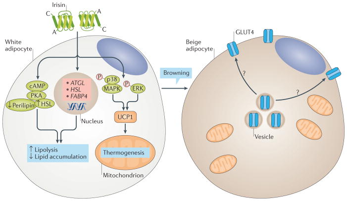
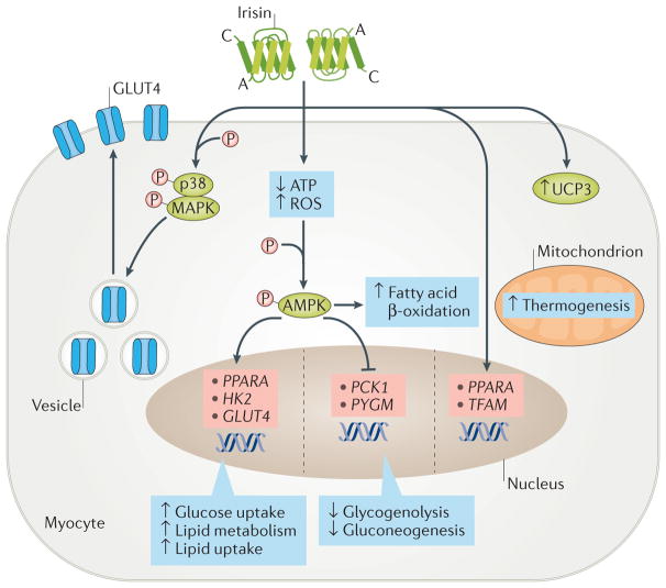
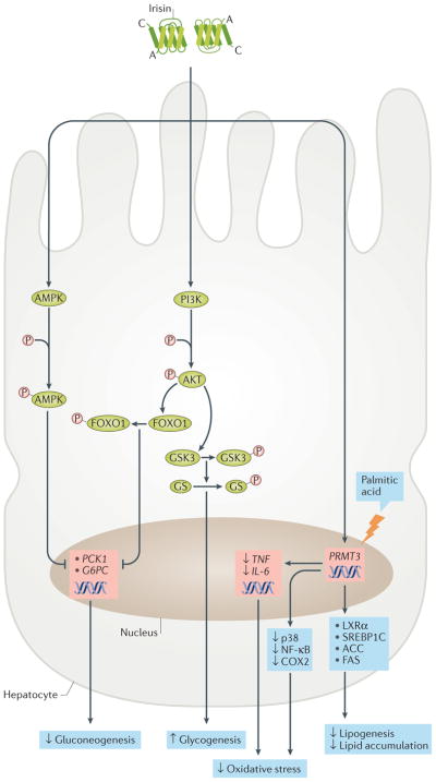
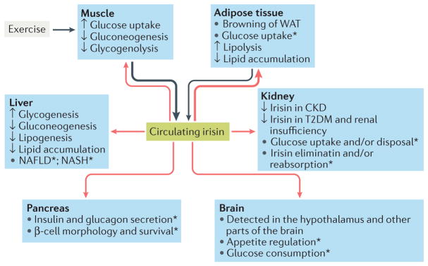
Irisin is a myokine that leads to increased energy expenditure by stimulating the 'browning' of white adipose tissue. In the first description of this hormone, increased levels of circulating irisin, which is cleaved from its precursor fibronectin type III domain-containing protein 5, were associated with improved glucose homeostasis by reducing insulin resistance. Consequently, several studies attempted to characterize the role of irisin in glucose regulation, but contradictory results have been reported, and even the existence of this hormone has been questioned. In this Review, we present the current knowledge on the physiology of irisin and its role in glucose homeostasis. We describe the mechanisms involved in the synthesis, secretion, circulation and regulation of irisin, and the controversies regarding the measurement of irisin. We also discuss the direct effects of irisin on glucose regulatory mechanisms in different organs, the indirect effects and interactions with other hormones, and the important open questions with regard to irisin in those organs. Finally, we present the results from animal interventional studies and from human clinical studies investigating the association of irisin with obesity, insulin resistance, type 2 diabetes mellitus and the metabolic syndrome.


Conflict of interest statement

Competing interests statement

The authors declare no competing interests.

Figures

Figure 1
Figure 1. Candidate signalling pathways of irisin in adipocytes
Irisin stimulates the browning of white adipose tissue (WAT) by inducing the expression of UCP1 gene and, consequently, of UCP1 protein via the p38 mitogen-activated protein kinase (MAPK) and extracellular-signal regulated kinase (ERK) pathway. Brown adipose tissue (BAT) has an increased number of mitochondria and increased energy expenditure due to elevated oxygen consumption, and accumulates smaller lipids than do white adipocytes. After cold exposure, BAT has higher glucose uptake and GLUT4 (also known as SLC2A4) expression than WAT. Irisin, which is also secreted from muscle after cold exposure, can stimulate browning; a possible involvement of irisin in glucose uptake and GLUT4 expression seems to be plausible but has not yet been investigated. Irisin also stimulates lipolysis via the cyclic AMP (cAMP)–protein kinase A (PKA)–perilipin–hormone-sensitive lipase (HSL) pathway and through the upregulation of the expression of PNPLA2 (also known as ATGL), HSL and FABP4.
Figure 2
Figure 2. Candidate signalling pathways of irisin in myocytes
Irisin can activate the AMP-activated protein kinase (AMPK) pathway by reducing intracellular ATP levels, or by increasing reactive oxygen species (ROS) or intracellular calcium concentrations. Activation of the AMPK pathway stimulates the expression of GLUT4 (also known as SLC2A4), HK2 and PPARA genes and inhibits the expression of PYGM and PCK1 (also known as PEPCKC). The high expression of GLUT4 and HK2, combined with the increased translocation of GLUT4 protein from the cytoplasm to the membrane (mainly via the p38 mitogen-activated protein kinase (MAPK) pathway), induces glucose uptake by myocytes. Conversely, inhibition of PYGM and PCK1 expression reduces glycogenolysis and gluconeogenesis. In addition, the increased expression of PPARA stimulates lipid metabolism. The irisin–AMPK pathway also increases fatty acid β-oxidation. Finally, irisin stimulates biogenesis in mitochondria by regulating the expression of PPARA and TFAM genes and of UCP3 protein.
Figure 3
Figure 3. Candidate signalling pathways of irisin in hepatocytes
Irisin reduces gluconeogenesis by downregulating the expression of PCK1 (also known as PEPCKC) and G6PC via AMP-activated protein kinase (AMPK) and the phosphoinositide 3-kinase (PI3K)–AKT–FOXO1 signalling pathway. In addition, it stimulates glycogenesis via the PI3K–AKT–glycogen synthase kinase 3 (GSK3)–glycogen synthase (GS) pathway. Irisin inhibits palmitic acid-induced lipogenesis and lipid accumulation, as well as oxidative stress, by downregulating expression of PRMT3. This leads to reduced expression of several lipogenic markers (for example, liver x receptor-α (LXRα), sterol regulatory element-binding protein 1C (SREBP1C), acetyl-CoA carboxylase (ACC) and fatty acid synthase (FAS)) and inflammatory markers (such as the genes TNF and IL6, and the proteins cyclooxygenase 2 (COX2), nuclear factor-κB (NF-κB) and p38 mitogen-activated protein kinase (MAPK)).
Figure 4
Figure 4. Effects of irisin on glucose homeostasis
Irisin is primarily secreted by muscle during exercise and secondarily by adipose tissue (black arrows). Irisin reaches different organs via the blood (red arrows), leading to changes in their handling of glucose and lipid homeostasis. The most important target of irisin is adipose tissue, where it stimulates the ‘browning’ of white adipose tissue (WAT). The effects of irisin on muscle, adipose tissue and liver favour states of normoglycaemia and normolipidaemia. *Although present in pancreas, muscle and brain, the role of irisin in these organs, as well as in kidney and liver (especially in nonalcoholic fatty liver disease (NAFLD) and nonalcoholic steatohepatitis (NASH)), has yet to be adequately investigated. CKD, chronic kidney disease; T2DM, type 2 diabetes mellitus.

Similar articles

Cited by

References

    1. Gerich JE. Physiology of glucose homeostasis. Diabetes Obes Metab. 2000;2:345–350. - PubMed
    1. Gregor MF, Hotamisligil GS. Inflammatory mechanisms in obesity. Annu Rev Immunol. 2011;29:415–445. - PubMed
    1. Shoelson SE, Lee J, Goldfine AB. Inflammation and insulin resistance. J Clin Invest. 2006;116:1793–1801. - PMC - PubMed
    1. Bostrom P, et al. A PGC1-α-dependent myokine that drives brown-fat-like development of white fat and thermogenesis. Nature. 2012;481:463–468. This is the first study to describe the existence of irisin and its role in thermogenesis in mice. - PMC - PubMed
    1. NCBI. Fibronectin type III domain-containing protein 5 precursor [Rattus norvegicus] NCBI; 2016. https://www.ncbi.nlm.nih.gov/protein/NP_001257910.1.

Publication types