. 2017 Feb 17:7:42781.
doi: 10.1038/srep42781.

Expression profiling of TRIM protein family in THP1-derived macrophages following TLR stimulation

Affiliations

Expression profiling of TRIM protein family in THP1-derived macrophages following TLR stimulation

Mei-Xiu Jiang et al. Sci Rep. .

Abstract

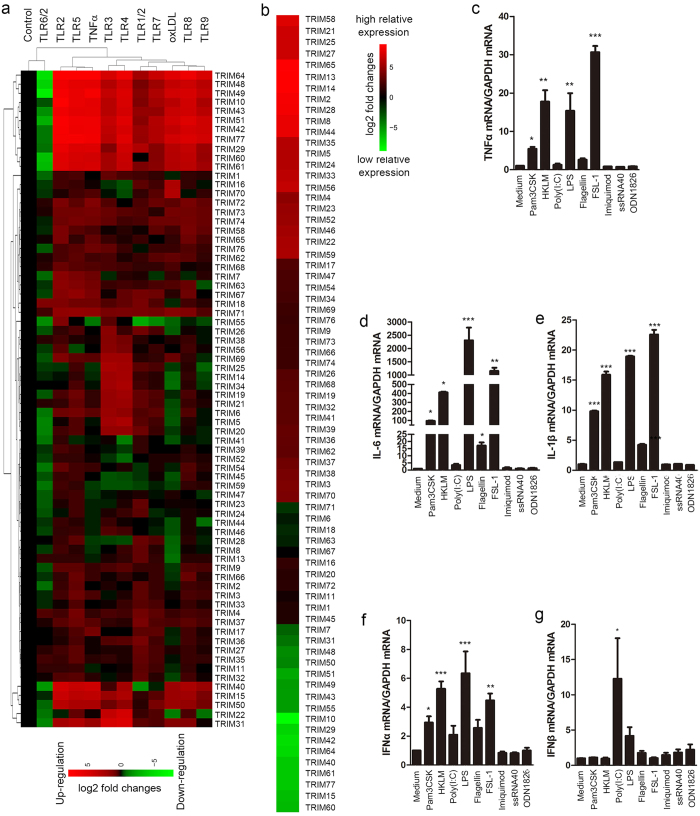
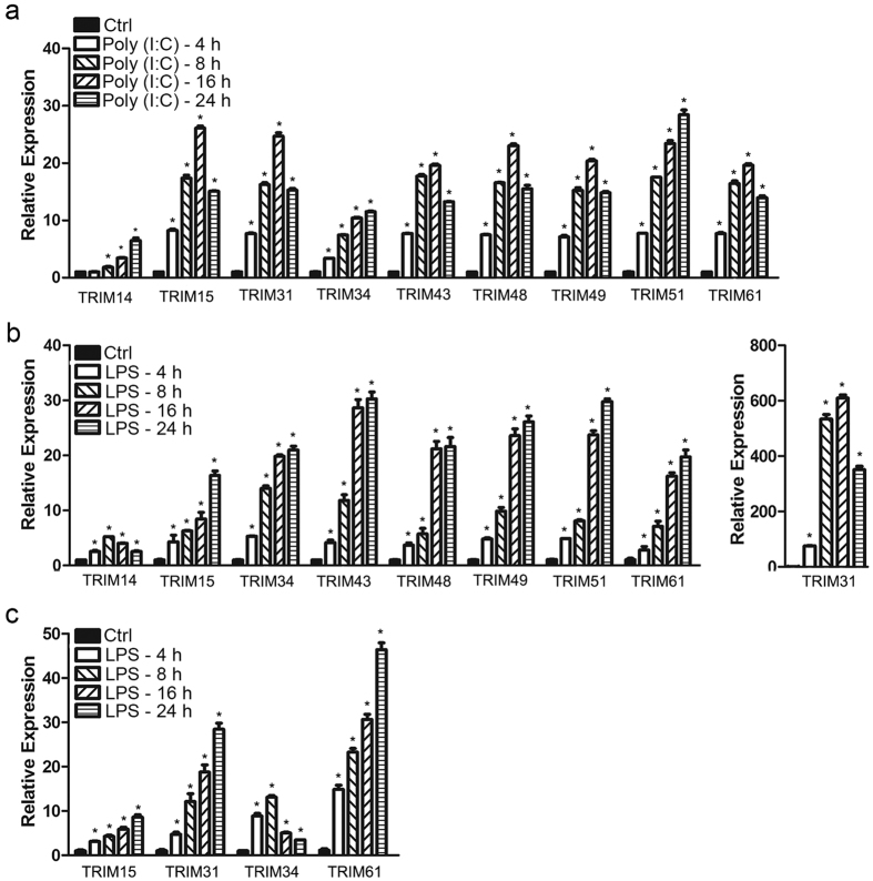
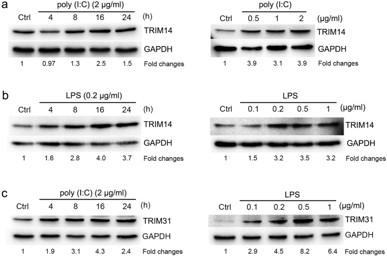
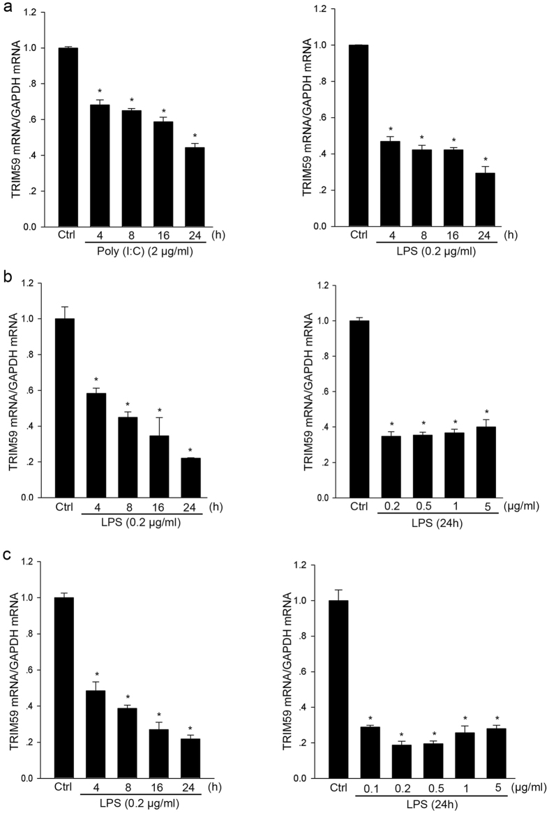
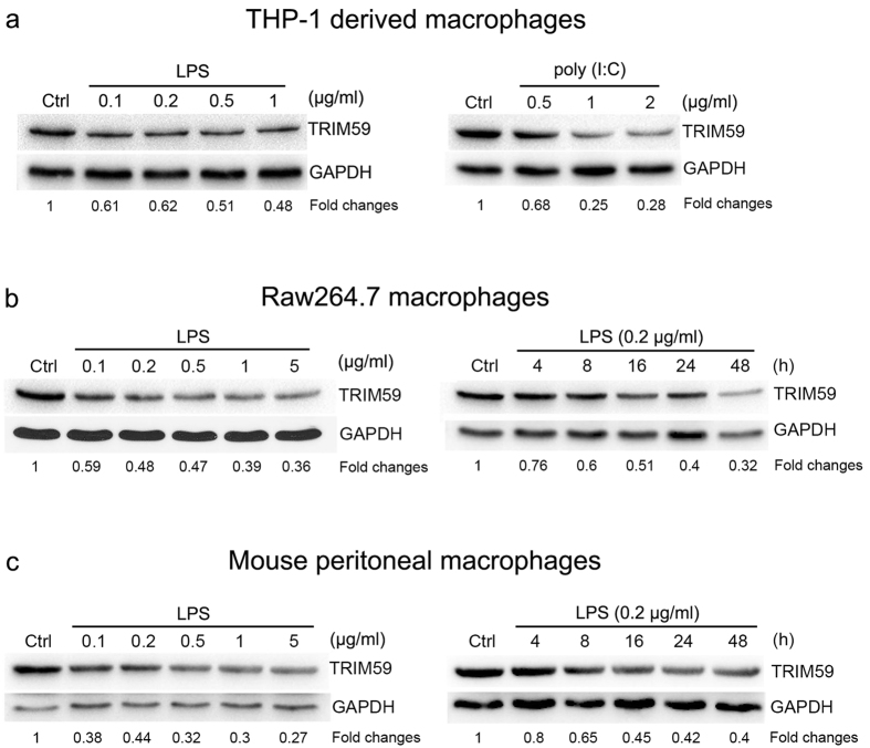
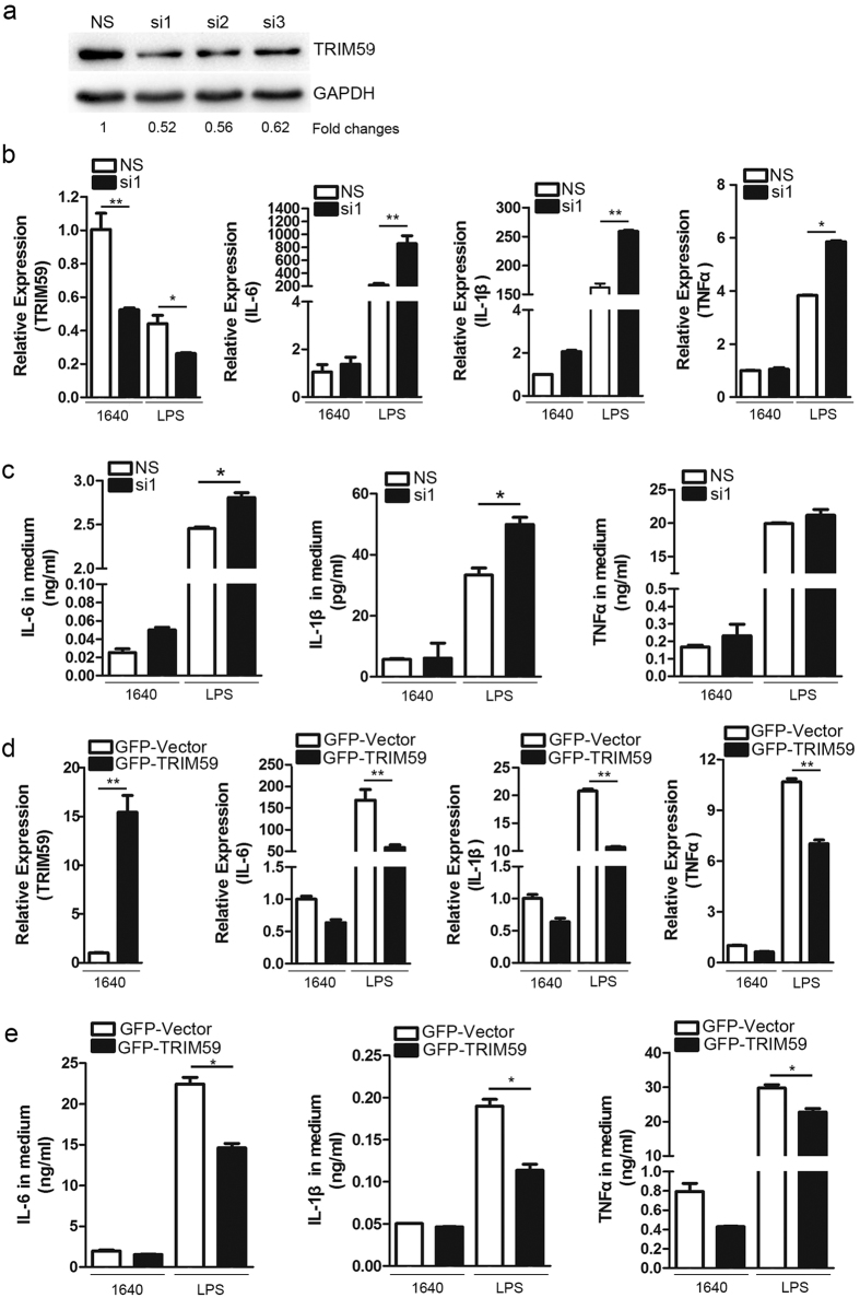
Activated macrophages play an important role in many inflammatory diseases including septic shock and atherosclerosis. However, the molecular mechanisms limiting macrophage activation are not completely understood. Members of the tripartite motif (TRIM) family have recently emerged as important players in innate immunity and antivirus. Here, we systematically analyzed mRNA expressions of representative TRIM molecules in human THP1-derived macrophages activated by different toll-like receptor (TLR) ligands. Twenty-nine TRIM members were highly induced (>3 fold) by one or more TLR ligands, among which 19 of them belong to TRIM C-IV subgroup. Besides TRIM21, TRIM22 and TRIM38 were shown to be upregulated by TLR3 and TLR4 ligands as previous reported, we identified a novel group of TRIM genes (TRIM14, 15, 31, 34, 43, 48, 49, 51 and 61) that were significantly up-regulated by TLR3 and TLR4 ligands. In contrast, the expression of TRIM59 was down-regulated by TLR3 and TLR4 ligands in both human and mouse macrophages. The alternations of the TRIM proteins were confirmed by Western blot. Finally, overexpression of TRIM59 significantly suppressed LPS-induced macrophage activation, whereas siRNA-mediated knockdown of TRIM59 enhanced LPS-induced macrophage activation. Taken together, the study provided an insight into the TLR ligands-induced expressions of TRIM family in macrophages.


Conflict of interest statement

The authors declare no competing financial interests.

Figures

Figure 1
Figure 1. Expression profiling of TRIM gene family in TLR-triggered macrophage activation.
THP1-derived macrophages were treated with the ligands for TLR1/2 (Pam3CSK, 0.2 μg/ml), TLR2 (HKLM, 2 × 108 cells/ml), TLR3 (Poly(I:C), 2 μg/ml), TLR4 (LPS, 0.2 μg/ml), TLR5 (Flagellin, 0.2 μg/ml), TLR6/2 (FSL-1, 0.2 μg/ml), TLR7 (Imiquimod, 0.2 μg/ml), TLR8 (ssRNA40, 0.2 μg/ml). TLR9 (ODN1826,1 μM), TNFα (10 ng/ml) and oxLDL (20 μg/ml) for 8 hours, respectively. The total RNAs were extracted for determining the mRNA expressions of 72 TRIM genes by qRT-PCR and normalized with GAPDH mRNA (n = 2). The relative gene log2 expression changes were analyzed by hierarchical clustering using Cluster2.11 software as described in “Methods” and presented as a heat map (a), green: Down-regulation of gene expression; black: No change; red: Up-regulation of gene expression. (b) Relative expression of TRIM genes in resting THP1-derived macrophages. Mean −ΔCt values were determined by subtracting GAPDH, and each TRIM gene was normalized to the median gene expression (TRIM67), and calculated 2−ΔΔCt. Resulting log2 values were represented as a heat map, green: low relative expression; black: median value; Red: high relative expression. The mRNA expression of TNFα, IL-6, IL-1β (c–e) and IFNα, IFNβ (f,g) were determined by qRT-PCR and normalized with GAPDH mRNA (n = 3, *p < 0.05 **p < 0.01; ***p < 0.001 vs Control).
Figure 2
Figure 2. Identification of a novel group of TRIM genes that were markedly induced by TLR3 and TLR4 ligands.
THP1-derived macrophages were treated with 2 μg/ml poly(I:C) (a) or 0.2 μg/ml LPS (b) in the indicated times; (c) Raw264.7 cells were treated with 0.2 μg/ml LPS for the indicated times. Total RNAs were extracted and the mRNA expressions of TRIM14, 15, 31, 34, 43, 48, 49, 51, 61 were determined by qRT-PCR and normalized with GAPDH mRNA. *Represents a P value < 0.01 compared with the control in the corresponding group (n = 3).
Figure 3
Figure 3. TRIM14 and TRIM31 proteins were induced by TLR3 and TLR4 ligands in dose- and time-dependent manners.
The cell lysates were extracted from THP1-derived macrophages and the TRIM14 protein (a,b) levels were determined by Western blot with stimulations of 2 μg/ml Poly(I:C) or 0.2 μg/ml LPS at the indicated times (a, left; b, left) or at the indicated concentrations for 16 hours (a, right; b, right), and the TRIM31 proteins (c) were examined with stimulation of 2 μg/ml Poly(I:C) in the indicated times (c, left) or with LPS stimulation at the indicated concentrations for 16 h (c, right). The fold changes of the protein expressions were showed at the bottom of each image and GAPDH was used as an internal control.
Figure 4
Figure 4. TRIM59 mRNA was down-regulated by TLR3 and TLR4 ligands in macrophages.
The total RNAs were extracted from various cells and the mRNA expressions of TRIM59 were determined by qRT-PCR and normalized with GAPDH mRNA. (a) THP1-derived macrophages were stimulated with 2 μg/ml Poly(I:C) (a, left) and 0.2 μg/ml LPS (a, right) in the indicated times; (b) Raw264.7 cells were treated with 0.2 μg/ml LPS in the indicated times (b, left), or with LPS at the indicated concentrations for 24 h (b, right); (c) Peritoneal macrophages isolated from male C57BL/6 mice were treated with 0.2 μg/ml LPS in the indicated times (c, left), or with LPS at the indicated concentrations for 24 h (c, right). *Represents a P value < 0.05 compared with the control in the corresponding group (n = 3).
Figure 5
Figure 5. TRIM59 protein was down-regulated by TLR3 and TLR4 ligands in macrophages.
The cell lysates were extracted from various cells and the expressions of TRIM59 proteins were determined by Western blot analysis and normalized with GAPDH protein. (a) THP1-derived macrophages were treated with 2 μg/ml Poly(I:C) (a, left) and 0.2 μg/ml LPS (a, right) in the indicated times; (b) Raw264.7 cells were treated with 0.2 μg/ml LPS in the indicated times (b, left), or with LPS at the indicated concentrations for 24 h (b, right); (c) Peritoneal macrophages isolated from male C57BL/6 mice were treated with 0.2 μg/ml LPS in the indicated times (c, left), or with LPS at the indicated concentrations for 24 h (c, right). The fold changes of the protein expression were showed at the bottom of each image and the GAPDH was used as an internal control.
Figure 6
Figure 6. LPS-induced expressions of proinflammatory cytokines were suppressed by TRIM59 overexpression or enhanced by TRIM59 knockdown in macrophages.
(a) Raw264.7 macrophages were transfected with 25 nM scrambled siRNA (NS) or TRIM59 siRNA1, 2, 3 for 2 days and the expressions of TRIM59 protein were determined by Western blot analysis and GAPDH was used as a control. Each experiment was repeated at least three times. (b) The cells were transfected with 25 nM scrambled siRNA (NS) or TRIM59 siRNA1 for 40 hours and then treated with 0.2 μg/ml LPS for 8 hours before mRNA extraction. The mRNA expressions of TRIM59 and proinflammatory cytokines including IL-6, IL-1β and TNFα were determined by qRT-PCR and normalized with GAPDH mRNA (n = 3, *p < 0.05; **p < 0.01). (c) The cells were transfected with 25 nM scrambled siRNA (NS) or TRIM59 siRNA1 for 24 hours and the cells were treated with 0.2 μg/ml LPS in serum-free medium for 24 hours. The concentrations of proinflammatory cytokines (IL-6, IL-1β and TNFα) in the medium were measured by ELISA (n = 3, *p < 0.05). (d) Peritoneal macrophages isolated from male C57BL/6 mice were transfected with 1.2 μg expression plasmid of GFP-TRIM59 and its control vector pEGFP-C2. After 40 hours, the cells were treated with 0.2 μg/ml LPS in serum-free medium for 8 hours. Total cellular RNAs were extracted and the mRNA expressions of TRIM59 and proinflammatory cytokines including IL-6, IL-1β and TNFα were determined by qRT-PCR and normalized with GAPDH mRNA (n = 3, **p < 0.01). (e) The peritoneal macrophages were transfected with 1.2 μg expression plasmid of GFP-TRIM59 and its control vector pEGFP-C2 for 24 hours and then the cells were treated with 0.2 μg/ml LPS for 24 hours, and the cytokine concentrations in the medium were measured by ELISA (n = 3, *p < 0.05).

References

    1. Barish G. D. et al.. A Nuclear Receptor Atlas: macrophage activation. Molecular endocrinology 19, 2466–2477, doi: 10.1210/me.2004-0529 (2005). - DOI - PubMed
    1. Burke B., Sumner S., Maitland N. & Lewis C. E. Macrophages in gene therapy: cellular delivery vehicles and in vivo targets. Journal of leukocyte biology 72, 417–428 (2002). - PubMed
    1. Yoneyama M. & Fujita T. Recognition of viral nucleic acids in innate immunity. Reviews in medical virology 20, 4–22, doi: 10.1002/rmv.633 (2010). - DOI - PubMed
    1. Versteeg G. A. et al.. The E3-ligase TRIM family of proteins regulates signaling pathways triggered by innate immune pattern-recognition receptors. Immunity 38, 384–398, doi: 10.1016/j.immuni.2012.11.013 (2013). - DOI - PMC - PubMed
    1. Takeuchi O. & Akira S. Pattern recognition receptors and inflammation. Cell 140, 805–820, doi: 10.1016/j.cell.2010.01.022 (2010). - DOI - PubMed

Publication types

LinkOut - more resources