. 2017 Feb 17:7:42897.
doi: 10.1038/srep42897.

In vitro and in vivo physiology of low nanomolar concentrations of Zn2+ in artificial cerebrospinal fluid

Affiliations

In vitro and in vivo physiology of low nanomolar concentrations of Zn2+ in artificial cerebrospinal fluid

Haruna Tamano et al. Sci Rep. .

Abstract

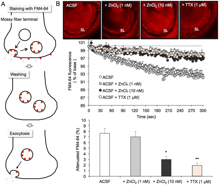
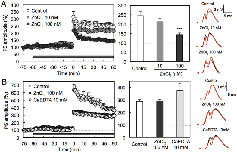
Artificial cerebrospinal fluid (ACSF), i.e., brain extracellular medium, which includes Ca2+ and Mg2+, but not other divalent cations such as Zn2+, has been used for in vitro and in vivo experiments. The present study deals with the physiological significance of extracellular Zn2+ in ACSF. Spontaneous presynaptic activity is suppressed in the stratum lucidum of brain slices from young rats bathed in ACSF containing 10 nM ZnCl2, indicating that extracellular Zn2+ modifies hippocampal presynaptic activity. To examine the in vivo action of 10 nM ZnCl2 on long-term potentiation (LTP), the recording region was perfused using a recording electrode attached to a microdialysis probe. The magnitude of LTP was not modified in young rats by perfusion with ACSF containing 10 nM ZnCl2, compared to perfusion with ACSF without Zn2+, but attenuated by perfusion with ACSF containing 100 nM ZnCl2. Interestingly, the magnitude of LTP was not modified in aged rats even by perfusion with ACSF containing 100 nM ZnCl2, but enhanced by perfusion with ACSF containing 10 mM CaEDTA, an extracellular Zn2+ chelator. The present study indicates that the basal levels of extracellular Zn2+, which are in the range of low nanomolar concentrations, are critical for synaptic activity and perhaps increased age-dependently.


Conflict of interest statement

The authors declare no competing financial interests.

Figures

Figure 1
Figure 1. Suppression of spontaneous mossy fiber exocytosis of young rats in the presence of 10 nM ZnCl2.
(A) Schematic illustration of mossy fiber exocytosis assessed by attenuation of FM4-64 fluorescence. (B) FM4-64 fluorescence images in the CA3 after loading FM4-64 into slices bathed in ACSF, ACSF containing 1 nM Zn2+, and ACSF containing 10 nM Zn2+, and FM4-64 fluorescence image in the CA3 after loading of FM4-64 into slices bathed in ACSF and transferring the slices to ACSF containing 1 μM TTX (time 0 sec) (upper). Each point and line (mean ± SEM) represents the changes in FM4-64 fluorescence in the stratum lucidum (SL) of brain slices bathed in ACSF (n = 17), 1 nM ZnCl2 (n = 5), 10 nM ZnCl2 (n = 8) and 1 μM TTX (n = 6) after loading FM4-64 (time 0), which is expressed as 100%. (n = 6) (middle). Each bar and line (the mean ± SEM) represents the rate of the decreased FM4-64 fluorescence at time 300 sec (lower). *p < 0.05; **p < 0.01, vs. ACSF (Tukey’s test).
Figure 2
Figure 2. No effect of 10 nM CuCl2 and 10 nM FeCl3 on spontaneous mossy fiber exocytosis of young rats.
FM4-64 fluorescence images in the CA3 after loading FM4-64 into slices bathed in ACSF and ACSF containing 10 nM Cu2+ or ACSF containing 10 nM Fe3+ (time 0 sec) (upper). Each point and line (mean ± SEM) represents the changes in FM4-64 fluorescence in the stratum lucidum (SL) of brain slices bathed in ACSF (n = 10), 10 nM CuCl2 (n = 6), and 10 nM FeCl3 (n = 6) after loading FM4-64 (time 0), which is expressed as 100%. (n = 6) (lower).
Figure 3
Figure 3. Intracellular Ca2+ imaging in the hippocampus of young rats with calcium orange AM.
Intracellular calcium orange fluorescence was imaged to estimate the basal levels of cytosolic Ca2+ in the hippocampus of brain slices bathed in ACSF (n = 7) and 10 nM ZnCl2 in ACSF (n = 7). SR, stratum radiatum; SL, stratum lucidum; DML. dentate molecular layer (left). Each bar and line (the mean ± SEM) represents fluorescent intensity in the SR, SL, and DML.
Figure 4
Figure 4. Action of extracellular Zn2+ and extracellular Zn2+ chelator in dentate gyrus LTP of young and aged rats.
(A) Recording region was perfused with ACSF in young rats and then 10 nM (n = 5) and 100 nM (n = 4) ZnCl2 in ACSF as shown by the shaded bar and HFS (10 trains of 20 pulses at 200 Hz separated by 1 s) was delivered at time 0 min (left). Each bar and line (mean ± SEM) represents the averaged PS amplitude of the last 10 min (time 50–60 min) (middle). ***p < 0.001, vs. control (ACSF) (n = 10) (Tukey’s test). Representative fEPSP recordings at time −70 (black line), −30 (grey line) and 50–60 min (red line) are shown (right). (B) Recording region was perfused with ACSF in aged rats and then 100 nM (n = 18) and 10 mM CaEDTA (n = 7) ZnCl2 in ACSF as shown by the shaded bar and HFS (10 trains of 20 pulses at 200 Hz separated by 1 s) was delivered at time 0 min (left). Each bar and line (mean ± SEM) represents the averaged PS amplitude of the last 10 min (time 50–60 min) (middle). *p < 0.05, vs. control (ACSF) (n = 26) (Tukey’s test). Representative fEPSP recordings at time −70 (black line), −30 (grey line) and 50–60 min (red line) are shown (right).

References

    1. Kim N. K. & Robinson H. P. Effects of divalent cations on slow unblock of native NMDA receptors in mouse neocortical pyramidal neurons. Eur. J. Neurosci. 34, 199–212 (2011). - PubMed
    1. Jones H. C. & Keep R. F. Brain fluid calcium concentration and response to acute hypercalcaemia during development in the rat. J. Physiol. 402, 579–593 (1988). - PMC - PubMed
    1. Neves G., Cooke S. F. & Bliss T. V. P. Synaptic plasticity, memory and the hippocampus: A neural network approach to causality. Nat. Rev. Neurosci. 9, 65–67 (2008). - PubMed
    1. Lisman J., Yasuda R. & Raghavachari S. Mechanisms of CaMKII action in long-term potentiation. Nat. Rev. Neurosci. 13, 169–182 (2012). - PMC - PubMed
    1. Alberdi E. et al.. Amyloid beta oligomers induce Ca2+ dysregulation and neuronal death through activation of ionotropic glutamate receptors. Cell Calcium 47, 264–272 (2010). - PubMed

LinkOut - more resources