. 2017 Feb 17;12(2):e0172125.
doi: 10.1371/journal.pone.0172125. eCollection 2017.

The p53 isoform delta133p53ß regulates cancer cell apoptosis in a RhoB-dependent manner

Affiliations

The p53 isoform delta133p53ß regulates cancer cell apoptosis in a RhoB-dependent manner

Nikola Arsic et al. PLoS One. .

Erratum in

Abstract

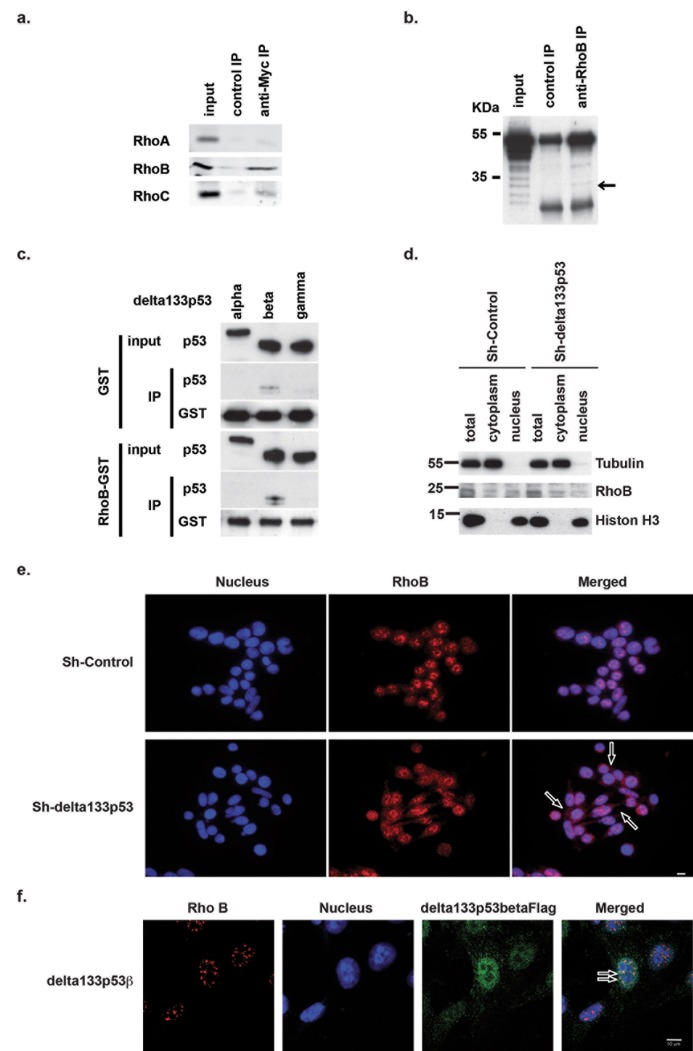
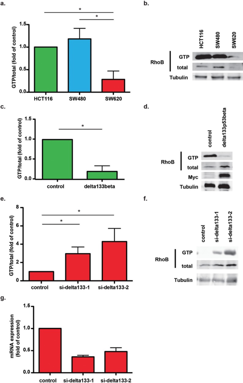
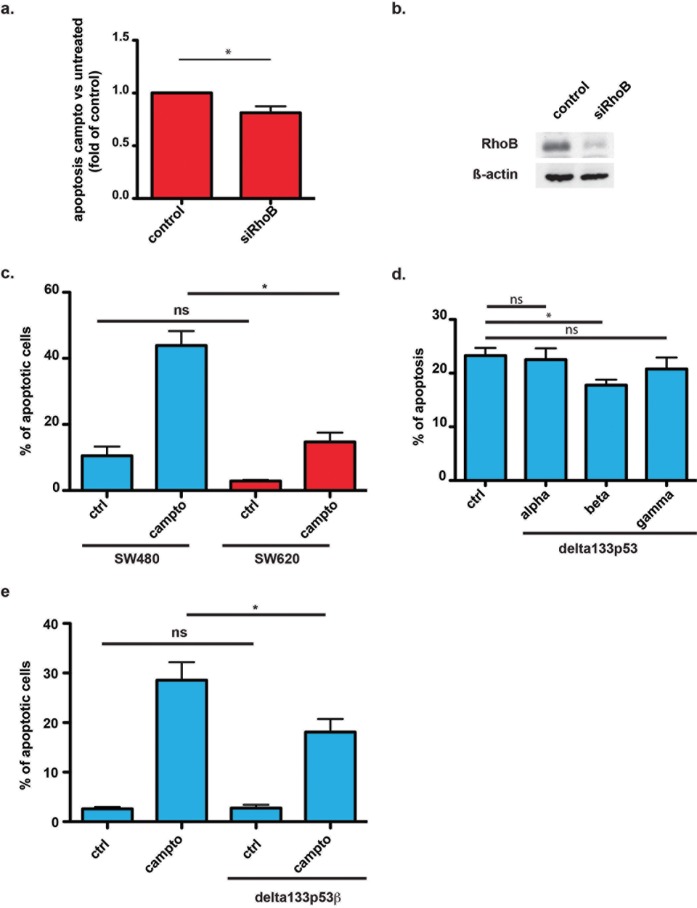
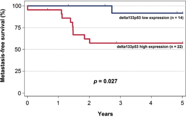
The TP53 gene plays essential roles in cancer. Conventionally, wild type (WT) p53 is thought to prevent cancer development and metastasis formation, while mutant p53 has transforming abilities. However, clinical studies failed to establish p53 mutation status as an unequivocal predictive or prognostic factor of cancer progression. The recent discovery of p53 isoforms that can differentially regulate cell cycle arrest and apoptosis suggests that their expression, rather than p53 mutations, could be a more clinically relevant biomarker in patients with cancer. In this study, we show that the p53 isoform delta133p53ß is involved in regulating the apoptotic response in colorectal cancer cell lines. We first demonstrate delta133p53ß association with the small GTPase RhoB, a well-described anti-apoptotic protein. We then show that, by inhibiting RhoB activity, delta133p53ß protects cells from camptothecin-induced apoptosis. Moreover, we found that high delta133p53 mRNA expression levels are correlated with higher risk of recurrence in a series of patients with locally advanced rectal cancer (n = 36). Our findings describe how a WT TP53 isoform can act as an oncogene and add a new layer to the already complex p53 signaling network.


Conflict of interest statement

Competing Interests: The authors have declared that no competing interests exist.

Figures

Fig 1
Fig 1. Delta133p53ß physically interacts with RhoB.
(a) Immunoblot analysis showing the specific co-immunoprecipitation of MYC-tagged delta133p53ß and endogenous RhoB or RhoC, to a lower extent, but not RhoA. (b) Immunoblot analysis showing the co-immunoprecipitation of endogenous RhoB and delta133p53ß with anti-RhoB antibodies. Arrow shows co-immunoprecipitation of delta133p53ß isoform. (c) In-vitro binding assay showing the direct interaction between recombinant RhoB-GST fusion protein and delta133p53ß, but not α or γ. (d) Cellular fractionation showing RhoB cytoplasmic re-localization in SW620 cells transfected with shRNAS against delta133p53 compared with shControl (mock-transfected cells). (e) Immunofluorescence analysis of RhoB localization in control SW620 cells (shControl) or after transfection with shdelta133p53. Arrows indicate examples of RhoB cytoplasmic localization. Scale bar: 10μm. (f) Confocal images showing the co-localization of RhoB and delta133p53ß in the nucleus of SW480 cells that overexpress delta133p53ß. Arrows indicate examples co-localization of RhoB and delta133p53ß in the nucleus. Scale bar: 10μm.
Fig 2
Fig 2. Delta133p53ß controls RhoB activity in CRC cell lines.
(a) RhoB activity in HCT116, SW480 and SW620 cells was assessed by using GTPase activity assays. Results are expressed as the fold change compared with HCT116 cells and represent the mean ± SEM of four independent experiments; *, p<0.05. (b) Representative immunoblot showing levels of total and GTP-bound RhoB in HCT116, SW480 and SW620 cells. (c) RhoB activity in mock-transfected (control) and delta133p53ß-overexpressing HCT116 cells. Results are expressed as the fold change compared with control and represent the mean ± SEM of three independent experiments; *, p<0.05. (d) Representative immunoblot showing MYC-tagged delta133p53ß expression in delta133p53ß-overexpressing and mock-transfected (control) HCT116 cells. (e) RhoB activity in mock-transfected (control) SW620 cells and following transfection with two different siRNAs against delta133p53. Results are expressed as the fold change compared with control cells and represent the mean ± SEM of four independent experiments; *, p<0.05. (f) Representative immunoblot showing the levels of total and GTP-bound RhoB in control and delta133p53-depleted SW620 cells. (g) Depletion of delta133p53 isoforms in SW620 cells after siRNA transfection was assessed by RT-quantitative PCR. Results are expressed as the fold change compared with mock-transfected SW620 cells (control) and represent the mean ± SEM of four independent experiments.
Fig 3
Fig 3. Delta133p53ß protects cancer cells from camptothecin-induced apoptosis.
(a) Apoptosis assay showing the sensitivity to 5μM camptothecin of mock-transfected (control) SW620 cells and after transfection with siRNAs against RhoB. Results are expressed as the apoptosis ratio of untreated versus camptothecin-treated cells and represent the mean ± SEM of four independent experiments; *, p<0.05. (b) Immunoblot showing RhoB downregulation in siRhoB-transfected SW620 cells compared with mock-transfected cells (control). (c) Apoptosis assay to test the sensitivity of SW480 and SW620 cells to 5μM camptothecin. Results are expressed as the percentage of apoptotic cells relative to all Hoechst-positive cells and represent the mean ± SEM of three independent experiments; *, p<0.05. (d) Apoptosis assay showing the sensitivity to 5μM camptothecin of mock-transfected (control) and SW480 cells that stably express delta133p53α, ß or γ. Results are expressed as the percentage of apoptotic cells relative to all Hoechst-positive cells and represent the mean ± SEM of three independent experiments; *, p<0.05. (e) Apoptosis assay showing the sensitivity to 5μM camptothecin of mock-transfected and SW480 cells that stably express delta133p53ß. Results are expressed as the percentage of apoptotic cells relative to all Hoechst-positive cells and represent the mean ± SEM of four independent experiments; *, p<0.05.
Fig 4
Fig 4. Kaplan–Meier curves of metastasis-free survival in the cohort of 36 patients with rectal cancer stratified according to delta133p53 expression (low/high).

References

    1. Vousden KH, Prives C (2009) Blinded by the Light: The Growing Complexity of p53. Cell 137: 413–431. 10.1016/j.cell.2009.04.037 - DOI - PubMed
    1. Gottlieb TM, Oren M (1998) p53 and apoptosis. Seminars in cancer biology 8: 359–368. - PubMed
    1. Symonds H, Krall L, Remington L, Saenz-Robles M, Lowe S, Jacks T, et al. (1994) p53-dependent apoptosis suppresses tumor growth and progression in vivo. Cell 78: 703–711. - PubMed
    1. Polyak K, Xia Y, Zweier JL, Kinzler KW, Vogelstein B (1997) A model for p53-induced apoptosis. Nature 389: 300–305. 10.1038/38525 - DOI - PubMed
    1. Soengas MS, Alarcon RM, Yoshida H, Giaccia AJ, Hakem R, Mak TW, et al. (1999) Apaf-1 and caspase-9 in p53-dependent apoptosis and tumor inhibition. Science 284: 156–159. - PubMed

MeSH terms