. 2017 Feb 17;13(2):e1006216.
doi: 10.1371/journal.ppat.1006216. eCollection 2017 Feb.

SUMO modification of a heterochromatin histone demethylase JMJD2A enables viral gene transactivation and viral replication

Affiliations

SUMO modification of a heterochromatin histone demethylase JMJD2A enables viral gene transactivation and viral replication

Wan-Shan Yang et al. PLoS Pathog. .

Abstract

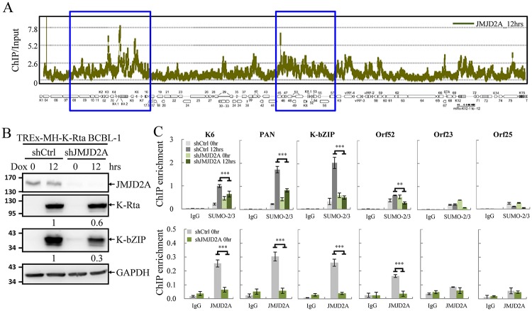
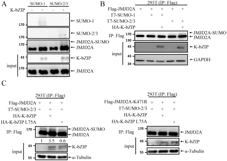
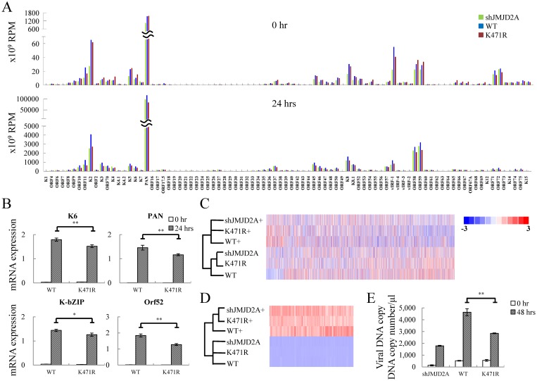
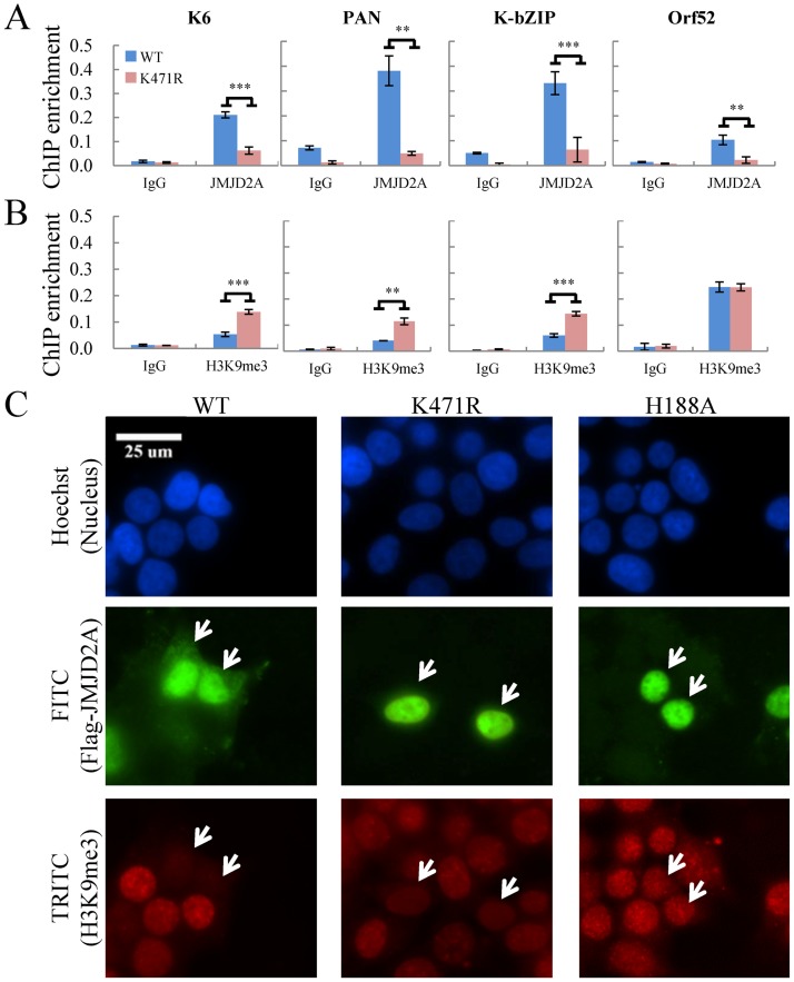
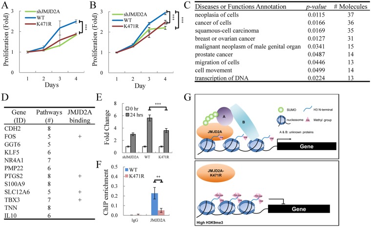
Small ubiquitin-like modifier (SUMO) modification of chromatin has profound effects on transcription regulation. By using Kaposi's sarcoma associated herpesvirus (KSHV) as a model, we recently demonstrated that epigenetic modification of viral chromatin by SUMO-2/3 is involved in regulating gene expression and viral reactivation. However, how this modification orchestrates transcription reprogramming through targeting histone modifying enzymes remains largely unknown. Here we show that JMJD2A, the first identified Jumonji C domain-containing histone demethylase, is the histone demethylase responsible for SUMO-2/3 enrichment on the KSHV genome during viral reactivation. Using in vitro and in vivo SUMOylation assays, we found that JMJD2A is SUMOylated on lysine 471 by KSHV K-bZIP, a viral SUMO-2/3-specific E3 ligase, in a SUMO-interacting motif (SIM)-dependent manner. SUMOylation is required for stabilizing chromatin association and gene transactivation by JMJD2A. These finding suggest that SUMO-2/3 modification plays an essential role in the epigenetic regulatory function of JMJD2A. Consistently, hierarchical clustering analysis of RNA-seq data showed that a SUMO-deficient mutant of JMJD2A was more closely related to JMJD2A knockdown than to wild-type. Our previous report demonstrated that JMJD2A coated and maintained the "ready to activate" status of the viral genome. Consistent with our previous report, a SUMO-deficient mutant of JMJD2A reduced viral gene expression and virion production. Importantly, JMJD2A has been implicated as an oncogene in various cancers by regulating proliferation. We therefore further analyzed the role of SUMO modification of JMJD2A in regulating cell proliferation. Interestingly, the SUMO-deficient mutant of JMJD2A failed to rescue the proliferation defect of JMJD2A knockdown cells. Emerging specific inhibitors of JMJD2A have been generated for evaluation in cancer studies. Our results revealed that SUMO conjugation mediates an epigenetic regulatory function of JMJD2A and suggests that inhibiting JMJD2A SUMOylation may be a novel avenue for anti-cancer therapy.


Conflict of interest statement

The authors have declared that no competing interests exist.

Figures

Fig 1
Fig 1. JMJD2A is required for efficient SUMO-2/3 enrichment on the viral genome during KSHV reactivation.
(A) ChIP-on-chip analysis of JMJD2A binding across the KSHV lytic genome. A JMJD2A ChIP assay was performed on TREx-MH-K-Rta BCBL-1 cells treated with 0.2 μg/ml doxycycline (Dox) for 12 hours (hrs). The genomic locations of KSHV ORFs are depicted below the histogram. The blue rectangular areas are locations that comprise high SUMO-2/3 levels after KSHV reactivation (Yang et al. 2015). (B) Total cell lysates (TCLs) from TREx-MH-K-Rta-shCtrl and -shJMJD2A BCBL-1 cells before and after Dox treatment (12 hrs) were immunoblotted with antibodies as indicated. Ratio is the relative signal of K-Rta or K-bZIP to GAPDH observed for Dox treatment at 12 hrs using TREx-MH-K-Rta-shCtrl BCBL-1 cells set as 1.0. (C) ChIP was performed with chromatin prepared from cells treated as described in (B) using rabbit IgG, anti-SUMO-2/3 (upper panel) and anti-JMJD2A (lower panel) antibodies. ChIP DNA was quantified by real-time quantitative PCR (qPCR) using primer pairs specific for promoter regions of KSHV K6, PAN, K-bZIP, Orf52, Orf23 and Orf25. (Data represent mean±SEM. n = 3. **p<0.01. ***p<0.005).
Fig 2
Fig 2. JMJD2A is SUMOylated at K471.
(A) Schematic representation of the domain structure of JMJD2A and the three putative SUMOylation sites. (B) in vitro SUMOylation assay using recombinant Flag-JMJD2A and SUMO isoforms as substrates. (C) in vivo SUMOylation assay was performed by transfecting 293T cells with plasmids expressing Flag-JMJD2A (0.4 μg) and T7-SUMO-1 or T7-SUMO-2 and -3 (1.2 μg). JMJD2A was immunoprecipitated (IP’d) with anti-Flag-M2 beads and analyzed by immunoblotting using anti-SUMO-1, anti-SUMO-2/3 and anti-JMJD2A antibodies (left panel). TCLs were immunoblotted with anti-SUMO-1, anti-SUMO-2/3 antibodies and anti-α-Tubulin (right panel). (D) Purified wild-type (WT) and different combinations of SUMOylation site mutants of Flag-JMJD2A were used for in vitro SUMOylation assay as described in (B). 4%-20% gradient SDS-PAGE was used to resolve JMJD2A and SUMOylated JMJD2A. (E) Flag-JMJD2A-WT or -K471R was expressed in 293T cells as described in (C) and IP’d using M2 beads. SUMOylated JMJD2A was analyzed with anti-JMJD2A antibody.
Fig 3
Fig 3. KSHV SUMO E3 ligase K-bZIP catalyses SUMO modification of JMJD2A.
(A) in vitro SUMOylation assay of JMJD2A was performed with the indicated combination of SUMO isoforms and K-bZIP. JMJD2A and SUMOylated JMJD2A were resolved by 4%-20% gradient SDS-PAGE and immunoblotted using anti-SUMO-1, anti-SUMO-2/3 and anti-JMJD2A antibodies. Input of JMJD2A and K-bZIP was detected by immunoblotting using specific antibodies as indicated. (B) in vivo SUMOylation assay was performed as described in Fig 2C, with co-transfection of Flag-JMJD2A (0.4 μg), T7-SUMO-1 or T7-SUMO-2 and -3 (0.1 μg), and WT K-bZIP (0.1 μg). 4%-20% gradient SDS-PAGE was used to resolve JMJD2A and SUMOylated JMJD2A. Immunoblotting was performed using anti-JMJD2A, anti-K-bZIP and anti-GAPDH antibodies. (C) Flag-K-bZIP-WT or -L75A were co-expressed in 293T cells with Flag-JMJD2A and T7-SUMO-2 and -3 as described in (B). SUMOylated JMJD2A and input proteins were detected by immunoblotting using specific antibodies as indicated. Ratio is the relative signal of JMJD2A-SUMO to total JMJD2A observed using signal in lane 1 set as 1.0.
Fig 4
Fig 4. JMJD2A SUMOylation is crucial for SUMO-2/3 enrichment on the KSHV genome during viral reactivation.
(A) TCLs from TREx-MH-K-Rta-shJMJD2A-Flag-JMJD2A-WT and -K471R BCBL-1 cells before and after 12 hrs Dox (0.2 μg/ml) treatment were immunoblotted with antibodies as indicated. Ratio is the relative signal of K-Rta or K-bZIP to α-Tubulin observed for Dox treatment at 24 hrs using TREx-MH-K-Rta-shJMJD2A-Flag-JMJD2A-WT BCBL-1 cells as 1.0. (B) ChIP-seq assay was performed with chromatin from cells treated as described in (A) using rabbit IgG and anti-SUMO-2/3 antibodies. Sequenced reads mapped to KSHV genome were normalized with total reads from the human genome. The histogram shows reads per million mapped reads (RPM) mapped across the KHSV genome (JMJD2A-WT, blue; -K471R, red). p = 2.27e-103 by Student’s-t test. The blue rectangles are high SUMO-2/3 regions as in Fig 1A. (C) ChIP-seq data was verified by real-time qPCR using primer pairs specific for promoter regions of KSHV K6, PAN, K-bZIP, Orf52, Orf23 and Orf25. (Data represent mean±SEM. n = 3. **p<0.01. ***p<0.005).
Fig 5
Fig 5. JMJD2A SUMOylation plays an essential role in KSHV viral gene transactivation and viral reactivation.
(A) RNA-seq was performed using total RNA from non-induced (0 hrs, upper panel) and 0.2 μg/ml Dox treated (24 hrs, lower panel) TREx-MH-K-Rta-shJMJD2A (green bars), -shJMJD2A-Flag-JMJD2A-WT (blue bars) and -K471R (red bars) BCBL-1 cells. One representative RNA-seq expression dataset of KSHV genes is presented as reads per million (RPM) mapped. (B) RT-qPCR verification of KSHV K6, PAN, K-bZIP and Orf52 expression in TREx-MH-K-Rta-shJMJD2A-Flag-JMJD2A-WT and -K471R BCBL-1 cells. (C and D) Heat map depicts hierarchical clustering of RNA-seq data of cellular (C) and KSHV (D) gene expression. (+, plus Dox 24h). (E) Supernatants from TREx-MH-K-Rta-shJMJD2A, -shJMJD2A-Flag-JMJD2A-WT and -K471R BCBL-1 cells treated as described in (A) for 48 hrs were collected, filtered, and the viral titers were determined by analyzing the virion-associated DNA levels using TaqMan qPCR. (Data represent mean±SEM. n = 3. **p<0.01.)
Fig 6
Fig 6. SUMOylation modulates the occupancy and demethylase activity of JMJD2A at target genes.
(A and B) ChIP was performed with chromatin prepared from TREx-MH-K-Rta-shJMJD2A-Flag-JMJD2A-WT and -K471R BCBL-1 cells using rabbit IgG, anti-JMJD2A (A) and anti-H3K9me3 (B) antibodies. ChIP DNA was quantified as described in Fig 1C. (C) Histone demethylase activity of Flag-tagged JMJD2A-WT, -K471R or -H188A protein in 293T cells was assessed by immunofluorescence staining (IF) using deconvolution fluorescence microscopy. Cells were fixed, stained with antibody specific for Flag (FITC) and H3K9me3 (TRITC), and mounted in Slow Fade Gold with DAPI. White arrows indicate cells transfected with Flag-JMJD2A-WT or its mutant.
Fig 7
Fig 7. JMJD2A SUMOylation affects the expression of cellular genes involved in cancer.
(A and B) WT JMJD2A but not its K471R mutant rescue proliferation of JMJD2A knockdown SLK (A) and BCBL-1 (B) cells. (A) SLK cells were sequentially infected with lentivirus expressing JMJD2A shRNA and Flag-tagged WT or K471R of JMJD2A. Cell proliferation was assessed by MTT assay. (B) Proliferation of TREx-MH-K-Rta-shJMJD2A, shJMJD2A-Flag-JMJD2A-WT and -K471R BCBL-1 cell lines was assessed by MTT assay. (C) Gene function analysis of cellular genes less upregulated in SUMOylation-deficient JMJD2A mutant (K471R) compared to JMJD2A-WT 24 hrs after KSHV reactivation. Gene list was shown in S7 Table. (D) Genes present in more than five identified functional pathways in (C). The plus (+) indicates genes with JMJD2A binding on the promoter region (transcription start site (TSS) ± 500bp). Gene list was shown in S8 Table. (E) RT-qPCR verification of TBX3 expression in TREx-MH-K-Rta-shJMJD2A, -shJMJD2A-Flag-JMJD2A-WT and -K471R BCBL-1 cells. (F) qPCR was performed with ChIP DNA from Fig 6A using primer pairs specific for promoter region of TBX3 (G) Schematic diagram illustrates the SUMO modification of JMJD2A in regulation gene transcription during KSHV reactivation.

References

    1. Shi Y, Lan F, Matson C, Mulligan P, Whetstine JR, et al. (2004) Histone demethylation mediated by the nuclear amine oxidase homolog LSD1. Cell 119: 941–953. 10.1016/j.cell.2004.12.012 - DOI - PubMed
    1. Klose RJ, Yamane K, Bae Y, Zhang D, Erdjument-Bromage H, et al. (2006) The transcriptional repressor JHDM3A demethylates trimethyl histone H3 lysine 9 and lysine 36. Nature 442: 312–316. 10.1038/nature04853 - DOI - PubMed
    1. Couture JF, Collazo E, Ortiz-Tello PA, Brunzelle JS, Trievel RC (2007) Specificity and mechanism of JMJD2A, a trimethyllysine-specific histone demethylase. Nat Struct Mol Biol 14: 689–695. 10.1038/nsmb1273 - DOI - PubMed
    1. Huang Y, Fang J, Bedford MT, Zhang Y, Xu RM (2006) Recognition of histone H3 lysine-4 methylation by the double tudor domain of JMJD2A. Science 312: 748–751. 10.1126/science.1125162 - DOI - PubMed
    1. Lee J, Thompson JR, Botuyan MV, Mer G (2008) Distinct binding modes specify the recognition of methylated histones H3K4 and H4K20 by JMJD2A-tudor. Nat Struct Mol Biol 15: 109–111. 10.1038/nsmb1326 - DOI - PMC - PubMed

Publication types

MeSH terms

Substances

LinkOut - more resources