

The combined action of mast cell chymase, tryptase and carboxypeptidase A3 protects against melanoma colonization of the lung
- PMID: 28212574
- PMCID: PMC5421910
- DOI: 10.18632/oncotarget.15339
The combined action of mast cell chymase, tryptase and carboxypeptidase A3 protects against melanoma colonization of the lung
Abstract
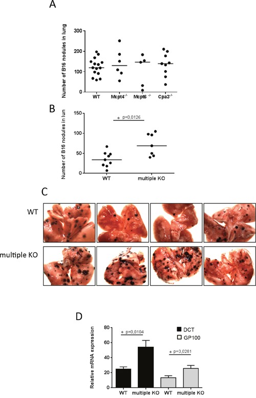
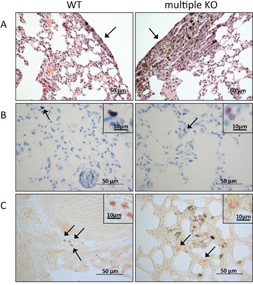
Mast cell secretory granules are densely packed with various bioactive mediators including proteases of chymase, tryptase and CPA3 type. Previous studies have indicated that mast cells can affect the outcome of melanoma but the contribution of the mast cell granule proteases to such effects has not been clear. Here we addressed this issue by assessing mice lacking either the chymase Mcpt4, the tryptase Mcpt6 or carboxypeptidase A3 (Cpa3), as well as mice simultaneously lacking all three proteases, in a model of melanoma dissemination from blood to the lung. Although mice with individual deficiency in the respective proteases did not differ significantly from wildtype mice in the extent of melanoma colonization, mice with multiple protease deficiency (Mcpt4/Mcpt6/Cpa3-deficient) exhibited a higher extent of melanoma colonization in lungs as compared to wildtype animals. This was supported by higher expression of melanoma-specific genes in lungs of Mcpt4/Mcpt6/CPA3-deficient vs. wildtype mice. Cytokine profiling showed that the levels of CXCL16, a chemokine with effects on T cell populations and NKT cells, were significantly lower in lungs of Mcpt4/Mcpt6/Cpa3-deficient animals vs. controls, suggesting that multiple mast cell protease deficiency might affect T cell or NKT cell populations. In line with this, we found that the Mcpt4/Mcpt6/Cpa3-deficiency was associated with a reduction in cells expressing CD1d, a MHC class 1-like molecule that is crucial for presenting antigen to invariant NKT (iNKT) cells. Together, these findings indicate a protective role of mast cell-specific proteases in melanoma dissemination, and suggest that this effect involves a CXCL16/CD1d/NKT cell axis.
Keywords: CD1d; carboxypeptidase A3; chymase; mast cells; tryptase.
Conflict of interest statement
The authors declare no conflicts of interest.
Figures


Similar articles
-
Mouse connective tissue mast cell proteases tryptase and carboxypeptidase A3 play protective roles in itch induced by endothelin-1.J Neuroinflammation. 2020 Apr 22;17(1):123. doi: 10.1186/s12974-020-01795-4. J Neuroinflammation. 2020. PMID: 32321525 Free PMC article.
-
Accumulation of intraepithelial mast cells with a unique protease phenotype in T(H)2-high asthma.J Allergy Clin Immunol. 2010 May;125(5):1046-1053.e8. doi: 10.1016/j.jaci.2010.03.003. J Allergy Clin Immunol. 2010. PMID: 20451039 Free PMC article.
-
Human mast cells arise from a common circulating progenitor.J Allergy Clin Immunol. 2013 Aug;132(2):463-9.e3. doi: 10.1016/j.jaci.2013.02.011. Epub 2013 Apr 9. J Allergy Clin Immunol. 2013. PMID: 23582567
-
The Role of Mast Cell Specific Chymases and Tryptases in Tumor Angiogenesis.Biomed Res Int. 2015;2015:142359. doi: 10.1155/2015/142359. Epub 2015 Jun 4. Biomed Res Int. 2015. PMID: 26146612 Free PMC article. Review.
-
The emerging role of mast cell proteases in asthma.Eur Respir J. 2019 Oct 31;54(4):1900685. doi: 10.1183/13993003.00685-2019. Print 2019 Oct. Eur Respir J. 2019. PMID: 31371445 Review.
Cited by
-
Carboxypeptidase A3-A Key Component of the Protease Phenotype of Mast Cells.Cells. 2022 Feb 6;11(3):570. doi: 10.3390/cells11030570. Cells. 2022. PMID: 35159379 Free PMC article. Review.
-
Exosome-mediated uptake of mast cell tryptase into the nucleus of melanoma cells: a novel axis for regulating tumor cell proliferation and gene expression.Cell Death Dis. 2019 Sep 10;10(9):659. doi: 10.1038/s41419-019-1879-4. Cell Death Dis. 2019. PMID: 31506436 Free PMC article.
-
Protective role of mouse mast cell tryptase Mcpt6 in melanoma.Pigment Cell Melanoma Res. 2020 Jul;33(4):579-590. doi: 10.1111/pcmr.12859. Epub 2020 Jan 19. Pigment Cell Melanoma Res. 2020. PMID: 31894627 Free PMC article.
-
Harnessing the Anti-Tumor Mediators in Mast Cells as a New Strategy for Adoptive Cell Transfer for Cancer.Front Oncol. 2022 Mar 31;12:830199. doi: 10.3389/fonc.2022.830199. eCollection 2022. Front Oncol. 2022. PMID: 35433433 Free PMC article. Review.
-
Thymic Stromal Lymphopoietin (TSLP) Is Cleaved by Human Mast Cell Tryptase and Chymase.Int J Mol Sci. 2024 Apr 5;25(7):4049. doi: 10.3390/ijms25074049. Int J Mol Sci. 2024. PMID: 38612858 Free PMC article.
References
-
- Wernersson S, Pejler G. Mast cell granules: armed for battle. Nature reviews. 2014;14:478–494. - PubMed
-
- Pejler G, Rönnberg E, Waern I, Wernersson S. Mast cell proteases: multifaceted regulators of inflammatory disease. Blood. 2010;115:4981–4990. - PubMed
-
- Pejler G, Åbrink M, Ringvall M, Wernersson S. Mast cell proteases. Adv Immunol. 2007;95:167–255. - PubMed
-
- Pejler G, Knight SD, Henningsson F, Wernersson S. Novel insights into the biological function of mast cell carboxypeptidase A. Trends Immunol. 2009;30:401–408. - PubMed
MeSH terms
Substances
LinkOut - more resources
Full Text Sources
Other Literature Sources
Medical
Research Materials
Miscellaneous
