Review
. 2017 Feb 17;15(1):10.
doi: 10.1186/s12964-017-0165-2.

c-Met expression and activity in urogenital cancers - novel aspects of signal transduction and medical implications

Affiliations
Review

c-Met expression and activity in urogenital cancers - novel aspects of signal transduction and medical implications

Ralf Hass et al. Cell Commun Signal. .

Abstract

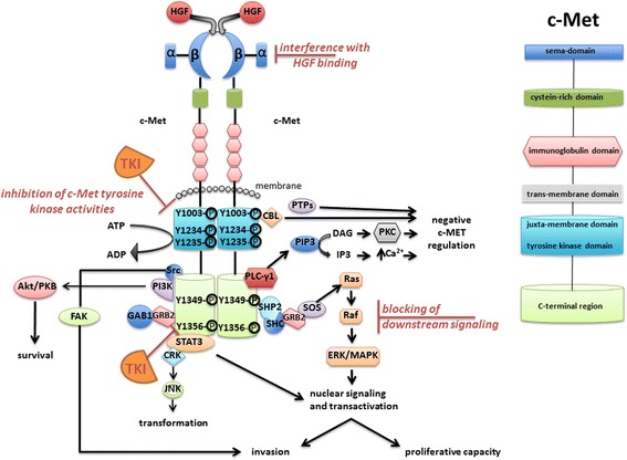
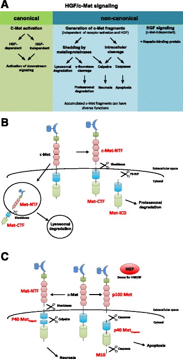
C-Met is a receptor tyrosine kinase with multiple functions throughout embryonic development, organogenesis and wound healing and is expressed in various epithelia. The ligand of c-Met is Hepatocyte Growth Factor (HGF) which is secreted among others by mesenchymal stroma/stem (MSC) cells.Physiological c-Met functions are centred around processes that underly cellular motility and invasive growth. Aberrant c-Met expression and activity is observed in numerous cancers and makes major contributions to cell malignancy. Importantly, HGF/c-Met signaling is crucial in the context of communication between cancer cells and the the tumor stroma.Here, we review recent findings on roles of dysregulated c-Met in urogenital tumors such as cancers of the urinary bladder, prostate, and ovary. We put emphasis on novel aspects of cancer-associated c-Met expression regulation on both, HGF-dependent and HGF-independent non-canonical mechanisms. Moreover, this review focusses on c-Met-triggered signalling with potential relevance for urogenital oncogenesis, and on strategies to specifically inhibit c-Met activity.

Keywords: Bladder, prostate and ovarian cancer; HGF/SF signalling; MSC; c-Met; c-Met inhibitors.


Figures

Fig. 1
Fig. 1
HGF/SF-mediated activation of c-Met and relayed downstream signalling. The c-Met receptor can be structured into distinct domains, including sema, cysteine-rich, immunoglobulin, trans-membrane, juxta-membrane, tyrosine kinase, and C-terminal region. Pharmacological intervention with activated c-Met signalling includes: (i) competitive interference with HGF/c-Met interaction, (ii) inhibition of the tyrosine kinase activity of c-Met with the use of tyrosine kinase inhibitors (TKI), or (iii) blocking of activated c-Met downstream signaling mediators. Accordingly, cell fate and development such as survival, transformation, cell motility, and proliferative capacity can be affected. This figure was adapted from Organ and Tsao, 2011 [2]
Fig. 2
Fig. 2
Pathways of c-Met signaling. a Overview of HGF/c-Met signaling via the canonical and non-canonical pathway. Canonical or “classical” HGF/Met signaling involves ligand-dependent and independent receptor activation which leads to the induction of downstream signaling cascades (left). Non-canonical HGF/c-Met signaling is independent of receptor activation. Generation of c-Met receptor fragments takes place under various cellular conditions such as apoptotic and necrotic stimuli as well as in the context of specific physiological circumstances. HGF is also able to exert signals independently of c-Met, e.g. upon interactions triggered by its heparin-binding domain. b Generation of c-Met fragments via shedding and cleavage by γ-secretase: Sheddases or metalloproteinases cleave full-length c-Met within its extracellular domain, resulting in different in a soluble extracellular N-terminal fragment (Met-NTF) and a membrane-associated C-terminal fragment (Met-CTF). Met-CTF can be further processed by the γ-secretase complex by presenilin-dependent intramembrane proteolysis (PS-RIP) into an intracellular domain (Met-ICD) which is routed to proteasomal degradation. Full-length membranous c-Met can also be internalized and cleaved by sheddases giving rise to Met-NTF and Met-CTF. These intracellularly generated c-Met fragments undergo lysosomal instead of proteasomal degradation. c Origin of c-Met fragments through intracellular cleavage by caspases and calpains: In response to apoptotic stimuli, c-Met is cleaved at two distinct sites in the intracellular domain by activated caspase-3, resulting in membrane-anchored p100 Met, a 40 kDa cytosolic p40 Met fragment and a small peptide (M10). Under necrotic conditions, c-Met is cleaved by metalloproteinases and further processed by calcium-independent proteases (calpains) instead of γ-secretase. The resulting product p40 Metcalpain differs from p40 Metcaspase by a few amino acid residues and is not able to promote apoptosis

References

    1. Rubin JS, Bottaro DP, Aaronson SA. Hepatocyte growth factor/scatter factor and its receptor, the c-met proto-oncogene product. Biochim Biophys Acta. 1993;1155:357–71. - PubMed
    1. Organ SL, Tsao MS. An overview of the c-MET signaling pathway. Ther Adv Med Oncol. 2011;3:S7–19. doi: 10.1177/1758834011422556. - DOI - PMC - PubMed
    1. Rong S, Segal S, Anver M, Resau JH, Vande Woude GF. Invasiveness and metastasis of NIH 3 T3 cells induced by Met-hepatocyte growth factor/scatter factor autocrine stimulation. Proc Natl Acad Sci U S A. 1994;91:4731–5. doi: 10.1073/pnas.91.11.4731. - DOI - PMC - PubMed
    1. Jeffers M, Rong S, Vande Woude GF. Hepatocyte growth factor/scatter factor-Met signaling in tumorigenicity and invasion/metastasis. J Mol Med (Berl) 1996;74:505–13. doi: 10.1007/BF00204976. - DOI - PubMed
    1. Sachs M, Weidner KM, Brinkmann V, Walther I, Obermeier A, Ullrich A, Birchmeier W. Motogenic and morphogenic activity of epithelial receptor tyrosine kinases. J Cell Biol. 1996;133:1095–107. doi: 10.1083/jcb.133.5.1095. - DOI - PMC - PubMed

Publication types

Substances

LinkOut - more resources