Review
. 2017 Jan 15;482(3):482-490.
doi: 10.1016/j.bbrc.2016.11.005. Epub 2017 Feb 3.

Elimination of the unnecessary: Intra- and extracellular signaling by anionic phospholipids

Affiliations
Review

Elimination of the unnecessary: Intra- and extracellular signaling by anionic phospholipids

Valerian E Kagan et al. Biochem Biophys Res Commun. .

Abstract

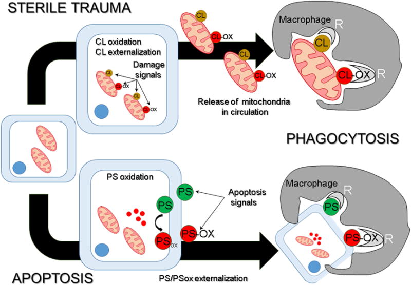
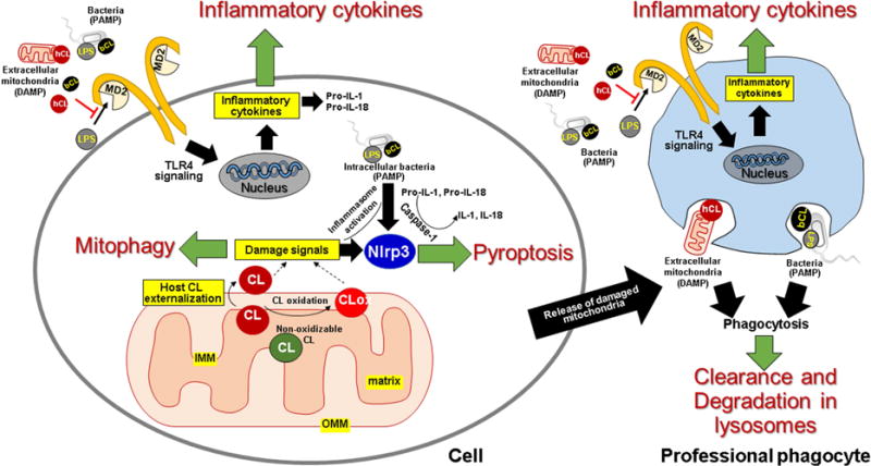
High fidelity of biological systems is frequently achieved by duplication of the essential intracellular machineries or, removal of the entire cell, which becomes unnecessary or even harmful in altered physiological environments. Carefully controlled removal of these cells, without damaging normal cells, requires precise signaling, and is critical to maintaining homeostasis. This review describes how two anionic phospholipids - phosphatidylserine (PS) and cardiolipin (CL) - residing in distinct compartments of the cell, signal removal of "the unnecessary" using several uniform principles. One of these principles is realized by collapse of inherent transmembrane asymmetry and the externalization of the signal on the outer membrane surface - mitochondria for CL and the plasma membrane for PS - to trigger mitophagy and phagocytosis, respectively. Release from damaged cells of intracellular structures with externalized CL or externalized PS triggers their elimination by phagocytosis. Another of these principles is realized by oxidation of polyunsaturated species of CL and PS. Highly specific oxidation of CL by cytochrome c serves as a signal for mitochondria-dependent apoptosis, while oxidation of externalized PS improves its effectiveness to trigger phagocytosis of effete cells.

Keywords: Apoptosis; Cardiolipin oxidation; Cardiolipin signaling; Mitophagy; Phagocytosis; Phosphatidylserine oxidation; Phosphatidylserine signaling.


Figures

Figure 1
Figure 1. CL and PS as elimination signals
There are at least two different ways in which PS and CL participate in elimination signaling. CL and PS are localized in spatially distinct membranes – the mitochondria and the plasma membrane, respectively - that sets apart their availability as beacons for clearance. During sterile trauma, mitochondria containing CL and peroxidized CL (CLox) are released by damaged cells. When encountered by macrophages, CL and CLox trigger phagocytosis with similar efficacies, whereby these membranes are engulfed, for subsequent degradation in lysosomes. Induction of apoptosis results in externalization of PS on the cell surface. Oxidative stress induced apoptosis is further characterized by lipid peroxidation that results in the presentation of both PS and PSox on the dying cells. While both PS species are targeted by macrophages for engulfment, the efficiency of PSox-dependent clearance is substantially higher than PS-dependent clearance.
Figure 2
Figure 2. Role of cardiolipins in homeostasis
Intra- or extracellular signaling by CL is triggered by its externalization to the mitochondrial surface. Intracellular CL: In healthy cells, CL is exclusively localized in the inner mitochondrial membrane (IMM) specifically on the inner leaflet of the IMM that faces the matrix. Damage to the mitochondria results in loss of this asymmetry and movement of the lipid to the outer leaflet of the IMM, to the inner leaflet of the outer mitochondrial membrane (OMM), and finally to the outer leaflet of the OMM. Additionally, oxidative stress results in peroxidation of oxidizable CL (having polyunsaturaed acyl chains) by cytochrome c, to produce CLox, which may also be presented at the mitochondrial surface as a damage signal. Once at the mitochondrial surface, CL in the presence of cytoplasmic LC3, signals clearance of the damaged organelle by mitophagy, thereby restoring intracellular homeostasis. Under pathological conditions, such as presence of intracellular pathogens or presence of intracellular pathogen associated molecular patterns (PAMPs) like lipopolysaccharide (LPS), mitochondrial CL and/or CLox promotes activation of the NLRP3 dependent inflammasome pathway that directs cell death by pyroptosis. Extracellular CL: Mitochondria released following cell damage provide human CL (hCL) as a source of extracellular damage associated molecular patterns (DAMPs), which challenge host cells, including professional phagocytes. Similarly, infections challenge these cells with bacterial CL (bCL) and LPS. Both hCL and bCL signal engulfment of mitochondrial and bacterial membranes by professional phagocytes for subsequent degradation in lysosomes. hCL and bCL also interfere with binding of LPS to MD2, thereby attenuating Toll-like receptor (TLR)-4 dependent inflammatory cytokine production. The attenuation of cytokine response and phagocytic clearance both contribute to restoration of homeostasis.

References

    1. Shi YB, Fu L, Hsia SC, Tomita A, Buchholz D. Thyroid hormone regulation of apoptotic tissue remodeling during anuran metamorphosis. Cell research. 2001;11:245–252. - PubMed
    1. Saunders JW., Jr Death in embryonic systems. Science (New York, NY) 1966;154:604–612. - PubMed
    1. Watson CJ. Involution: apoptosis and tissue remodelling that convert the mammary gland from milk factory to a quiescent organ. Breast cancer research: BCR. 2006;8:203. - PMC - PubMed
    1. Wu J, Carlock C, Zhou C, Nakae S, Hicks J, Adams HP, Lou Y. IL-33 is required for disposal of unnecessary cells during ovarian atresia through regulation of autophagy and macrophage migration. Journal of immunology (Baltimore, Md: 1950) 2015;194:2140–2147. - PMC - PubMed
    1. Cecconi F, Alvarez-Bolado G, Meyer BI, Roth KA, Gruss P. Apaf1 (CED-4 homolog) regulates programmed cell death in mammalian development. Cell. 1998;94:727–737. - PubMed

LinkOut - more resources