Review
. 2017 Apr:45:24-30.
doi: 10.1016/j.ceb.2017.01.003. Epub 2017 Feb 16.

Focal adhesion kinase signaling in unexpected places

Affiliations
Review

Focal adhesion kinase signaling in unexpected places

Elizabeth G Kleinschmidt et al. Curr Opin Cell Biol. 2017 Apr.

Abstract

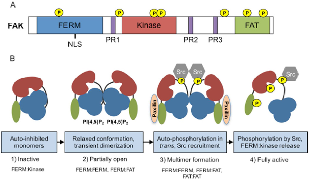
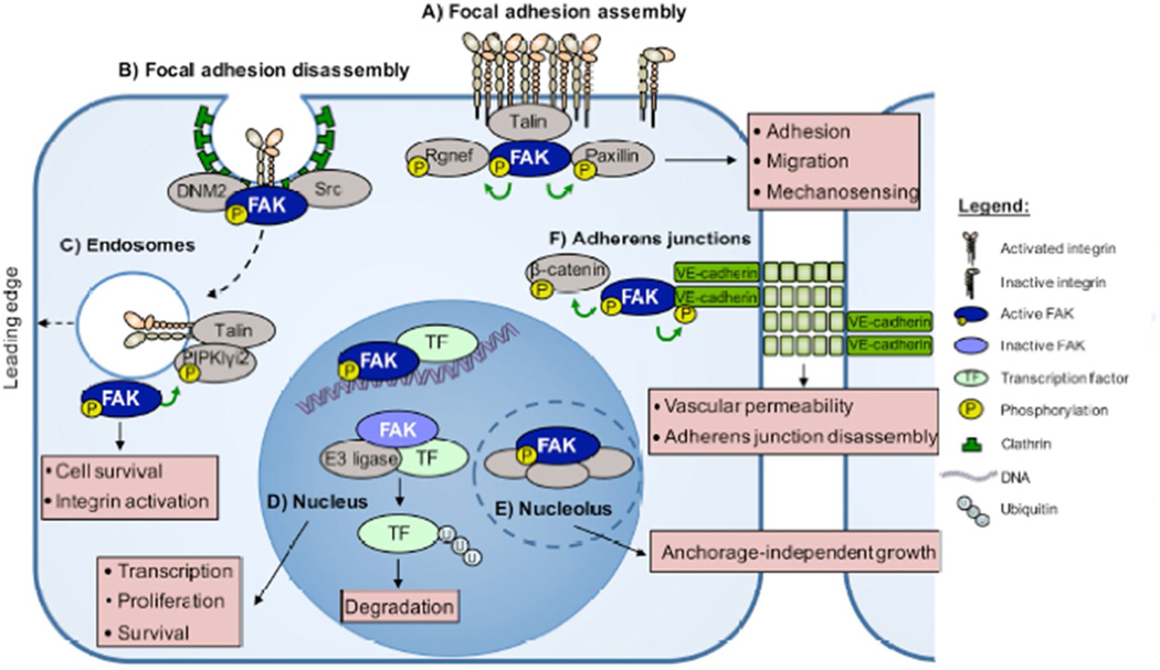
Focal adhesion kinase (FAK) is a cytoplasmic protein-tyrosine kinase first identified at extracellular matrix and integrin receptor cell adhesion sites and is a key regulator of cell movement. FAK is activated by a variety of stimuli. Herein, we discuss advances in conformational-associated FAK activation and dimerization mechanisms. Additionally, new roles have emerged for FAK signaling at cell adhesions, adherens junctions, endosomes, and the nucleus. In light of these new findings, we review how FAK activation at these sites is connected to the regulation of integrin recycling-activation, vascular permeability, cell survival, and transcriptional regulation, respectively. Studies uncovering FAK signaling connections in unexpected places within cells have yielded important new regulatory insights in cell biology.


Figures

Figure 1
Figure 1
Schematic of FAK domains and mechanisms leading to activation. A) FAK contains an amino-terminal protein band 4.1-ezrin-radixin-moesin (FERM) homology domain (blue), a central kinase domain (red), and a carboxy-terminal focal adhesion targeting (FAT) domain (green). These domains are connected by linker regions containing proline-rich regions (PR1–3, purple). Location of phosphorylation sites and a nuclear localization sequence (NLS) is shown. B) Proposed mechanism of FAK activation. 1) Inactive FAK exists as an auto-inhibited monomer, a conformation maintained by interactions between the FERM (blue) and kinase (red) domains. 2) FERM binding to phosphatidylinositol-4,5-biphosphate [PI(4,5)P2] induces a relaxed conformation, FAK clustering, and transient FERM:FERM and FAT:FAT dimerization. 3) Transient FERM:FERM and FAT:FAT dimerization allows FAK autophosphorylation at Y397 in trans (yellow). A FERM:FAT (FAT shown in green) interaction can be reinforced by paxillin at focal adhesions. Y397 FAK phosphorylation creates a binding site for the SH2 domain of Src. 4) Src further phosphorylates FAK at multiple sites inducing full FAK activation by a conformational shift that includes FERM:kinase release.
Figure 2
Figure 2. FAK functions throughout the cell
A) Proteins such as Rgnef, talin, and paxillin contribute to FAK localization at sites of integrin clustering. Activated FAK phosphorylates Rgnef and paxillin, promoting the assembly of focal adhesions. B) FAK binds dynamin-2 (DNM2), triggering focal adhesion disassembly and integrin endocytosis. FAK at adhesions regulates cell adhesion, migration, and mechanosensing. C) FAK binds to and is activated at endosomes. FAK phosphorylation of PIPKIγi2 results in talin recruitment, which in turn maintains the active conformation of endosome-associated integrins. FAK-endosome signaling facilitates the polarized reassembly of activated integrins at the leading edge of migrating cells. FAK at endosomes facilitates integrin activation and cell survival. D) In the nucleus, activated FAK binds to transcription factors (TFs) to modulate gene expression. Inactive FAK in the nucleus functions as a scaffold to facilitate transcription factor turnover via enhanced ubiquitination by complexing with different E3 ligases. E) In the nucleolus, activated FAK complexes with proteins important in promoting stem cell phenotypes. FAK in the nucleus and nucleolus alters transcription, survival, and anchorage-independent cell growth. F) Active FAK localizes to adherens junctions (AJs) and directly binds VE-cadherin in response to vascular endothelial growth factor stimulation of endothelial cells. FAK phosphorylates β-catenin and VE-cadherin, triggering AJ disassembly. Right, depiction of figure elements.

References

    1. Yoon H, Dehart JP, Murphy JM, Lim ST. Understanding the roles of FAK in cancer: inhibitors, genetic models, and new insights. J Histochem Cytochem. 2015;63:114–128. - PMC - PubMed
    1. Roy-Luzarraga M, Hodivala-Dilke K. Molecular Pathways: Endothelial Cell FAK-A Target for Cancer Treatment. Clin Cancer Res. 2016;22:3718–3724. - PMC - PubMed
    1. Sulzmaier FJ, Jean C, Schlaepfer DD. FAK in cancer: mechanistic findings and clinical applications. Nat Rev Cancer. 2014;14:598–610. - PMC - PubMed
    1. Weis SM, Lim ST, Lutu-Fuga KM, Barnes LA, Chen XL, Gothert JR, Shen TL, Guan JL, Schlaepfer DD, Cheresh DA. Compensatory role for Pyk2 during angiogenesis in adult mice lacking endothelial cell FAK. J Cell Biol. 2008;181:43–50. - PMC - PubMed
    1. Lim ST, Miller NL, Nam JO, Chen XL, Lim Y, Schlaepfer DD. Pyk2 inhibition of p53 as an adaptive and intrinsic mechanism facilitating cell proliferation and survival. J Biol Chem. 2010;285:1743–1753. - PMC - PubMed

Substances