Discovery, Diagnosis, and Etiology of Craniofacial Ciliopathies
- PMID: 28213462
- PMCID: PMC5585846
- DOI: 10.1101/cshperspect.a028258
Discovery, Diagnosis, and Etiology of Craniofacial Ciliopathies
Abstract
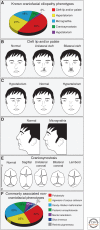
Seventy-five percent of congenital disorders present with some form of craniofacial malformation. The frequency and severity of these malformations makes understanding the etiological basis crucial for diagnosis and treatment. A significant link between craniofacial malformations and primary cilia arose several years ago with the determination that ∼30% of ciliopathies could be primarily defined by their craniofacial phenotype. The link between the cilium and the face has proven significant, as several new "craniofacial ciliopathies" have recently been diagnosed. Herein, we reevaluate public disease databases, report several new craniofacial ciliopathies, and propose several "predicted" craniofacial ciliopathies. Furthermore, we discuss why the craniofacial complex is so sensitive to ciliopathic dysfunction, addressing tissue-specific functions of the cilium as well as its role in signal transduction relevant to craniofacial development. As a whole, these analyses suggest a characteristic facial phenotype associated with craniofacial ciliopathies that can perhaps be used for rapid discovery and diagnosis of similar disorders in the future.
Copyright © 2017 Cold Spring Harbor Laboratory Press; all rights reserved.
Figures


Similar articles
-
The emerging face of primary cilia.Genesis. 2011 Apr;49(4):231-46. doi: 10.1002/dvg.20728. Epub 2011 Apr 1. Genesis. 2011. PMID: 21305689 Free PMC article. Review.
-
Ciliopathic micrognathia is caused by aberrant skeletal differentiation and remodeling.Development. 2021 Feb 15;148(4):dev194175. doi: 10.1242/dev.194175. Development. 2021. PMID: 33589509 Free PMC article.
-
Unmasking the ciliopathies: craniofacial defects and the primary cilium.Wiley Interdiscip Rev Dev Biol. 2015 Nov-Dec;4(6):637-53. doi: 10.1002/wdev.199. Epub 2015 Jul 14. Wiley Interdiscip Rev Dev Biol. 2015. PMID: 26173831 Review.
-
Craniofacial Ciliopathies Reveal Specific Requirements for GLI Proteins during Development of the Facial Midline.PLoS Genet. 2016 Nov 1;12(11):e1006351. doi: 10.1371/journal.pgen.1006351. eCollection 2016 Nov. PLoS Genet. 2016. PMID: 27802276 Free PMC article.
-
Cilia-dependent GLI processing in neural crest cells is required for tongue development.Dev Biol. 2017 Apr 15;424(2):124-137. doi: 10.1016/j.ydbio.2017.02.021. Epub 2017 Mar 9. Dev Biol. 2017. PMID: 28286175 Free PMC article.
Cited by
-
Neural crest cells utilize primary cilia to regulate ventral forebrain morphogenesis via Hedgehog-dependent regulation of oriented cell division.Dev Biol. 2017 Nov 15;431(2):168-178. doi: 10.1016/j.ydbio.2017.09.026. Epub 2017 Sep 21. Dev Biol. 2017. PMID: 28941984 Free PMC article.
-
Loss of intraflagellar transport 140 in osteoblasts cripples bone fracture healing.Fundam Res. 2022 Sep 19;5(4):1795-1803. doi: 10.1016/j.fmre.2022.09.006. eCollection 2025 Jul. Fundam Res. 2022. PMID: 40777780 Free PMC article.
-
Primary cilia deficiency in neural crest cells models anterior segment dysgenesis in mouse.Elife. 2019 Dec 17;8:e52423. doi: 10.7554/eLife.52423. Elife. 2019. PMID: 31845891 Free PMC article.
-
Ciliary and extraciliary Gpr161 pools repress hedgehog signaling in a tissue-specific manner.Elife. 2021 Aug 4;10:e67121. doi: 10.7554/eLife.67121. Elife. 2021. PMID: 34346313 Free PMC article.
-
Identification of a heterogeneous and dynamic ciliome during embryonic development and cell differentiation.Development. 2023 Apr 15;150(8):dev201237. doi: 10.1242/dev.201237. Epub 2023 Apr 27. Development. 2023. PMID: 36971348 Free PMC article.
References
-
- Abzhanov A, Protas M, Grant RB, Grant PR, Tabin CJ. 2004. Bmp4 and morphological variation of beaks in Darwin’s finches. Science 305: 1462–1465. - PubMed
-
- Baker K, Beales P. 2009. Making sense of cilia in disease: The human ciliopathies. Am J Med Genet 151C: 281–295. - PubMed
-
- Basch ML, Bronner-Fraser M. 2006. Neural crest inducing signals. Adv Exp Med Biol 589: 24–31. - PubMed
-
- Beales PL, Bland E, Tobin JL, Bacchelli C, Tuysuz B, Hill J, Rix S, Pearson CG, Kai M, Hartley J, et al. 2007. IFT80, which encodes a conserved intraflagellar transport protein, is mutated in Jeune asphyxiating thoracic dystrophy. Nat Genet 39: 727–729. - PubMed
Publication types
MeSH terms
Substances
Grants and funding
LinkOut - more resources
Full Text Sources
Other Literature Sources
Medical
Research Materials
