. 2017 Apr 1;312(4):L568-L578.
doi: 10.1152/ajplung.00117.2016. Epub 2017 Feb 17.

Hypoxia induces arginase II expression and increases viable human pulmonary artery smooth muscle cell numbers via AMPKα1 signaling

Affiliations

Hypoxia induces arginase II expression and increases viable human pulmonary artery smooth muscle cell numbers via AMPKα1 signaling

Jianjing Xue et al. Am J Physiol Lung Cell Mol Physiol. .

Abstract

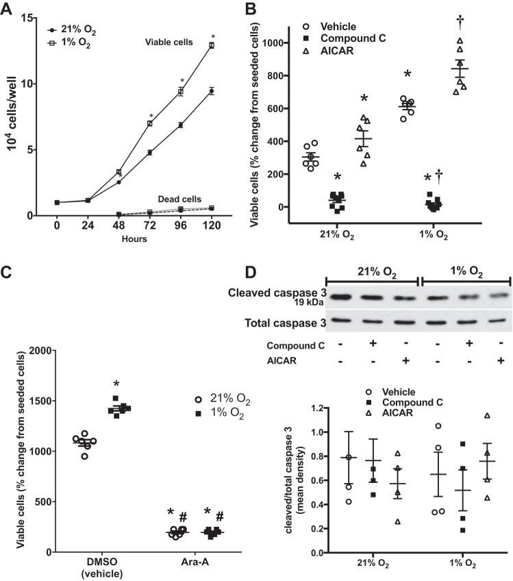
Pulmonary artery smooth muscle cell (PASMC) proliferation is one of the hallmark features of hypoxia-induced pulmonary hypertension. With only supportive treatment options available for this life-threatening disease, treating and preventing the proliferation of PASMCs is a viable therapeutic option. A key promoter of hypoxia-induced increases in the number of viable human PASMCs is arginase II, with attenuation of viable cell numbers following pharmacologic inhibition or siRNA knockdown of the enzyme. Additionally, increased levels of arginase have been demonstrated in the pulmonary vasculature of patients with pulmonary hypertension. The signaling pathways responsible for the hypoxic induction of arginase II in PASMCs, however, remain unknown. Hypoxia is a recognized activator of AMPK, which is known to be expressed in human PASMCs (hPASMCs). Activation of AMPK by hypoxia has been shown to promote cell survival in PASMCs. In addition, pharmacologic agents targeting AMPK have been shown to attenuate chronic hypoxia-induced pulmonary hypertension in animal models. The present studies tested the hypothesis that hypoxia-induced arginase II expression in hPASMCs is mediated through AMPK signaling. We found that pharmacologic inhibitors of AMPK, as well as siRNA knockdown of AMPKα1, prevented hypoxia-induced arginase II. The hypoxia-induced increase in viable hPASMC numbers was also prevented following both pharmacologic inhibition and siRNA knockdown of AMPK. Furthermore, we demonstrate that overexpression of AMPK induced arginase II protein expression and viable cells numbers in hPASMCs.

Keywords: l-arginine; pulmonary hypertension; pulmonary vasculature; vascular remodeling; vascular smooth muscle cells.


Figures

Fig. 1.
Fig. 1.
Hypoxia activates AMPK. Human pulmonary artery smooth muscle cells (PASMCs) were exposed to 21% O2 (normoxia) or 1% O2 (hypoxia) for 20 and 30 min. Protein was assayed for phosphorylated-AMPK (p-AMPK) by Western blot analysis and was quantified by densitometry. A: representative Western blots are shown for p-AMPK, total AMPK, and β-actin. B: data are shown as fold change ± SE after 20 and 30 min of hypoxia relative to normoxia control. *Significant difference, hypoxia vs. normoxia, P < 0.01; n = 3.
Fig. 2.
Fig. 2.
Pharmacologic inhibition of AMPK prevents hypoxia-induced arginase II protein expression, whereas AMPK activation increases arginase II protein expression. Human PASMCs were treated with the AMPK inhibitor Compound C, the cell-permeable AMPK activator AICAR, or vehicle (DMSO for the AMPK inhibitors or dH2O for AMPK activator, N1-(β-d-ribofuranosyl)-5-aminoimidazole-4-carboxamide (AICAR), and incubated in 21% O2 (normoxia) or 1% O2 (hypoxia) for 48 h. Protein was assayed for arginase II protein expression by Western blot analysis and was quantified by densitometry. A: representative Western blots are shown for arginase II and β-actin. B: data are shown as a fold change ± SE relative to vehicle-treated, normoxia-exposed human PASMC controls (n = 4). *Significant difference compared with vehicle-treated normoxia, P < 0.05. †Significant difference compared with vehicle-treated hypoxia, P < 0.0001.
Fig. 3.
Fig. 3.
AMPK overexpression induces arginase II expression and increases viable hPASMC numbers. Human PASMCs were seeded on six-well plates and transfected with an empty pCMV6-Entry vector with in-frame MYC/DDK tags or pCMV6-Entry vector with in-frame MYC/DDK tags containing AMPKα1, AMPKα2, or AMPKα1/2. The transfected hPASMCs were placed in 21% O2 (normoxia) or 1% O2 (hypoxia) for 48 h then protein was analyzed by Western blot analysis. Protein was assayed for myc and arginase II expression by Western blot analysis and was quantified by densitometry. Representative Western blots are shown for myc (A) and arginase II and β-actin (B). Data are shown as fold-change ± SE relative to empty vector control (vControl) (n = 4–6) (C). Human PASMCs were seeded on six-well plates and transfected with an empty pCMV6-Entry vector with in-frame MYC/DDK tags or pCMV6-Entry vector with in-frame MYC/DDK tags containing AMPKα1, AMPKα2, or AMPKα(1+2). The transfected hPASMCs were seeded onto six-well plates at 104 cells per well and incubated for 120 h in normoxia or hypoxia, and viable cell numbers were determined using trypan blue exclusion. D: data are shown as viable cell numbers as a percentage change from the number of seeded cells (n = 6). *Significant difference compared with vControl, P < 0.01. †Significant difference compared with hypoxia, vControl, P < 0.05.
Fig. 4.
Fig. 4.
AMPKα1 knockdown prevents hypoxia-induced arginase II protein expression. Human PASMCs were transfected with AMPKα isoform-specific siRNA or scramble siRNA for 24 h, and then exposed to 21% O2 (normoxia) or 1% O2 (hypoxia) for 48 h. Protein was assayed for AMPKα1, AMPKα2, or arginase II protein expression by Western blot and was quantified by densitometry. Representative Western blots are shown for AMPKα1 (A), AMPKα2 (B), and arginase II and β-actin (C). Data are shown as means ± SE relative to normoxia, scramble siRNA-treated controls (n = 3–10). *Significant difference compared with normoxia, scramble siRNA-treated, P < 0.05. †Significant difference, arginase II vs. hypoxia, scramble siRNA-treated, P < 0.0001.
Fig. 5.
Fig. 5.
Knockdown of AMPKα1, but not AMPKα2, attenuates hypoxic activation of AMPK. Human PASMCs were treated with AMPKα isoform-specific siRNA or scramble siRNA for 24 h, and then exposed to 1% O2 (hypoxia) for 48 h. Protein was assayed for p-AMPK, total AMPK, p-ACC, or total ACC protein expression by Western blot and was quantified by densitometry. Representative Western blots are shown for p-AMPK, total AMPK (A), p-ACC, total ACC, and β-actin (B). Data are shown as fold-change ± SE relative to scramble siRNA-treated controls (n = 3–7). *Significant difference, compared with scramble siRNA-treated, P < 0.01. †Significant difference, compared with siAMPKα1-treated, P < 0.05.
Fig. 6.
Fig. 6.
Pharmacologic inhibition of AMPK decreases the number of viable hPASMCs, whereas AMPK activation enhances hypoxia-induced increase in viable cell numbers with no effect on apoptosis. Human PASMCs were seeded on six-well plates at a density of 104 cells per well, and the cells were incubated for 120 h in 21% O2 (normoxia) or 1% O2 (hypoxia), and viable cell numbers were determined at each 24-h interval using trypan blue exclusion. A: numbers of dead cells were counted beginning at the 48-h time point. Data are shown as viable or dead cell numbers per well (n = 3 for each time-point). Human PASMCs were treated with Compound C, AICAR, vehicle (B) or Ara-A or vehicle (C) (DMSO for the AMPK inhibitors or dH2O for AICAR), incubated in normoxia or hypoxia, and viable cell numbers were determined using trypan blue exclusion. Data are shown as viable cell numbers as a percent change from the number of seeded cells (n = 6–9). Human PASMCs were treated with Compound C, AICAR, or vehicle, and incubated in normoxia or hypoxia for 48 h. Protein was assayed for cleaved caspase 3 expression by Western blot analysis and quantified by densitometry. Representative Western blots are shown for cleaved caspase 3 and total caspase 3. Data are shown as fold change ± SE, relative to normoxia, vehicle-treated hPASMC controls (n = 4) (D). *Significant difference, compared with normoxia, vehicle-treated, P < 0.05. †Significant difference from hypoxia, vehicle-treated, P < 0.0001.
Fig. 7.
Fig. 7.
siRNA knockdown of AMPK prevents hypoxia-induced increase in viable hPASMC numbers. Human PASMCs were transfected with AMPKα isoform-specific siRNA or scramble siRNA. The siRNA-transfected cells were then seeded on six-well plates at a density of 104 cells per well and incubated for 120 h in 21% O2 (normoxia) or 1% O2 (hypoxia), and viable cell numbers were determined using trypan blue exclusion. A: data are shown as viable cell numbers as a percentage change from number of seeded cells (n = 7). Human PASMCs were transfected with AMPKα isoform-specific siRNA or scramble siRNA and incubated in normoxia or hypoxia for 48 h. Protein was assayed for cleaved caspase 3 expression by Western blot analysis and was quantified by densitometry. B: representative Western blots are shown for cleaved caspase 3 and total caspase 3. Data are shown as fold-change ± SE relative to normoxia, scramble siRNA-treated hPASMC controls (n = 3–5) (C). D: alternatively, LDH cytotoxicity assay was performed. *Significant difference, compared with normoxia, scramble siRNA-treated, P < 0.05. †Significant difference from hypoxia, scramble siRNA-treated, P < 0.0001. ‡Significant difference, hypoxia siAMPKα2 or siAMPKα1/2 compared with normoxia siAMPKα2 or siAMPKα1/2, P < 0.0001.
Fig. 8.
Fig. 8.
siRNA knockdown of arginase II, in part, inhibits viable cell numbers in AMPK-activated, hypoxic hPASMCs. Human PASMCs were transfected with siRNA against arginase II. The transfected hPASMCs were then seeded onto six-well plates at a density of 104 cells per well and incubated with AICAR or vehicle (dH2O) for 120 h in 21% O2 (normoxia) or 1% O2 (hypoxia), and viable cell numbers were determined using trypan blue exclusion. Data are shown as fold change of viable cell numbers relative to normoxia, scramble siRNA-transfected hPASMCs. *Significant difference, compared with normoxia, scramble siRNA-treated, P < 0.05. †Significant difference from hypoxia, scramble siRNA-treated, P < 0.0005. ‡Significant difference, compared with siArgII.

References

    1. Ban HS, Xu X, Jang K, Kim I, Kim BK, Lee K, Won M. A novel malate dehydrogenase 2 inhibitor suppresses hypoxia-inducible factor-1 by regulating mitochondrial respiration. PLoS One 11: e0162568, 2016. doi:10.1371/journal.pone.0162568. - DOI - PMC - PubMed
    1. Bradford MM. A rapid and sensitive method for the quantitation of microgram quantities of protein utilizing the principle of protein-dye binding. Anal Biochem 72: 248–254, 1976. doi:10.1016/0003-2697(76)90527-3. - DOI - PubMed
    1. Cantó C, Auwerx J. AMP-activated protein kinase and its downstream transcriptional pathways. Cell Mol Life Sci 67: 3407–3423, 2010. doi:10.1007/s00018-010-0454-z. - DOI - PMC - PubMed
    1. Chen B, Calvert AE, Cui H, Nelin LD. Hypoxia promotes human pulmonary artery smooth muscle cell proliferation through induction of arginase. Am J Physiol Lung Cell Mol Physiol 297: L1151–L1159, 2009. doi:10.1152/ajplung.00183.2009. - DOI - PubMed
    1. Chen B, Calvert AE, Meng X, Nelin LD. Pharmacologic agents elevating cAMP prevent arginase II expression and proliferation of pulmonary artery smooth muscle cells. Am J Respir Cell Mol Biol 47: 218–226, 2012. doi:10.1165/rcmb.2011-0015OC. - DOI - PMC - PubMed