. 2017 May 1;312(5):L609-L624.
doi: 10.1152/ajplung.00531.2016. Epub 2017 Feb 17.

Central role of T helper 17 cells in chronic hypoxia-induced pulmonary hypertension

Affiliations

Central role of T helper 17 cells in chronic hypoxia-induced pulmonary hypertension

Levi D Maston et al. Am J Physiol Lung Cell Mol Physiol. .

Abstract

Inflammation is a prominent pathological feature in pulmonary arterial hypertension, as demonstrated by pulmonary vascular infiltration of inflammatory cells, including T and B lymphocytes. However, the contribution of the adaptive immune system is not well characterized in pulmonary hypertension caused by chronic hypoxia. CD4+ T cells are required for initiating and maintaining inflammation, suggesting that these cells could play an important role in the pathogenesis of hypoxic pulmonary hypertension. Our objective was to test the hypothesis that CD4+ T cells, specifically the T helper 17 subset, contribute to chronic hypoxia-induced pulmonary hypertension. We compared indices of pulmonary hypertension resulting from chronic hypoxia (3 wk) in wild-type mice and recombination-activating gene 1 knockout mice (RAG1-/-, lacking mature T and B cells). Separate sets of mice were adoptively transferred with CD4+, CD8+, or T helper 17 cells before normoxic or chronic hypoxic exposure to evaluate the involvement of specific T cell subsets. RAG1-/- mice had diminished right ventricular systolic pressure and arterial remodeling compared with wild-type mice exposed to chronic hypoxia. Adoptive transfer of CD4+ but not CD8+ T cells restored the hypertensive phenotype in RAG1-/- mice. Interestingly, RAG1-/- mice receiving T helper 17 cells displayed evidence of pulmonary hypertension independent of chronic hypoxia. Supporting our hypothesis, depletion of CD4+ cells or treatment with SR1001, an inhibitor of T helper 17 cell development, prevented increased pressure and remodeling responses to chronic hypoxia. We conclude that T helper 17 cells play a key role in the development of chronic hypoxia-induced pulmonary hypertension.

Keywords: CD4 T cells; SR1001; inflammation; interleukin-6; retinoid-related orphan receptor-γτ.


Figures

Fig. 1.
Fig. 1.
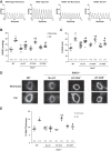
CD4+ T cells contribute to chronic hypoxia (CH)-induced pulmonary hypertension (PH). A: representative right ventricular (RV) systolic pressure (RVSP) traces from isoflurane-anesthetized wild-type (WT) and RAG1−/− mice without adoptive transfer (AT) or AT of enriched CD4+ or CD8+ T cells exposed to 21 days of CH or normoxia, as indicated. B: summary of peak RVSP data. C: %RV to left ventricle plus septum (%RV/LV + S) weight. Values are means ± SE; n = no. of animals. *P < 0.05 vs. normoxia; #P < 0.05 vs. CH WT; &P < 0.05 vs. CH RAG1−/−. No AT, analyzed by 2-way ANOVA, followed by multiple-comparison Student-Newman-Keuls test. D: representative images of α-smooth muscle actin-labeled pulmonary artery sections with external diameters <150 µm from normoxic and CH (21 days) WT and RAG1−/− mice receiving either no adoptive transfer or adoptive transfer with enriched CD4+ or CD8+ T cells; scale bar, 100 μm. E: %wall thickness of pulmonary arteries from the groups described in A. Values are means ± SE; n = no. of animals; at least 10 arteries/animal were measured; *P < 0.05 vs. normoxia; #P < 0.05 vs. WT CH; &P < 0.05 vs. No AT CH, analyzed by 2-way ANOVA, followed by multiple comparisons Student-Newman-Keuls test.
Fig. 2.
Fig. 2.
CD4+ cell depletion attenuates the development of CH-induced PH. Anti-CD4 or control antibody (2 mg/kg) given once/wk, starting 1 wk before CH (21 days). A: representative flow cytometry plots of splenocytes labeled for CD3 (gated), CD4, and CD8. B: peak RVSP. C: Fulton’s index. D: %wall thickness. E: hematocrit. Values are means ± SE. *P < 0.05 vs. normoxia; #P < 0.05 vs. CH control (n = no. of animals, and in D at least 10 arteries/animal were measured; 2-way ANOVA followed by multiple-comparison Student-Newman-Keuls test).
Fig. 3.
Fig. 3.
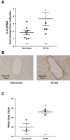
CH increases lung IL-6 levels. A: lungs from mice exposed to either normoxia or 5 days of CH (CH 5d) were homogenized and IL-6 mRNA levels determined by real-time PCR, with β-actin used as an endogenous control. B: representative images of immunohistochemical detection of IL-6 in lung sections. C: summary analysis data of the mean gray value of the medial layer of anti-IL-6-stained pulmonary artery sections with external diameters of 30–80 μm. Values are means ± SE. *P < 0.05 vs. normoxia; n = 3 mice/group, 5–12 arteries/mouse, analyzed by unpaired t-test. ●, Normoxia; ■, chronic hypoxia.
Fig. 4.
Fig. 4.
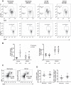
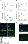
CH leads to an increase in lung T helper 17 (TH17) cells without T regulatory cells being affected. Lungs were removed from vehicle- and SR1001 (25 mg·kg−1·day sc−1)-treated mice after 5 days of normoxic or CH (CH 5d) exposure. Single-cell suspensions from the lungs were labeled with anti-CD3, anti-CD4, and anti-IL-17A or FoxP3. A: representative scatter plots. Cells were gated based on forward and side scatter and CD3 CD4. B: summary of the %CD4+ IL-17A+ or CD4+ FoxP3+ cells relative to total CD3+ cells normalized to the average of the vehicle-treated normoxic group of each set of experiments. C: representative scatter plots of CD3+, CD4+ T helper cells from lungs from mice exposed to normoxia or 5 days of CH. D: summary of the %CD3+ T cells relative to total cells and CD3+, CD4+, and T helper cells relative to total CD3+ cells. Values are means ± SE; n = no. of animals, *P < 0.05 vs. normoxia vehicle; #P < 0.05 vs. CH vehicle, analyzed by 2-way ANOVA, followed by multiple-comparison Student-Newman-Keuls test.
Fig. 5.
Fig. 5.
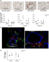
Inhibition of TH17 cell development attenuates CH-induced increases in perivascular T cells. SR1001 was delivered daily by sc injection (25 mg·kg−1·day−1) for the duration of normoxic or CH exposure. A: representative images from normoxic and CH (5 days) mice treated with or without SR1001 in which perivascular T cells (CD3+ cells) were quantified. Arrows depict CD3+ cells. Scale bar, 100 µm. B: summary of the no. of perivascular CD3+ cells from mice exposed to 2, 5, or 21 days of normoxia or CH treated with or without SR1001. The perivascular region was defined as external to vessel media and internal to vessel adventitia. C: representative images of lung sections from vehicle-treated mice exposed to 21 days of CH colabeled with anti-IL17A (green) and anti-CD3 (red). Elastic lamina is shown in blue. Arrows depict double-positive cells. Scale bar, 50 µm. D: summary of the no. of CD3+ cells in the lung parenchyma of mice exposed to 5 days of normoxia or CH treated with or without SR1001. Values are means ± SE. *P < 0.05 vs. normoxia vehicle; #P < 0.05 vs. vehicle CH; n = no. of animals; at least 5–15 arteries (<150 µm outer diameter/mouse) were measured and analyzed by 2-way ANOVA, followed by multiple comparisons Student-Newman-Keuls test.
Fig. 6.
Fig. 6.
Inhibition of TH17 cell development attenuates CH-induced PH. SR1001 was delivered sc (25 mg·kg−1·day−1) via an implantable osmotic pump. A: RVSP following 21 days of normoxia or CH. B: Fulton’s index. C: pulmonary arterial wall thickness measured in small pulmonary arteries (<150 µm) labeled for α-smooth muscle actin. D: hematocrit. E: no. of Ki-67+ cells/small pulmonary artery outer diameter. Ki-67 is a marker of cell proliferation. Ki-67+ cells were detected by immunofluorescence microscopy in the arterial wall (shown in red). The elastic lamina in the arterial wall is shown in blue. Nuclei are shown in green. CH 5d, 5 days of CH. F: representative images of lung sections labeled with anti-cleaved caspase 3, which is an apoptosis marker. Scale bar, 40 µm. Arrows depict positive cells expressing activated caspase 3 in lung parenchyma. Values are means ± SE; n = no. of animals, and in C and D at least 10 arteries/animal were measured. *P < 0.05 vs. normoxia vehicle; #P < 0.05 vs. normoxic SR1001, analyzed by 2-way ANOVA, followed by multiple-comparison Student-Newman-Keuls test.
Fig. 7.
Fig. 7.
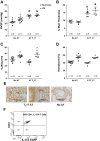
The adoptive transfer of TH17 cells causes spontaneous PH in RAG1−/− mice. CD4+ T cells were purified from IL-17A enhanced green fluorescent protein (EGFP) mice and cultured under TH17-polarizing conditions. TH17 cells were purified by fluorescence-activated cell sorting and injected into RAG1−/− mice. After 14 days, mice remained in normoxia or were exposed to CH for 21 days and compared with mice that did not receive TH17 cells. A: RVSP. B: %wall thickness. C: Fulton’s index. D: hematocrit. E: representative images of lung sections immunostained for CD3 from mice receiving adoptive transfer or a RAG1−/− mouse that received no adoptive transfer. Scale bars, 100 µm. F: flow cytometric analysis of TH17 cells from the spleens of mice that received an adoptive transfer. Values are means ± SE; n = no. of animals. *P < 0.05 vs. normoxia; #P < 0.05 vs. normoxia No AT; &P < 0.05 vs. CH No AT. Analyzed by 2-way ANOVA, followed by multiple comparisons Student-Newman-Keuls test.
Fig. 8.
Fig. 8.
Inhibition of TH17 cell polarization reverses CH-induced PH. Wild-type mice were exposed to CH for 21 days, followed by administration of SR1001 (25 mg·kg−1·day sc−1) or vehicle for an additional 14 days in CH. A: RVSP. B: Fulton’s index. C: %wall thickness. D: no. of CD3+ cells/vessel diameter (µm). Values are means ± SE; n = no. of animals, and in C and D at least 10 arteries/animal were measured. *P < 0.05 vs. vehicle, analyzed by unpaired t-test.
Fig. 9.
Fig. 9.
Total lung macrophage numbers are not affected by CH exposure. Lungs were removed from mice following 5 days of CH exposure. Single-cell suspensions from the lungs were incubated with anti-CD45, anti-F4/80, and anti-CD11b to label macrophages. A: representative scatter plots. Cells were gated based on forward and side scatter and CD45. B: summary of %F4/80+ CD11c+ cells relative to total CD45+ cells normalized to the average of the vehicle-treated normoxic group of each set of experiments. Values are means ± SE; n = no. of animals, analyzed by 2-way ANOVA, followed by multiple-comparison Student-Newman-Keuls test.
Fig. 10.
Fig. 10.
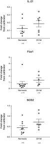
CH does not affect lung IL-21, Fizz1, or NOS2 mRNA levels. IL-21, Fizz1 (macrophage type 2 marker), and NOS2 (macrophage type 1 marker) transcripts were measured by real-time PCR in lungs from WT mice exposed to normoxia or 5 days of CH. Values are means ± SE; n = no. of animals, analyzed by unpaired t-test.
Fig. 11.
Fig. 11.
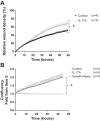
IL-17A stimulates migration but does not affect mouse pulmonary arterial smooth muscle cell number in culture. A: %wound density in the presence or absence of IL-17A (100 ng/ml). B: confluency density normalized to time 0 in the presence or absence of IL-17A (100 ng/ml) in serum-starved cells. Growth medium was used a positive control. *P < 0.05, cells from n = 4 mice, 2-way repeated-measures ANOVA, followed by multiple-comparison Student-Newman-Keuls test.

References

    1. Akdis CA, Akdis M. Mechanisms and treatment of allergic disease in the big picture of regulatory T cells. J Allergy Clin Immunol 123: 735–746, 2009. doi: 10.1016/j.jaci.2009.02.030. - DOI - PubMed
    1. Artis D, Spits H. The biology of innate lymphoid cells. Nature 517: 293–301, 2015. doi: 10.1038/nature14189. - DOI - PubMed
    1. Ball MK, Waypa GB, Mungai PT, Nielsen JM, Czech L, Dudley VJ, Beussink L, Dettman RW, Berkelhamer SK, Steinhorn RH, Shah SJ, Schumacker PT. Regulation of hypoxia-induced pulmonary hypertension by vascular smooth muscle hypoxia-inducible factor-1α. Am J Respir Crit Care Med 189: 314–324, 2014. doi: 10.1164/rccm.201302-0302OC. - DOI - PMC - PubMed
    1. Bettelli E, Korn T, Oukka M, Kuchroo VK. Induction and effector functions of T(H)17 cells. Nature 453: 1051–1057, 2008. doi: 10.1038/nature07036. - DOI - PMC - PubMed
    1. Beurel E, Harrington LE, Jope RS. Inflammatory T helper 17 cells promote depression-like behavior in mice. Biol Psychiatry 73: 622–630, 2013. doi: 10.1016/j.biopsych.2012.09.021. - DOI - PMC - PubMed

Publication types

MeSH terms