. 2017 Mar 31;292(13):5555-5570.
doi: 10.1074/jbc.M116.760751. Epub 2017 Feb 17.

Identification of Regions in the Spt5 Subunit of DRB Sensitivity-inducing Factor (DSIF) That Are Involved in Promoter-proximal Pausing

Affiliations

Identification of Regions in the Spt5 Subunit of DRB Sensitivity-inducing Factor (DSIF) That Are Involved in Promoter-proximal Pausing

Yijun Qiu et al. J Biol Chem. .

Abstract

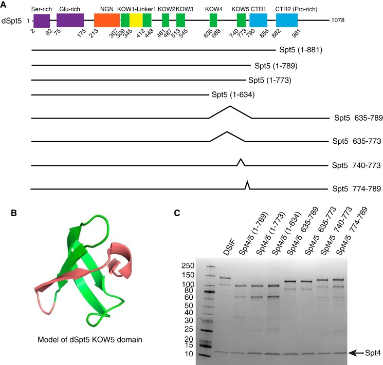
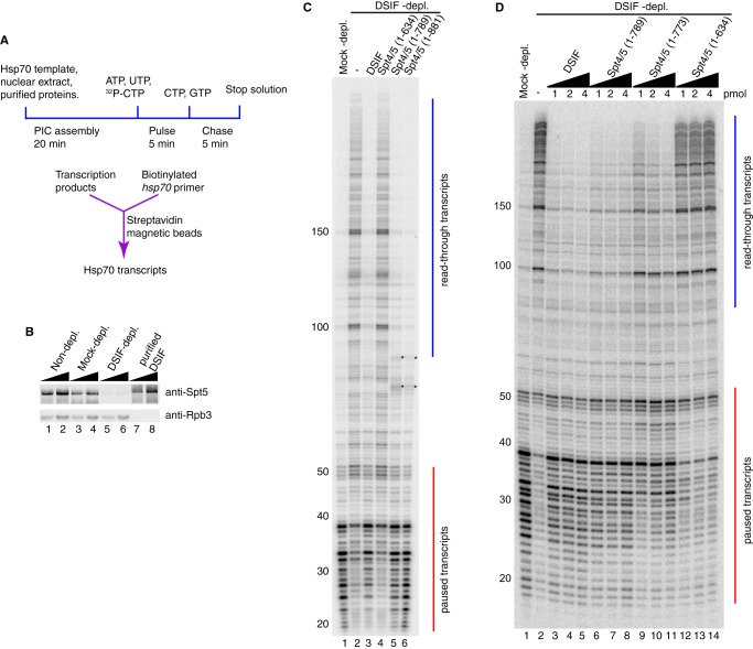
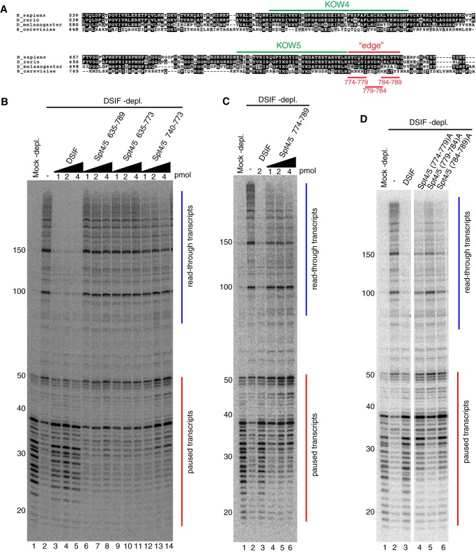
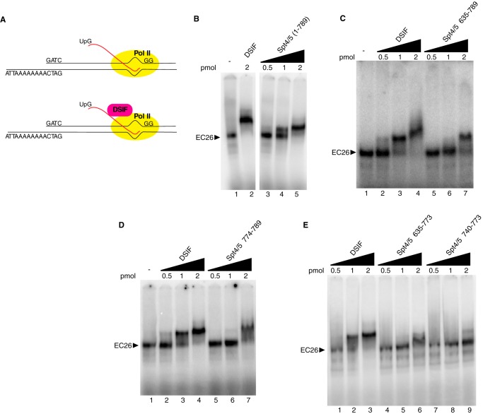
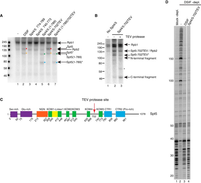
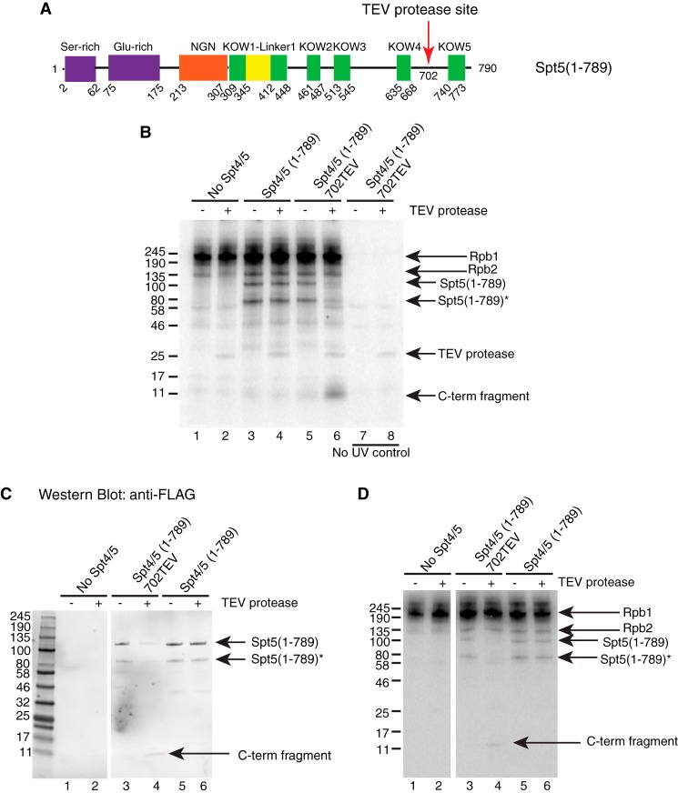
DRB sensitivity-inducing factor (DSIF or Spt4/5) is a conserved transcription elongation factor that both inhibits and stimulates transcription elongation in metazoans. In Drosophila and vertebrates, DSIF together with negative elongation factor (NELF) associates with RNA polymerase II during early elongation and causes RNA polymerase II to pause in the promoter-proximal region of genes. The mechanism of how DSIF establishes pausing is not known. We constructed Spt5 mutant forms of DSIF and tested their capacity to restore promoter-proximal pausing to DSIF-depleted Drosophila nuclear extracts. The C-terminal repeat region of Spt5, which has been implicated in both inhibition and stimulation of elongation, is dispensable for promoter-proximal pausing. A region encompassing KOW4 and KOW5 of Spt5 is essential for pausing, and mutations in KOW5 specifically shift the location of the pause. RNA cross-linking analysis reveals that KOW5 directly contacts the nascent transcript, and deletion of KOW5 disrupts this interaction. Our results suggest that KOW5 is involved in promoter-proximal pausing through contact with the nascent RNA.

Keywords: DSIF; KOW domain; RNA polymerase II; RNA-protein interaction; Spt4/5; mutagenesis; promoter-proximal pausing; transcription elongation factor; transcription regulation.


Conflict of interest statement

The authors declare that they have no conflicts of interest with the contents of this article

Figures

FIGURE 1.
FIGURE 1.
Design of Spt5 mutants and purification of DSIF derivatives. A, diagram showing domains of Drosophila Spt5 and some of the Spt5 mutants. The region encompassing KOW4, KOW5, and the CTR were subjected to mutagenesis. B, the structure of dSpt5 KOW5 domain modeled by Phyre2 (59) using the NMR structure of hSpt5 KOW5 domain (PDB code 2E70) as a template, displayed in PyMOL (version 1.7.4, Schrödinger, LLC). The green part corresponds to the KOW motif spanning from amino acid 740 to 773. The red part corresponds to the “edge” of KOW5 spanning from amino acid 774 to 789. C, DSIF with different Spt5 derivatives was expressed in E. coli cells and purified through two affinity columns, binding Spt4-His and Spt5-FLAG subunits, respectively. The largest polypeptide corresponds to full-length Spt5, and the next largest polypeptide is an Spt5 breakdown product caused by loss of part of the N terminus. Purified proteins were analyzed on a 4–20% SDS-polyacrylamide gel and stained with Coomassie Blue.
FIGURE 2.
FIGURE 2.
In vitro transcription with different versions of DSIF reveals a region in Spt5 important for promoter-proximal pausing. A, diagram of the in vitro transcription assay. Normal and mutant DSIF proteins were added to nuclear extracts depleted of endogenous DSIF and then incubated with a plasmid containing the hsp70 promoter. Transcripts were radiolabeled by a pulse-chase procedure and purified by hybridizing to biotinylated hsp70 oligonucleotides that were then captured by streptavidin magnetic beads. Hsp70 transcripts were analyzed on a 10% polyacrylamide gel containing 8 m urea. B, Western blotting analysis of nuclear extracts and recombinant DSIF probed with antiserum against Spt5. Lanes 1–6 show two amounts each of non-depleted extract, mock-depleted extract, and DSIF-depleted extract, corresponding to one-eighth or one-fourth of nuclear extract used in each in vitro transcription reaction. Lanes 7 and 8 correspond to 0.125 and 0.25 pmol of recombinant DSIF. Rpb3 is a subunit of RNA polymerase II. C and D, DSIF with different Spt5 mutations was compared with normal DSIF for pausing activity. Red vertical lines from +20 to +50 indicate paused transcripts. Blue vertical lines from +100 and beyond indicate read-through transcripts. C, truncation mutants Spt4/5(1–881), Spt4/5(1–789), and Spt4/5(1–635) were first tested. Spt4/5(1–881) and Spt4/5(1–789) restored pausing similarly as normal DSIF. Spt4/5(1–635) failed to restore pausing. The asterisks in lanes 5 and 6 indicate contaminating radiolabeled nucleic acids generated by the extract independently of Pol II (7). The results shown in C are representative of three independent experiments. D, 1, 2, and 4 pmol of Spt4/5(1–789), Spt4/5(1–773), and Spt4/5(1–635) were further tested. The amount of protein used in each reaction is shown above each lane. Spt4/5(1–789) restored pausing. Spt4/5(1–635) failed to restore pausing. Spt4/5(1–773) partially restored pausing and shifted the pause sites downstream. The results shown in D are representative of two independent experiments.
FIGURE 3.
FIGURE 3.
Mutations in KOW5 shift the pause site downstream. A, alignment of the amino acid sequences of Spt5 from four species was performed with Clustal Omega (60). The region encompassing KOW4 and KOW5 is shown. The similarity is displayed by BoxShade (version 3.21), with black or gray background indicating identical or similar residues, respectively. KOW4 and KOW5 represent the initially defined KOW motif. Additional amino acids that form a Tudor-like domain with the KOW5 motif are noted as the “edge” of the KOW5 domain. The alanine substitutions in the edge region are shown in red. B–D, in vitro transcription of hsp70 was done as described in the legend to Fig. 2. B, 1, 2, or 4 pmol of each protein was used as indicated above each lane. Δ635–789 and Δ635–773 failed to restore pausing with all three amounts. Δ740–773 partially restored pausing and shifted the pause location downstream. The addition of more protein increased the intensity of paused transcripts but did not shift them back to their normal location. C, 1, 2, or 4 pmol of Δ774–789 was used as indicated above each lane. 2 pmol of normal DSIF was used as a positive control. Δ774–789 partially restored pausing and shifted the pause sites downstream. The addition of more protein increased the intensity of paused transcripts but did not shift them back to the normal pause location. The results shown in B and C are representative of two independent experiments. D, three alanine substitution mutants in the edge of KOW5 (A) were tested along with normal DSIF for pausing activity. 2 pmol of each protein was used. All three mutants partially restored pausing and shifted the pause sites downstream. Mutant (779–784)A shows a more severe defect in pausing compared with the other two mutants. The results shown in D are representative of three independent experiments.
FIGURE 4.
FIGURE 4.
DSIF mutants defective in pausing have lower binding affinity for elongation complexes. A, elongation complexes were formed by initiating transcription with purified Pol II, UpG, ATP, UTP, radioactive CTP, and O-methyl-GTP on a tailed template with a 26-nucleotide G-less cassette. After the complexes were stalled at the end of the G-less cassette, they were incubated with increasing amounts of purified normal or mutant DSIF proteins and then analyzed on 4% native gels. B–E, normal or mutant DSIF was added to the stalled EC at 0.5, 1, and 2 pmol to compare their affinity with EC26. DSIF shifted more than 50% of EC26 at 1 pmol and 100% of EC26 at 2 pmol. Spt4/5(1–789) showed similar affinity as DSIF. Spt4/5Δ635–789 did not shift EC26 at 1 pmol and partially shifted it at 2 pmol. Spt5Δ774–789 shifted most of EC26 at 2 pmol, forming an indistinct band. Spt5Δ635–773 and Spt5Δ740–773 partially shifted EC26 at 2 pmol. The results shown are representative of two independent experiments.
FIGURE 5.
FIGURE 5.
UV cross-linking suggests that KOW5 is important for the contact with the nascent RNA. Elongation complexes were generated as described in Fig. 4 except that UTP was replaced with photoreactive bromo-UTP. After UV treatment, the samples were digested with DNase I and RNase A. Proteins were analyzed by 7% Tris acetate PAGE. A, UV cross-linking in the absence (lane 1) or presence of various Spt4/5 derivatives (lanes 2–7). Cross-linked Rpb1 (245 kDa) and Rpb2 (135 kDa) are detected in every lane. The addition of DSIF results in a cross-linked band just above Rpb2, corresponding to full-length Spt5 (lane 2, red dot). The addition of Spt4/5(1–789) results in a cross-linked band at 100 kDa (lane 5, green dot). The addition of mutant Δ774–789 or Δ740–773 did not result in cross-linked Spt5 (lanes 3 and 4). The orange dot in lane 2 and blue dot in lane 5 identify the breakdown products of Spt5 derivatives that are evident in Fig. 1C. Spt5–702TEV and Spt5(1–789)702TEV cross-linked in the same way as the corresponding proteins lacking the TEV site (compare lanes 6 and 7 with lanes 2 and 5). B, TEV digestion of cross-linked Spt5–702TEV reveals cross-linking of RNA on both sides of the 702TEV site. Proteins were digested with TEV protease after UV cross-linking and before nuclease treatment. Lanes 2 and 3 show the cross-linked products before and after TEV digestion, respectively. After digestion, two bands at 100 kDa and around 46 kDa corresponding to predicted N-terminal and C-terminal fragments of Spt5–702TEV were detected (lane 3). The band between 58 and 80 kDa in lane 3 (asterisk) is likely to be the N-terminal fragment from the breakdown product marked as Spt5–702TEV*. C, schematic showing the location of a TEV protease site inserted between KOW4 and KOW5 domains in full-length Spt5. D, in vitro transcription assay indicates that Spt4/5–702TEV has pausing activity similar to that of normal DSIF.
FIGURE 6.
FIGURE 6.
UV cross-linking indicates that KOW5 directly contacts with the nascent RNA. A UV-cross-linking assay was performed as described in the legend to Fig. 5. For TEV-digested samples, TEV protease was added 1 h before nuclease treatment. Proteins were analyzed by 4–20% SDS-PAGE. A, diagram showing the location of a TEV protease site inserted between the KOW4 and KOW5 domains in CTR-deleted Spt5. B, TEV digestion following UV cross-linking of the CTR-deleted Spt5 with the 702TEV site. Before TEV digestion, cross-linked Spt5(1–789) and its breakdown product were detected in lanes 3 and 5. After digestion, a ∼10 kDa band corresponding to the predicted C-terminal fragment of Spt5(1–789)702TEV was detected in lane 6 but not in lane 4. The 10 kDa band was not detected in samples without UV treatment (lanes 7 and 8). It is not known why the TEV protease is labeled with radioactivity even in the absence of UV treatment. C and D, after UV cross-linking and TEV digestion, the products were separated on a 4–20% SDS-polyacrylamide gel and transferred to a nitrocellulose membrane for Western blotting. The blot was probed with FLAG antibody and later exposed to a phosphor screen to detect radiolabeled proteins. C, Western blotting image showing that the 10-kDa digestion fragment, as well as the full-length and breakdown versions of Spt5(1–789), is FLAG-tagged. D, phosphor image showing proteins that are cross-linked to radiolabeled RNA.
FIGURE 7.
FIGURE 7.
Mutations in KOW4-5 region of Spt5 fail to rescue the lethality caused by Spt5 RNAi in flies. A, Western blotting analysis for ubiquitous expression of Spt5 derivatives in flies. Whole fly samples were prepared from pupae of the matings between Spt5 derivative lines and an actin-Gal4 driver line (BDSC 4414). BDSC 4414 alone was used as a negative control that does not ectopically express Spt5 derivatives. FLAG antibody detects ectopically expressed Spt5 derivatives. Spt5 antibody detects both endogenous and ectopically expressed Spt5. M1BP antibody was used to detect M1BP as a loading control. B, Western blotting analysis for expression of Spt5 derivatives in salivary glands. Glands were isolated from third instar larvae of the matings between Spt5 derivative lines and a salivary gland Gal4 driver line (BDSC 1824). BDSC 1824 alone was used as a negative control. Rpb3 antibody was used to detect Rpb3 as a loading control. C, fly rescue assay for Spt5 derivatives. Spt5 lines with a Gal4-regulated Spt5 RNAi transgene or the Spt5 RNAi line alone were mated to actin-Gal4/CyO (BDSC 4414). In total, 158, 258, 157, 136, and 136 adult flies were generated from Spt5 RNAi, Spt5 WT, Spt5 Δ774–789, Spt5 Δ635–789, and Spt5 (779–784)A, respectively. The percentage of straight wing flies and curly wing flies in the progeny were calculated. Spt5 WT generated more than 30% of straight wing flies. Spt5 Δ635–789 generated less than 1.5% straight wing flies. Spt5 RNAi, Spt5 Δ774–789, and Spt5 (779–784)A generated no straight wing flies.
FIGURE 8.
FIGURE 8.
KOW4-5 deletion causes reduced affinity of Spt5 for chromosomes. A, immunofluorescence micrographs of polytene chromosomes of larvae from normal and mutant Spt5 lines mated to the salivary gland driver line BDSC 1824. Signal from FLAG staining is shown in the red channel. Signal from Rpb3 staining is shown in the green channel. DNA staining is shown in the blue channel. All four Spt5 derivatives associated with chromosomes and co-localized with Pol II at many loci. The exposure time was optimized for each channel in each image. B, direct comparison between Spt5 WT and Spt5Δ635–789 on the same slide. MSL-2 staining for the X chromosome of male larvae was used to distinguish males containing one Spt5 transgene and females containing the other Spt5 transgene. A field with chromosomes from both lines was selected for the pictures in the first row. The Spt5 WT and Spt5Δ635–789 chromosomes are outlined with green and red boxes, respectively. Magnified views of the chromosomes in the first row are displayed in the second and third rows. Spt5Δ635–789 showed significantly weaker FLAG signal than Spt5 WT regardless of sex. C, ChIP analysis on the hsp70 promoter. Spt5 WT and Spt5Δ635–789 were mated to salivary gland Gal4 driver line BDSC1824, and glands were prepared for ChIP with FLAG, Rpb3, and Spt5 antibodies. Glands from the BDSC1824 line, which does not express FLAG-tagged protein, was used as a negative control. All three lines showed similar enrichment for Rpb3 and total Spt5. The ChIP signal for FLAG-Spt5 WT was well above background or pre-immune control, whereas the ChIP signal for FLAG-Spt5Δ635–789 was near background. Three or more biological replicates were used for each ChIP experiment. Error bars, S.E.

References

    1. Muse G. W., Gilchrist D. A., Nechaev S., Shah R., Parker J. S., Grissom S. F., Zeitlinger J., and Adelman K. (2007) RNA polymerase is poised for activation across the genome. Nat. Genet. 39, 1507–1511 - PMC - PubMed
    1. Zeitlinger J., Stark A., Kellis M., Hong J.-W., Nechaev S., Adelman K., Levine M., and Young R. A. (2007) RNA polymerase stalling at developmental control genes in the Drosophila melanogaster embryo. Nat. Genet. 39, 1512–1516 - PMC - PubMed
    1. Core L. J., Waterfall J. J., and Lis J. T. (2008) Nascent RNA sequencing reveals widespread pausing and divergent initiation at human promoters. Science 322, 1845–1848 - PMC - PubMed
    1. Lee C., Li X., Hechmer A., Eisen M., Biggin M. D., Venters B. J., Jiang C., Li J., Pugh B. F., and Gilmour D. S. (2008) NELF and GAGA factor are linked to promoter-proximal pausing at many genes in Drosophila. Mol. Cell. Biol. 28, 3290–3300 - PMC - PubMed
    1. Nechaev S., Fargo D. C., dos Santos G., Liu L., Gao Y., and Adelman K. (2010) Global analysis of short RNAs reveals widespread promoter-proximal stalling and arrest of Pol II in Drosophila. Science 327, 335–338 - PMC - PubMed

MeSH terms

LinkOut - more resources