. 2017 Apr 17;83(9):e03389-16.
doi: 10.1128/AEM.03389-16. Print 2017 May 1.

Accurate Quantification of Laminarin in Marine Organic Matter with Enzymes from Marine Microbes

Affiliations

Accurate Quantification of Laminarin in Marine Organic Matter with Enzymes from Marine Microbes

Stefan Becker et al. Appl Environ Microbiol. .

Abstract

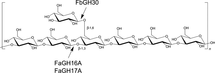
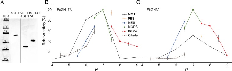
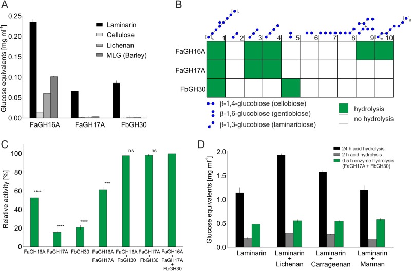
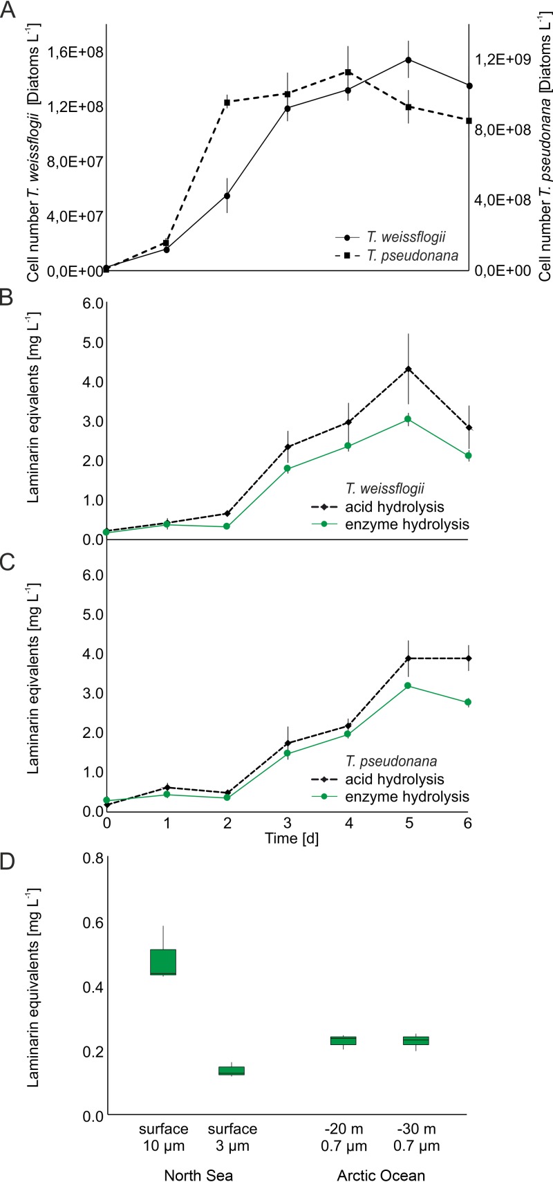
Marine algae produce a variety of glycans, which fulfill diverse biological functions and fuel the carbon and energy demands of heterotrophic microbes. A common approach to analysis of marine organic matter uses acid to hydrolyze the glycans into measurable monosaccharides. The monosaccharides may be derived from different glycans that are built with the same monosaccharides, however, and this approach does not distinguish between glycans in natural samples. Here we use enzymes to digest selectively and thereby quantify laminarin in particulate organic matter. Environmental metaproteome data revealed carbohydrate-active enzymes from marine flavobacteria as tools for selective hydrolysis of the algal β-glucan laminarin. The enzymes digested laminarin into glucose and oligosaccharides, which we measured with standard methods to establish the amounts of laminarin in the samples. We cloned, expressed, purified, and characterized three new glycoside hydrolases (GHs) of Formosa bacteria: two are endo-β-1,3-glucanases, of the GH16 and GH17 families, and the other is a GH30 exo-β-1,6-glucanase. Formosa sp. nov strain Hel1_33_131 GH30 (FbGH30) removed the β-1,6-glucose side chains, and Formosa agariphila GH17A (FaGH17A) and FaGH16A hydrolyzed the β-1,3-glucose backbone of laminarin. Specificity profiling with a library of glucan oligosaccharides and polysaccharides revealed that FaGH17A and FbGH30 were highly specific enzymes, while FaGH16A also hydrolyzed mixed-linked glucans with β-1,4-glucose. Therefore, we chose the more specific FaGH17A and FbGH30 to quantify laminarin in two cultured diatoms, namely, Thalassiosira weissflogii and Thalassiosira pseudonana, and in seawater samples from the North Sea and the Arctic Ocean. Combined, these results demonstrate the potential of enzymes for faster, stereospecific, and sequence-specific analysis of select glycans in marine organic matter.IMPORTANCE Marine algae synthesize substantial amounts of the glucose polymer laminarin for energy and carbon storage. Its concentrations, rates of production by autotrophic organisms, and rates of digestion by heterotrophic organisms remain unknown. Here we present a method based on enzymes that hydrolyze laminarin and enable its quantification even in crude substrate mixtures, without purification. Compared to the commonly used acid hydrolysis, the enzymatic method presented here is faster and stereospecific and selectively cleaves laminarin in mixtures of glycans, releasing only glucose and oligosaccharides, which can be easily quantified with reducing sugar assays.

Keywords: algal bloom; carbon cycle; diatoms; glycobiology; glycoside hydrolase; laminarin; laminarinase; marine microbes; organic matter; β-glucan.


Figures

FIG 1
FIG 1
Laminarin structure and enzyme activities. The linear storage glucan consists of a β-1,3-d-glucose polysaccharide with β-1,6-linked monomer side chains. The characterization of three enzymes from marine Bacteroidetes, belonging to the GH16, GH17, and GH30 families, presented here showed that the GH30 enzyme hydrolyzed the β-1,6-linked side chain and the GH17 and GH16 enzymes hydrolyzed the β-1,3-d-linked main chain of laminarin.
FIG 2
FIG 2
Recombinant glycoside hydrolases from marine Bacteroidetes showing greatest activity in MOPS buffer at neutral pH. (A) SDS-PAGE analysis of purified enzymes. One gel is shown. Unimportant lanes were intentionally omitted, as indicated by vertical lines. Approximately 0.5 μg of each protein was loaded on the gel. All proteins were run on the same gel, and lanes were spliced for clarity. (B and C) Enzymatic rates were measured with 0.1% (wt/vol) laminarin, which was hydrolyzed by 100 nM (∼5.0 μg ml−1) purified FaGH17A (B) or FbGH30 (C) at 37°C for 30 min in 50 mM buffer. The greatest activity rate was observed in MOPS buffer at pH 7.0 and was set as the 100% reference value. MES, morpholineethanesulfonic acid; MMT, malic acid-MES-Tris base.
FIG 3
FIG 3
Glycoside hydrolases showing different levels of specificity for laminarin and related glucans. (A) Activity tests with glucan polysaccharides and oligosaccharides containing β-1,3, β-1,4, and β-1,6 linkages, based on the PAHBAH reducing sugar assay. Shown are mean and standard deviation (SD) values from three technical replicates. (B) Enzyme specificity tested with defined oligosaccharide substrates, using thin-layer chromatography. The results are presented as a heatmap (see Fig. S3 in the supplemental material). The substrates at 0.1% (wt/vol) were hydrolyzed for 30 min at 37°C by 100 nM (∼5 μg ml−1) purified enzyme in PBS buffer at pH 7.5. (C) Mixtures of FbGH30 and FaGH17A or FbGH30 and FaGH16A, showing greater activity than the individual enzymes. The highest activity level with all three enzymes was set to 100%, and all other samples were compared to that value. Laminarin at 0.1% (wt/vol) was hydrolyzed for 30 min at 37°C by 100 nM (∼5.0 μg ml−1) of each purified enzyme in PBS buffer at pH 7.5, and hydrolysis was measured with the PAHBAH assay. Shown are mean and SD values from three technical replicates. ****, P < 0.0001; ***, P < 0.001, independent two-sample Student's t test. ns, not significant. (D) Comparison of hydrolysis yields of enzymatic, partial acid, and total acid hydrolysis of different polysaccharides. Lichenan, carrageenan, and mannan at 0.1% (wt/vol) were added to 0.1% (wt/vol) laminarin and were hydrolyzed for 30 min at 37°C with 100 nM purified enzyme (∼5 μg ml−1 of FaGH16A, FaGH17A, or FbGH30) in 50 mM MOPS buffer. Boiling for 5 min at 100°C stopped the reaction. Partial acid hydrolysis was conducted for 2 h at 20°C with 50 mM H2SO4. Total acid hydrolysis was carried out for 24 h at 100°C with 1 M HCl. The reaction mixtures were analyzed with the PAHBAH assay. All experiments were carried out in triplicate. Shown are mean and SD values from three technical replicates.
FIG 4
FIG 4
Laminarin quantification in Thalassiosira weissflogii and T. pseudonana laboratory cultures and in environmental samples. (A) Numbers of diatoms cultured in modified ESAW/HESNW medium at 15°C, with a 12-h/12-h light/dark cycle, for 6 days. (B) Quantification of laminarin extracted from T. weissflogii by acid (dashed black line) or enzyme (solid green line) hydrolysis. (C) Quantification of laminarin extracted from T. pseudonana by acid (dashed black line) or enzyme (solid green line) hydrolysis. (D) Laminarin contents of particulate organic matter, which was concentrated by filtering seawater from the North Sea near Helgoland (spring 2009) and from the Arctic Ocean near Svalbard (summer 2015) and enzymatically hydrolyzed. Water-soluble extracts were hydrolyzed for 30 min at 37°C with 100 nM (∼5.0 μg ml−1) FaGH17A and FbGH30, with 1 mg ml−1 BSA, in 50 mM MOPS buffer at pH 7. Alternatively, the extracted polysaccharides were hydrolyzed twice for 2 h at 20°C with 50 mM H2SO4, with shaking at 1,500 rpm. The products were quantified with the PAHBAH reducing sugar assay. The calibration curve was prepared with laminarin hydrolyzed with enzyme or acid, and the reducing sugar signals were measured as described above (see Fig. S5).

References

    1. Field C, Behrenfeld M, Randerson J, Falkowski P. 1998. Primary production of the biosphere: integrating terrestrial and oceanic components. Science 281:237–240. doi: 10.1126/science.281.5374.237. - DOI - PubMed
    1. Myklestad S. 1974. Production of carbohydrates by marine planktonic diatoms. I. Comparison of nine different species in culture. J Exp Mar Biol Ecol 15:261–274.
    1. Painter TJ. 1983. Algal polysaccharides, p 195–285. In Aspinall GO. (ed), The polysaccharides. Academic Press, New York, NY.
    1. Senni K, Pereira J, Gueniche F, Delbarre-Ladrat C, Sinquin C, Ratiskol J, Godeau G, Fischer AM, Helley D, Colliec-Jouault S. 2011. Marine polysaccharides: a source of bioactive molecules for cell therapy and tissue engineering. Mar Drugs 9:1664–1681. doi: 10.3390/md9091664. - DOI - PMC - PubMed
    1. Gal A, Wirth R, Kopka J, Fratzl P, Faivre D, Scheffel A. 2016. Macromolecular recognition directs calcium ions to coccolith mineralization sites. Science 353:590–593. doi: 10.1126/science.aaf7889. - DOI - PubMed

Publication types

MeSH terms