. 2018 Mar;28(2):225-233.
doi: 10.1111/bpa.12496. Epub 2017 Apr 6.

Diagnostic red flags: steroid-treated malignant CNS lymphoma mimicking autoimmune inflammatory demyelination

Affiliations

Diagnostic red flags: steroid-treated malignant CNS lymphoma mimicking autoimmune inflammatory demyelination

Alonso Barrantes-Freer et al. Brain Pathol. 2018 Mar.

Abstract

The presence of inflammation and demyelination in a central nervous system (CNS) biopsy points towards a limited, yet heterogeneous group of pathologies, of which multiple sclerosis (MS) represents one of the principal considerations. Inflammatory demyelination has also been reported in patients with clinically suspected primary central nervous system lymphoma (PCNSL), especially when steroids had been administered prior to biopsy acquisition. The histopathological changes induced by corticosteroid treatment can range from mild reduction to complete disappearance of lymphoma cells. It has been proposed that in the absence of neoplastic B cells, these biopsies are indistinguishable from MS, yet despite the clinical relevance, no histological studies have specifically compared the two entities. In this work, we analyzed CNS biopsies from eight patients with inflammatory demyelination in whom PCNSL was later histologically confirmed, and compared them with nine well defined early active multiple sclerosis lesions. In the patients with steroid-treated PCNSL (ST-PCNSL) the interval between first and second biopsy ranged from 3 to 32 weeks; all of the patients had received corticosteroids before the first, but not the second biopsy. ST-PCNSL patients were older than MS patients (mean age: ST-PCNSL: 62 ± 4 years, MS: 30 ± 2 years), and histological analysis revealed numerous apoptoses, patchy and incomplete rather than confluent and complete demyelination and a fuzzy lesion edge. The loss of Luxol fast blue histochemistry was more profound than that of myelin proteins in immunohistochemistry, and T cell infiltration in ST-PCNSL exceeded that in MS by around fivefold (P = 0.005). Our data indicate that in the presence of extensive inflammation and incomplete, inhomogeneous demyelination, the neuropathologist should refrain from primarily considering autoimmune inflammatory demyelination and, even in the absence of lymphoma cells, instigate close clinical follow-up of the patient to detect recurrent lymphoma.

Keywords: corticosteroids; demyelination; diffuse large B cell lymphoma; inflammation; multiple sclerosis.


Conflict of interest statement

The authors do not report any conflict of interest.

Figures
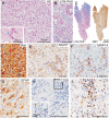
Figure 1
Figure 1
Histology of a frontal brain lesion biopsy obtained under systemic corticosteroid therapy 5 months before histological confirmation of a diffuse large B cell lymphoma. A. Representative HE microphotograph showing marked hypercellularity with extensive infiltration of macrophages and mononuclear cells, loosening of the tissue matrix and scattered apoptoses (inset, arrowhead). B. LFB histochemistry revealing extensive, but in part incomplete demyelination. C. The reduction in myelin density is more apparent in LFB/PAS histochemistry (left) than in MBP IHC (right). D. Bielschowsky silver impregnation indicates a distended but preserved axonal lattice. E. KiM1P IHC showing a prominent infiltration of macrophages with a perivascular as well as diffuse distribution in the lesion. F. Numerous macrophages express MRP14 indicating recent invasion. G. GFAP IHC demonstrating reactive gliosis. H. Few, mostly perivascular, CD20+ B lymphocytes without evident pleomorphic features (inset) are observed. I. Abundant perivascular and parenchymal infiltration of CD3+ T lymphocytes. Biel: Bielschowsky silver impregnation. CD = cluster of differentiation; IHC = immunhistochemistry; MRP14 = migration inhibitory factor‐related protein 14; MBP = myelin basic protein; GFAP = glial fibrillary acidic protein. Unless explicitly stated scale bars = 100 µm.
Figure 2
Figure 2
Characteristics of demyelination in ST‐PCNSL and MS. High magnification images of tissue sections stained with LFB/PAS. A and C–E. ST‐PCNSL lesions showing incomplete and patchy demyelination. Note the sparse and coarse seemingly swollen or disrupted myelin sheaths adjacent to and within areas of frank demyelination. The vessel walls appear thickened and exhibit an intense PAS staining. Numerous macrophages containing PAS‐positive degradation products can be observed. B and F–H. MS lesions show confluent, near complete myelin loss with no or only few residual myelin sheaths. Unlike ST‐PCNSL lesions, no evident thickening or intense PAS positivity of the vessel walls can be observed. LFB/PAS = Luxol fast blue/Periodic acid Schiff. Scale bars = 100 µm.
Figure 3
Figure 3
The extent of lymphocytic infiltration is a key histological hallmark in the differential diagnosis of ST‐PCNSL and MS. Representative microphotographs of CD3 IHC showing a dense lymphocyte infiltration (CD3, brown) in ST‐PCNSL (A and C,D) as compared with MS (B and E,F). Note the predominantly perivascular localization of T cells in MS (B and E,F) and the more diffuse, but also in part perivascular distribution in ST‐PCNSL (A and C,D). Scale bars = 100 µm.

References

    1. Absinta M, Sati P, Reich DS (2016) Advanced MRI and staging of multiple sclerosis lesions. Nat Rev Neurol 12:358–368. - PMC - PubMed
    1. Alderson L, Fetell MR, Sisti M, Hochberg F, Cohen M, Louis DN (1996) Sentinel lesions of primary CNS lymphoma. J Neurol Neurosurg Psychiatry 60:102–105. - PMC - PubMed
    1. Brecher K, Hochberg FH, Louis DN, de la Monte S, Riskind P (1998) Case report of unusual leukoencephalopathy preceding primary CNS lymphoma. J Neurol Neurosurg Psychiatry 65:917–920. - PMC - PubMed
    1. Brück W, Brunn A, Klapper W, Kuhlmann T, Metz I, Paulus W, Deckert M (2013) Differenzialdiagnose lymphoider Infiltrate im Zentralnervensystem. Der Pathologe 34:186–197. - PubMed
    1. Brück W, Porada P, Poser S, Rieckmann P, Hanefeld F, Kretzschmarch HA, Lassmann H (1995) Monocyte/macrophage differentiation in early multiple sclerosis lesions. Ann Neurol 38:788–796. - PubMed

Publication types

MeSH terms

Substances