

Dental Pulp Tissue Regeneration Using Dental Pulp Stem Cells Isolated and Expanded in Human Serum
- PMID: 28216268
- PMCID: PMC5797986
- DOI: 10.1016/j.joen.2016.11.018
Dental Pulp Tissue Regeneration Using Dental Pulp Stem Cells Isolated and Expanded in Human Serum
Abstract
Introduction: Dental pulp-derived stem cells (DPSCs) have the potential to regenerate dentin and dental pulp tissue because of their differentiation capacity and angiogenic properties. However, for regenerative approaches to gain regulatory and clinical acceptance, protocols are needed to determine more feasible ways to cultivate DPSCs, namely, without the use of xenogeneic-derived components (animal sera) and exogenous growth factors.
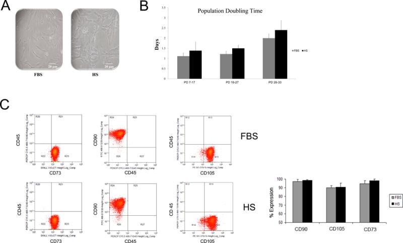
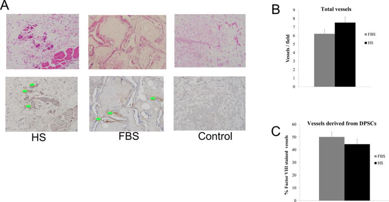
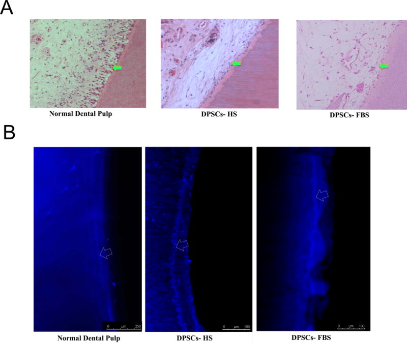
Methods: In this study, human DPSCs were isolated from third molars and expanded in standard culture conditions containing fetal bovine serum (DPSCs-FBS) or conditions containing human serum (DPSCs-HS). After cell characterization and evaluation of their angiogenic secretome, DPSCs were seeded in tooth slice/scaffolds and implanted subcutaneously into immunodeficient mice. After 30 days, tooth slices were retrieved and evaluated for dental pulp tissue regeneration. Immunohistochemistry and confocal microscopy were used to quantify blood vessel formation and evaluate predentin and dentin formation.
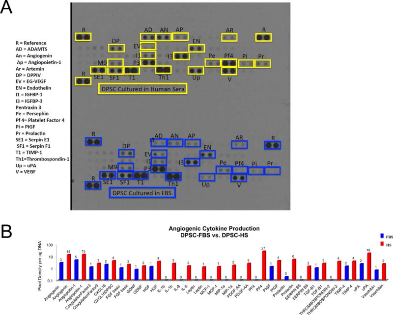
Results: After culture, DPSCs-HS produced concentrations of angiogenic growth factors equivalent to DPSCs-FBS. Additionally, in DPSCs-HS, several angiogenic factors were produced in at least 1-fold higher concentrations than in DPSCs-FBS. In vivo, it was determined that DPSCs-HS produced a robust angiogenic response and regeneration of dentin equivalent to DPSCs-FBS.
Conclusions: These findings show that DPSCs can be isolated and expanded to clinical scale numbers in media devoid of animal serum or exogenous growth factors and still maintain their pulp regenerative properties. The implications of these findings are significant for further development of clinical protocols using DPSCs in cell therapies.
Keywords: Angiogenesis; cell therapy; dental pulp stem cells; dentin; pulp tissue engineering.
Copyright © 2016 American Association of Endodontists. Published by Elsevier Inc. All rights reserved.
Conflict of interest statement
Acknowledgement: The authors deny any conflicts of interest
Figures
References
-
- Hara K, Yamada Y, Nakamura S, Umemura E, Ito K, Ueda M. Potential characteristics of stem cells from human exfoliated deciduous teeth compared with bone marrow-derived mesenchymal stem cells for mineralized tissue-forming cell biology. Journal of endodontics. 2011;37(12):1647–1652. - PubMed
-
- Sakai VT, Zhang Z, Dong Z, Neiva KG, Machado MA, Shi S, et al. SHED differentiate into functional odontoblasts and endothelium. Journal of dental research. 2010;89(8):791–796. - PubMed
-
- Osathanon T, Sawangmake C, Nowwarote N, Pavasant P. Neurogenic differentiation of human dental pulp stem cells using different induction protocols. Oral diseases. 2013 - PubMed
MeSH terms
Substances
Grants and funding
LinkOut - more resources
Full Text Sources
Other Literature Sources
Medical
