. 2017 Feb 3:8:32.
doi: 10.3389/fphar.2017.00032. eCollection 2017.

Feedback Activation of Basic Fibroblast Growth Factor Signaling via the Wnt/β-Catenin Pathway in Skin Fibroblasts

Affiliations

Feedback Activation of Basic Fibroblast Growth Factor Signaling via the Wnt/β-Catenin Pathway in Skin Fibroblasts

Xu Wang et al. Front Pharmacol. .

Abstract

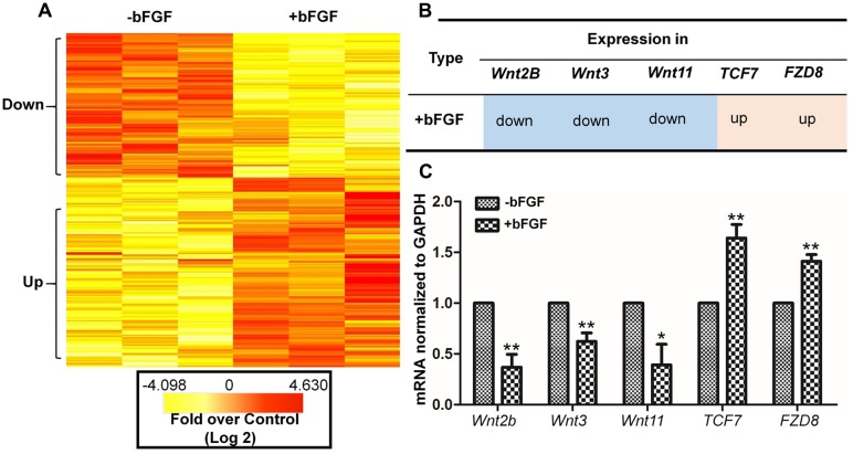
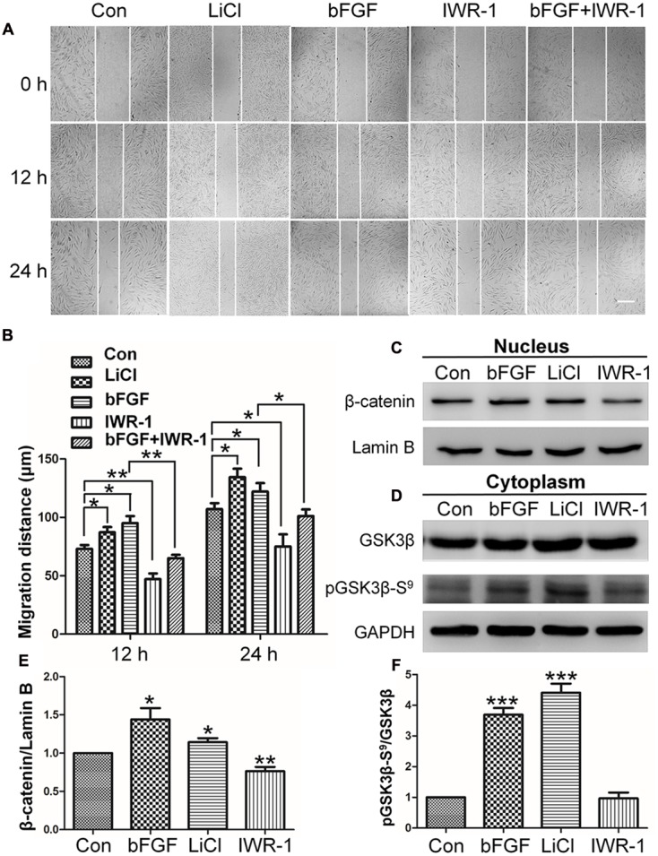
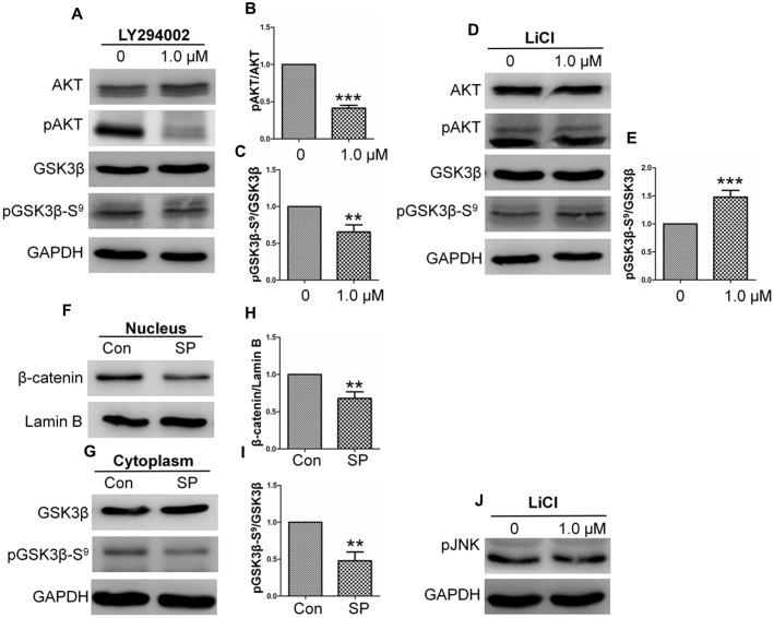
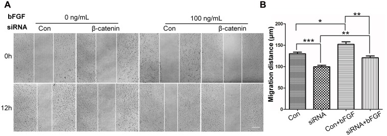
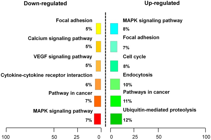
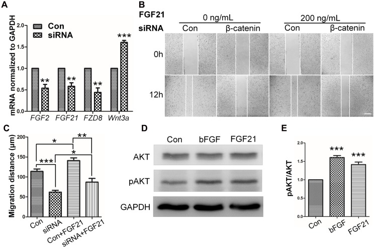
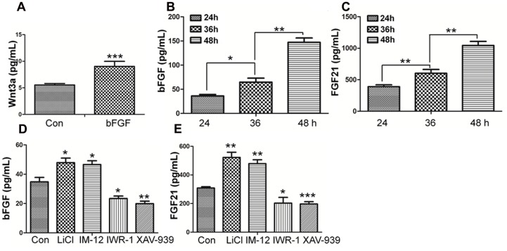
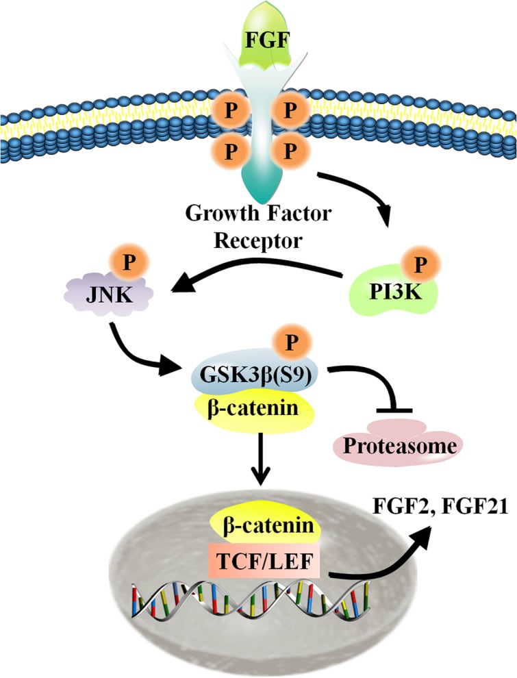
Skin wound healing is a complex process requiring the coordinated behavior of many cell types, especially in the proliferation and migration of fibroblasts. Basic fibroblast growth factor (bFGF) is a member of the FGF family that promotes fibroblast migration, but the underlying molecular mechanism remains elusive. The present RNA sequencing study showed that the expression levels of several canonical Wnt pathway genes, including Wnt2b, Wnt3, Wnt11, T-cell factor 7 (TCF7), and Frizzled 8 (FZD8) were modified by bFGF stimulation in fibroblasts. Enzyme-linked immunosorbent assay (ELISA) analysis also showed that Wnt pathway was activated under bFGF treatment. Furthermore, treatment of fibroblasts with lithium chloride or IWR-1, an inducer and inhibitor of the Wnt signaling pathway, respectively, promoted and inhibited cell migration. Also, levels of cytosolic glycogen synthase kinase 3 beta phosphorylated at serine9 (pGSK3β Ser9) and nuclear β-catenin were increased upon exposure to bFGF. Molecular and biochemical assays indicated that phosphoinositide 3-kinase (PI3K) signaling activated the GSK3β/β-catenin/Wnt signaling pathway via activation of c-Jun N-terminal kinase (JNK), suggesting that PI3K and JNK act at the upstream of β-catenin. In contrast, knock-down of β-catenin delayed fibroblast cell migration even under bFGF stimulation. RNA sequencing analysis of β-catenin knock-down fibroblasts demonstrated that β-catenin positively regulated the transcription of bFGF and FGF21. Moreover, FGF21 treatment activated AKT and JNK, and accelerated fibroblast migration to a similar extent as bFGF does. In addition, ELISA analysis demonstrated that both of bFGF and FGF21 were auto secretion factor and be regulated by Wnt pathway stimulators. Taken together, our analyses define a feedback regulatory loop between bFGF (FGF21) and Wnt signaling acting through β-catenin in skin fibroblasts.

Keywords: GSK3β; Wnt signaling pathway; bFGF; cell migration; transcriptome; β-catenin.


Figures

FIGURE 1
FIGURE 1
Basic fibroblast growth factor-regulated Wnt signaling genes in skin fibroblasts. (A) Heat map representation of genes whose expression levels were altered after 1 h of bFGF (100 ng/mL) treatment in human fibroblasts. Gene expression is shown by a pseudocolor scale, with yellow denoting low levels of gene expression, and red denoting high levels of gene expression (P < 0.05). (B) bFGF-regulated Wnt-related genes were classified into up-regulated and down-regulated clusters. (C) qRT-PCR was performed to monitor the mRNA levels of FZD8, Wnt2b, Wnt11, Wnt3, and TCF7. GAPDH was used as an internal control. Data represent mean values ± the SE (n = 5 replicates; P < 0.05, ∗∗P < 0.01 versus the untreated control group; Student’s t-test).
FIGURE 2
FIGURE 2
Effects of LiCl, bFGF, and IWR-1 treatment on cell migration, nuclear β-catenin, and GSK3β phosphorylation levels in skin fibroblasts. (A) A wound healing assay was performed after treatment with LiCl (1.0 μM), bFGF (100 ng/mL), IWR-1 (1.0 μM), or bFGF (100 ng/mL) plus IWR-1 (1.0 μM). The wounded cell monolayers were photographed at 0, 12, and 24 h after treatment. White vertical lines indicate the borders of the wound area. Bar = 500 μm. (B) Cell migration distances shown in (A) were measured and plotted (P < 0.05, ∗∗P < 0.01; Student’s t-test). (C) Nuclear β-catenin levels were altered upon treatment with bFGF, LiCl, or IWR-1. Preparation of nuclear lysates and Western blots were performed at 30 min (bFGF) or 1 h after each treatment. Nuclear lysates (20 μg of total protein) were loaded into each lane of the gel, electrophoresed, and transferred to a PVDF membrane. (D) Phosphorylation levels of GSK3β at Ser9 were altered by treatment with bFGF, LiCl, and IWR-1. The cells were stimulated with bFGF (100 ng/mL), LiCl (1 μM), or IWR-1 (1 μM) for 30 min or 1 h, and cell lysates (20 μg) were loaded into each lane of a SDS-polyacrylamide gel, electrophoresed, and transferred to a PVDF membrane. Densitometry data for β-catenin (C) or pGSK3β Ser9 (D) from the blot shown in (E,F) were normalized to those of Lamin B or GSK3β. The results are presented as fold changes relative to control fibroblasts grown in 5.5 mM glucose-containing DMEM. Data represent mean values ± the SE (n = 5 independent experiments; P < 0.05, ∗∗P < 0.01, ∗∗∗P < 0.001; Student’s t-test). Con, control.
FIGURE 3
FIGURE 3
Basic fibroblast growth factor activates the GSK3β/β-catenin/WNT signaling pathway via the PI3K/JNK pathway. Human skin fibroblasts were treated for 1 h with the specific PI3K inhibitor, Ly294002 (1.0 μM), the GSK3β inhibitor, LiCl (1.0 μM), or solvent control. (A) Total cell lysates were then prepared and analyzed via Western blotting. Akt Ser473 and GSK3β Ser9 phosphorylation levels were blocked by Ly294002. (D) GSK3β Ser9 phosphorylation was increased by treatment with LiCl. (F) Nuclear β-catenin levels were increased by treatment with the JNK inhibitor, SP600125. (G) Phosphorylation levels of GSK3β at Ser9 were reduced by treatment with SP600125 for 1 h. (J) JNK phosphorylation was unchanged by LiCl treatment. Densitometry was performed on 3–4 Western blots (each representative of an independent experiment) per condition. Densitometry data for pAKT (B) or pGSK3β Ser9 (C) from the blots shown in (A) were normalized to those of AKT or GSK3β. Densitometry data for pGSK3β Ser9 (E) from the blot shown in (D) were normalized to those of GSK3β. Densitometry data for nuclear β-catenin (H) from the blot shown in (F) were normalized to those of Lamin B. Densitometry data for pGSK3β Ser9 (I) from the blot shown in (F) were normalized to those of GSK3β. The results are presented as fold changes relative to control fibroblasts grown in 5.5 mM glucose-containing DMEM. Data represent mean values ± the SE (n = 5 independent experiments; P < 0.05, ∗∗P < 0.01, ∗∗∗P < 0.001; Student’s t-test). Con, control.
FIGURE 4
FIGURE 4
Effect of siRNA-mediated inhibition of β-catenin in NIH3T3 cell migration. (A) A wound healing assay was performed in cells transfected with scrambled control siRNA or β-catenin siRNA with or without bFGF (100 ng/mL). The wounded cell monolayers were photographed at 0 and 12 h after the treatment. White vertical lines indicate the borders of the wound area. Bar = 500 μm. (B) Cell migration distances shown in (A) were measured and plotted (P < 0.05, ∗∗P < 0.01, ∗∗∗P < 0.001; Student’s t-test). Con, control.
FIGURE 5
FIGURE 5
Pathway analysis of β-catenin-regulating genes. β-catenin suppression-regulating genes were classified into an up-regulated cluster and a down-regulated cluster.
FIGURE 6
FIGURE 6
Role of FGF and β-catenin in fibroblast migration and downstream regulations. (A) qRT-PCR was performed to monitor the mRNA levels of FGF2, FGF21, FZD8, and Wnt3a. GAPDH was used as an internal control. Data represent mean values ± the SE of five replicates (∗∗P < 0.01, ∗∗∗P < 0.0001 versus the untreated control group; Student’s t-test). (B) A wound healing assay was performed with or without FGF21 treatment (200 ng/mL), and the wounded cell monolayers were photographed 0 and 12 h later. White vertical lines indicate the borders of the wound area. Bar = 500 μm. (C) Cell migration distances were measured based on the data shown in (B). Data represent mean values ± the SE of five replicates (P < 0.05, ∗∗P < 0.01, ∗∗∗P < 0.0001; Student’s t-test). (D) Western blotting was performed to analyze the protein levels of pAKT after 30 min treatment of fibroblasts with bFGF (100 ng/mL) or FGF21 (200 ng/mL). Densitometry data for pAKT (E) from the blot shown in (D) were normalized to those of AKT. All experiments were performed 24 h after application of 5 μg/mL mitomycin-C (a cell proliferation inhibitor). Data represent mean values ± the SE (n = 5 independent experiments; ∗∗∗P < 0.0001 versus untreated control; Student’s t-test). Con, control.
FIGURE 7
FIGURE 7
Effect of bFGF on the secretion of Wnt3a and the Effects of normal condition and Wnt pathway agonists and antagonists on the secretion of bFGF and FGF21 in fibroblasts by ELISA. (A) The cultured cells were treated with 100 ng/mL bFGF for 24 h, and the cell supernatants were collected to detect Wnt3a-secreted level. The results are presented the secretion of Wnt3a in fibroblasts is increased upon bFGF stimulation for 24 h. (B,C) The cells were cultured normally, and the cultured supernatants were collected after 24, 36, 48 h, respectively. The results are presented the release of bFGF and FGF21 in fibroblasts grown in 5.5 mM glucose-containing DMEM for 24 h. (D,E) The cultured cells were treated with 1.0 μM LiCl, 0.5 μM IM-12, 1.0 IWR-1, 0.5 μM XAV-939 or vehicle for 24 h, and the cell supernatants were collected to test the bFGF and FGF21 release. Elisa analysis showed that the secretion of bFGF and FGF21 in fibroblasts is up regulated upon LiCl and IM-12 stimulation for 24 h, while down regulated upon IWR-1 and XAV-939. Data represent mean values ± the SE (n = 5 independent experiments; P < 0.05, ∗∗P < 0.01, ∗∗∗P < 0.001; Student’s t-test). Con, control.
FIGURE 8
FIGURE 8
Schematic representation of the crosstalk between the Wnt and FGF signaling pathways in fibroblasts. For additional explanation, please see text.

Similar articles

Cited by

References

    1. Aberle H., Bauer A., Stappert J., Kispert A., Kemler R. (1997). beta-catenin is a target for the ubiquitin-proteasome pathway. EMBO J. 16 3797–3804. 10.1093/emboj/16.13.3797 - DOI - PMC - PubMed
    1. Aman A., Piotrowski T. (2008). Wnt/β-catenin and fgf signaling control collective cell migration by restricting chemokine receptor expression. Dev. Cell 11 749–761. 10.1016/j.devcel.2008.10.002 - DOI - PubMed
    1. Amini-Nik S., Cambridge E., Yu W., Guo A., Whetstone H., Nadesan P., et al. (2014). β-catenin–regulated myeloid cell adhesion and migration determine wound healing. J. Clin. Invest. 124 2599–2610. 10.1172/JCI62059 - DOI - PMC - PubMed
    1. Azimian-Zavareh V., Hossein G., Janzamin E. (2012). Effect of lithium chloride and antineoplastic drugs on survival and cell cycle of androgen-dependent prostate cancer LNCap cells. Indian J. Pharmacol. 44 714–721. 10.4103/0253-7613.103265 - DOI - PMC - PubMed
    1. Badman M. K., Pissios P., Kennedy A. R., Koukos G., Flier J. S., Maratos-Flier E. (2007). Hepatic fibroblast growth factor 21 is regulated by PPARalpha and is a key mediator of hepatic lipid metabolism in ketotic states. Cell Metab. 5 426–437. 10.1016/j.cmet.2007.05.002 - DOI - PubMed

LinkOut - more resources