Review
. 2017 Feb 3:8:45.
doi: 10.3389/fimmu.2017.00045. eCollection 2017.

High Endothelial Venules and Other Blood Vessels: Critical Regulators of Lymphoid Organ Development and Function

Affiliations
Review

High Endothelial Venules and Other Blood Vessels: Critical Regulators of Lymphoid Organ Development and Function

Ann Ager. Front Immunol. .

Abstract

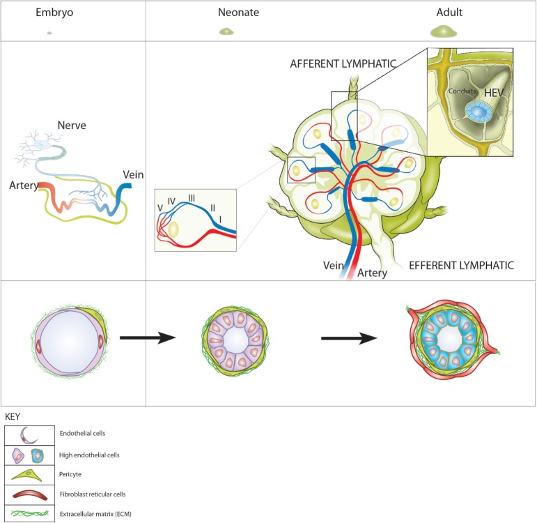
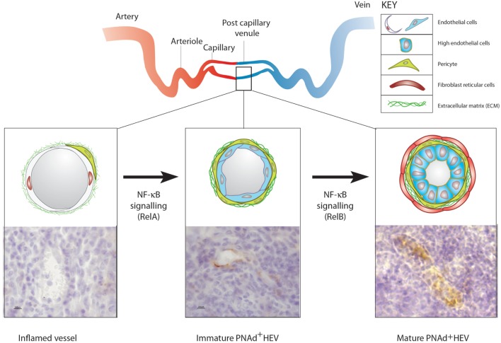
The blood vasculature regulates both the development and function of secondary lymphoid organs by providing a portal for entry of hemopoietic cells. During the development of lymphoid organs in the embryo, blood vessels deliver lymphoid tissue inducer cells that initiate and sustain the development of lymphoid tissues. In adults, the blood vessels are structurally distinct from those in other organs due to the requirement for high levels of lymphocyte recruitment under non-inflammatory conditions. In lymph nodes (LNs) and Peyer's patches, high endothelial venules (HEVs) especially adapted for lymphocyte trafficking form a spatially organized network of blood vessels, which controls both the type of lymphocyte and the site of entry into lymphoid tissues. Uniquely, HEVs express vascular addressins that regulate lymphocyte entry into lymphoid organs and are, therefore, critical to the function of lymphoid organs. Recent studies have demonstrated important roles for CD11c+ dendritic cells in the induction, as well as the maintenance, of vascular addressin expression and, therefore, the function of HEVs. Tertiary lymphoid organs (TLOs) are HEV containing LN-like structures that develop inside organized tissues undergoing chronic immune-mediated inflammation. In autoimmune lesions, the development of TLOs is thought to exacerbate disease. In cancerous tissues, the development of HEVs and TLOs is associated with improved patient outcomes in several cancers. Therefore, it is important to understand what drives the development of HEVs and TLOs and how these structures contribute to pathology. In several human diseases and experimental animal models of chronic inflammation, there are some similarities between the development and function of HEVs within LN and TLOs. This review will summarize current knowledge of how hemopoietic cells with lymphoid tissue-inducing, HEV-inducing, and HEV-maintaining properties are recruited from the bloodstream to induce the development and control the function of lymphoid organs.

Keywords: blood vessels; ectopic lymphoid structures; high endothelial venules; lymph nodes; mucosal addressin; peripheral node addressin; tertiary lymphoid organs.


Figures

Figure 1
Figure 1
Blood vessels and high endothelial venules (HEVs) in the development of lymph nodes (LNs). Top: LN development represented as relative sizes of (left) embryonic and (right) neonatal and adult LNs. Middle (left): in the embryo, neuronal stimulation induces retinoic acid-dependent expression of CXCL13 by mesenchymal cells that can be reverse transcytosed and presented on the inner surface of embryonic blood vessels. (Right) In LNs of adults, the HEV network extends from the cortical/paracortical junction adjacent to B cell follicles to large collecting veins in the hilar region gradually increasing in diameter from the smallest, order V venules to the largest order I venules (left hand inset). HEV connect directly to afferent lymphatics via fibroblast reticular cell-coated conduits that form the supporting internal scaffold on which lymphocytes and antigen presenting cells crawl during immunosurveillance (right hand insert). This enables the recruitment of fetal liver-derived CXCR5+ α4β7 integrin-expressing lymphoid tissue inducer cells to the sites where LN develop. (Right) At birth, structurally distinct MAdCAM-1-expressing HEV lined with cuboidal endothelial cells and supported by a thickened fibronectin containing basal lamina are visible. During the first weeks of life, MAdCAM-1-expressing HEV recruit CD11c+ neonatal migratory dendritic cells mobilized from the intestinal lamina propria, undergo a switch from MAdCAM-1 to peripheral node addressin expression and become ensheathed by fibroblast reticular cells. The vascular addressin switch is followed by rapid growth and a concomitant increase in cellularity and expansion in LN.
Figure 2
Figure 2
The development of ectopic high endothelial venules (HEVs). Top: diagrammatic representation of blood vasculature in non-lymphoid organs and the location of postcapillary venules. Bottom (left): blood vessels in acutely inflamed tissues do not express vascular addressins but are able to recruit activated lymphoid cells. (Middle) TNFα, LTα, or LTαβ expressing T or NK cells recruited by inflamed blood vessels induce the expression of peripheral node addressin (PNAd) (and/or MAdCAM-1) in blood vessels lined with flat endothelial cells (ECs) by stimulating classical NF-κB (RelA) signaling in ECs. (Right) Sustained contact between LTαβ expressing activated lymphoid cells and PNAd expressing flat EC induces LTβR-dependent non-canonical NF-κB (RelB) signaling in ECs, which allows the full maturation of PNAd-expressing HEV lined with cuboidal ECs filled with transmigrating lymphocytes. It is not known if the recruitment of fibroblast reticular cells into the perivascular sheath surrounding HEV is driven by LTβR signaling in ECs or perivascular cells or both.

References

    1. Masopust D, Schenkel JM. The integration of T cell migration, differentiation and function. Nat Rev Immunol (2013) 13(5):309–20.10.1038/nri3442 - DOI - PubMed
    1. Fletcher AL, Malhotra D, Turley SJ. Lymph node stroma broaden the peripheral tolerance paradigm. Trends Immunol (2011) 32(1):12–8.10.1016/j.it.2010.11.002 - DOI - PMC - PubMed
    1. Girard JP, Moussion C, Forster R. HEVs, lymphatics and homeostatic immune cell trafficking in lymph nodes. Nat Rev Immunol (2012) 12(11):762–73.10.1038/nri3298 - DOI - PubMed
    1. Drayton DL, Liao S, Mounzer RH, Ruddle NH. Lymphoid organ development: from ontogeny to neogenesis. Nat Immunol (2006) 7(4):344–53.10.1038/ni1330 - DOI - PubMed
    1. Bombardieri M, Barone F, Humby F, Kelly S, McGurk M, Morgan P, et al. Activation-induced cytidine deaminase expression in follicular dendritic cell networks and interfollicular large B cells supports functionality of ectopic lymphoid neogenesis in autoimmune sialoadenitis and MALT lymphoma in Sjogren’s syndrome. J Immunol (2007) 179(7):4929–38.10.4049/jimmunol.179.7.4929 - DOI - PubMed