

Bidirectional ventricular tachycardia in cardiac sarcoidosis
- PMID: 28217233
- PMCID: PMC5300862
- DOI: 10.1016/j.joa.2016.05.003
Bidirectional ventricular tachycardia in cardiac sarcoidosis
Abstract
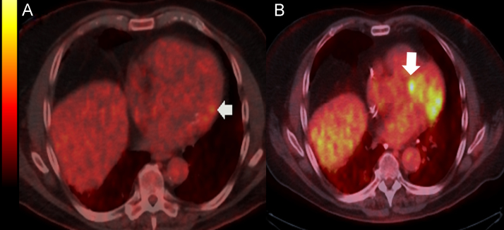
A 73-year-old man with history of pulmonary sarcoidosis was found to have runs of non-sustained bidirectional ventricular tachycardia (BVT) with two different QRS morphologies on a Holter monitor. Cardiac magnetic resonance delayed gadolinium imaging revealed a region of patchy mid-myocardial enhancement within the left ventricular basal inferolateral myocardium. An 18-fluorodeoxyglucose positron emission tomography (FDG-PET) showed increased uptake in the same area, consistent with active sarcoid, with no septal involvement. Follow-up FDG-PET one year later showed disease progression with new septal involvement. Cardiac sarcoidosis, characterized by myocardial inflammation and interstitial fibrosis that can lead to conduction system disturbance and macro re-entrant arrhythmias, should be considered in differential diagnosis of BVT. BVT may indicate septal involvement with sarcoidosis before the lesions are large enough to be detected radiologically.
Keywords: Arrhythmias; BVT, Bidirectional ventricular tachycardia; Cardiac sarcoidosis; ICD, Implantable cardioverter defibrillator; Magnetic resonance tomography; PET, Positron emission tomography; PVC, Premature ventricular contraction; Positron emission tomography; VT, Ventricular tachycardia.; Ventricular tachycardia.
Figures


References
-
- Sung R.K., Kim A.M., Tseng Z.H. Diagnosis and ablation of multiform fascicular tachycardia. J. Cardiovasc. Electrophysiol. 2013;24:297–304. - PubMed
-
- Naruse Y., Sekiguchi Y., Nogami A. Systematic treatment approach to ventricular tachycardia in cardiac sarcoidosis. Circ. Arrhythm. Electrophysiol. 2014;7:407–413. - PubMed
-
- Banba K., Kusano K.F., Nakamura K. Relationship between arrhythmogenesis and disease activity in cardiac sarcoidosis. HeartRhythm. 2007;4:1292–1299. - PubMed
-
- Jefic D., Joel B., Good E. Role of radiofrequency catheter ablation of ventricular tachycardia in cardiac sarcoidosis: report from a multicenter registry. HeartRhythm. 2009;6:189–195. - PubMed
LinkOut - more resources
Full Text Sources
Other Literature Sources
