. 2017 Feb 8;9(4):191-208.
doi: 10.4254/wjh.v9.i4.191.

Regulation of hepatic microRNA expression by hepatocyte nuclear factor 4 alpha

Affiliations

Regulation of hepatic microRNA expression by hepatocyte nuclear factor 4 alpha

Hong Lu et al. World J Hepatol. .

Abstract

Aim: To uncover the role of hepatocyte nuclear factor 4 alpha (HNF4α) in regulating hepatic expression of microRNAs.

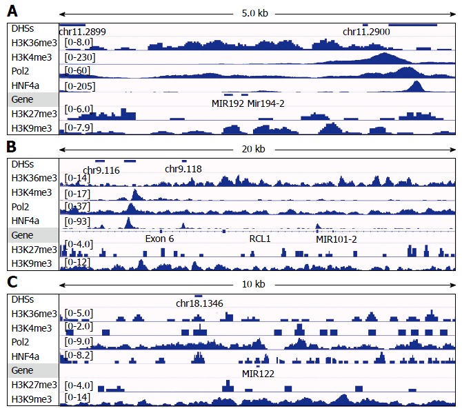
Methods: Microarray and real-time PCR were used to determine hepatic expression of microRNAs in young-adult mice lacking Hnf4α expression in liver (Hnf4α-LivKO). Integrative genomics viewer software was used to analyze the public chromatin immunoprecipitation-sequencing datasets for DNA-binding of HNF4α, RNA polymerase-II, and histone modifications to loci of microRNAs in mouse liver and human hepatoma cells. Dual-luciferase reporter assay was conducted to determine effects of HNF4α on the promoters of mouse and human microRNAs as well as effects of microRNAs on the untranslated regions (3'UTR) of two genes in human hepatoma cells.

Results: Microarray data indicated that most microRNAs remained unaltered by Hnf4α deficiency in Hnf4α-LivKO mice. However, certain liver-predominant microRNAs were down-regulated similarly in young-adult male and female Hnf4α-LivKO mice. The down-regulation of miR-101, miR-192, miR-193a, miR-194, miR-215, miR-802, and miR-122 as well as induction of miR-34 and miR-29 in male Hnf4α-LivKO mice were confirmed by real-time PCR. Analysis of public chromatin immunoprecipitation-sequencing data indicates that HNF4α directly binds to the promoters of miR-101, miR-122, miR-194-2/miR-192 and miR-193, which is associated with histone marks of active transcription. Luciferase reporter assay showed that HNF4α markedly activated the promoters of mouse and human miR-101b/miR-101-2 and the miR-194/miR-192 cluster. Additionally, miR-192 and miR-194 significantly decreased activities of luciferase reporters for the 3'UTR of histone H3F3 and chromodomain helicase DNA binding protein 1 (CHD1), respectively, suggesting that miR-192 and miR-194 might be important in chromosome remodeling through directly targeting H3F3 and CHD1.

Conclusion: HNF4α is essential for hepatic basal expression of a group of liver-enriched microRNAs, including miR-101, miR-192, miR-193a, miR-194 and miR-802, through which HNF4α may play a major role in the post-transcriptional regulation of gene expression and maintenance of the epigenome in liver.

Keywords: Hepatocyte nuclear factor 4 alpha; Human; Knockout; Liver; Mice; miR-101; miR-122; miR-192; miR-194; miR-802.


Conflict of interest statement

Conflict-of-interest statement: The authors have no conflict of interest to declare.

Figures

Figure 1
Figure 1
Heat map and unsupervised hierarchical clustering of hepatic microRNAs in male and female Hnf4a-LivKO mice. The heat map diagram shows the results of the 2-way hierarchical clustering of microRNAs and samples. Each row represents a microRNA and each column represents a pooled liver sample. The microRNA clustering tree is shown on the left, and the sample clustering tree appears at the top. The color scale shown at the top illustrates the relative expression level of a microRNA across all samples: Red color represents an expression level above mean, blue color represents expression lower than the mean. The clustering is performed on log2(Hy3/Hy5) ratios which passed the filtering criteria on variation across samples; LogMedianDRatios differencies > 0.58, corresponding to 50% differential expression. WTM: Wild-type male; WTF: Wild-type female; KOM: Knockout male; KOF: Knockout female.
Figure 2
Figure 2
Hepatic microRNA expression in young-adult male mice with liver-specific deletion of Hnf4a (Hnf4a-LivKO) (A-D). microRNAs in total RNA from livers of Hnf4a-LivKO and wild-type (WT) control mice (n = 5-6) were determined by miRCURY LNA™ Universal RT microRNA PCR (Exiqon). Mean ± SE. aP < 0.05 compared to WT control. HNF4α: Hepatocyte nuclear factor 4 alpha.
Figure 3
Figure 3
Analysis of DNAse-I hypersensitive sites as well as DNA-binding of HNF4α, RNA polymerase II (Pol2), and methylated histones to loci of miR-122a (A), miR-194-2/miR-192 (B), miR-101b (C), miR-193 (D) and miR-802 (E) in wildtype mouse liver. DNA-binding of HNF4α to these microRNA loci in the mouse small intestine (HNF4α _Int) was compared to those in the mouse liver (HNF4α_Liv). Data of DHSs (determined by DNAse-seq) and DNA-binding of proteins (determined by ChIP-seq) were retrieved from the public database of GEO DataSets and visualized in the IGV software. The peak values/ranges for each mark were shown in square brackets or under the line mark. DHSs: DNAse-I hypersensitive sites; HNF4α: Hepatocyte nuclear factor 4 alpha; H3K36me3: H3 trimethylation at lysine-36; H3K4me3: H3 trimethylation at lysine-4; H3K27me3: H3 trimethylation at lysine-27; H3K9me3: H3 trimethylation at lysine-9; Pol2: Polymerase 2; HNF4α: Hepatocyte nuclear factor 4 alpha; ChIP-seq: Chromatin immunoprecipitation-sequencing; IGV: Integrative genomics viewer.
Figure 4
Figure 4
Analysis of DNAse-I hypersensitive sites as well as DNA-binding of HNF4α, RNA polymerase II (Pol2), and methylated histones to loci of miR-194-1/miR-215 (A), miR-26a-1 (B), miR-26a-2 (C) and miR-26b (D) in wildtype mouse liver. DNA-binding of HNF4α to these microRNA loci in the mouse small intestine (HNF4α _Int) was compared to those in the mouse liver (HNF4α_Liv). Data of DHSs (determined by DNAse-seq) and DNA-binding of proteins (determined by ChIP-seq) were retrieved from the public database of GEO DataSets and visualized in the IGV software. The peak values/ranges for each mark were shown in square brackets or under the line mark. DHSs: DNAse-I hypersensitive sites; H3K36me3: H3 trimethylation at lysine-36; H3K4me3: H3 trimethylation at lysine-4; H3K27me3: H3 trimethylation at lysine-27; H3K9me3: H3 trimethylation at lysine-9; Pol2: Polymerase 2; HNF4α: Hepatocyte nuclear factor 4 alpha; ChIP-seq: Chromatin immunoprecipitation-sequencing; IGV: Integrative genomics viewer.
Figure 5
Figure 5
Analysis of DNAse-I hypersensitive sites as well as DNA-binding of HNF4α, RNA polymerase II (Pol2), and methylated histones to loci of miR-29a/miR-29b (A), miR-124a-1 (B), miR-124a-2 (C), miR-124a-3 (D) and miR-134 (E) in wildtype mouse liver. DNA-binding of HNF4α to these microRNA loci in the mouse small intestine (HNF4α _Int) was compared to those in the mouse liver (HNF4α_Liv). Data of DHSs (determined by DNAse-seq) and DNA-binding of proteins (determined by ChIP-seq) were retrieved from the public database of GEO DataSets and visualized in the IGV software. The peak values/ranges for each mark were shown in square brackets or under the line mark. DHSs: DNAse-I hypersensitive sites; H3K36me3: H3 trimethylation at lysine-36; H3K4me3: H3 trimethylation at lysine-4; H3K27me3: H3 trimethylation at lysine-27; H3K9me3: H3 trimethylation at lysine-9; Pol2: Polymerase 2; HNF4α: Hepatocyte nuclear factor 4 alpha; ChIP-seq: Chromatin immunoprecipitation-sequencing; IGV: Integrative genomics viewer.
Figure 6
Figure 6
Analysis of DNAse-I hypersensitive sites as well as DNA-binding of HNF4α, RNA polymerase II (Pol2), and methylated histones to loci of miR-194-2/miR-192 (A), miR-101-2 (B) and miR-122 (C) in human hepatoma HepG2 cells. Data of DHSs (determined by DNAse-seq) and DNA-binding of proteins (determined by ChIP-seq) were retrieved from the public database of GEO DataSets and visualized in the IGV software. The peak values/ranges for each mark were shown in square brackets or under the line mark. DHSs: DNAse-I hypersensitive sites; H3K36me3: H3 trimethylation at lysine-36; H3K4me3: H3 trimethylation at lysine-4; H3K27me3: H3 trimethylation at lysine-27; H3K9me3: H3 trimethylation at lysine-9; Pol2: Polymerase 2; HNF4α: Hepatocyte nuclear factor 4 alpha; ChIP-seq: Chromatin immunoprecipitation-sequencing; IGV: Integrative genomics viewer.
Figure 7
Figure 7
Activation of mouse (A) and human (B-D) miR-194-2/miR-192 promoter by HNF4α. Human hepatoma HepG2 cells were transfected with firefly luciferase vectors containing wild-type and mutant miR-194-2 promoter, pRL-CMV, and an expression vector for HNF4α/HNF1α. Dual-luciferase reporter assay was conducted 24 h after transfection. The y-axis represents relative luciferase activity for microRNA promoter normalized by the renilla luciferase. n = 4, Mean ± SE. aP < 0.05 compared to vector control; cP < 0.05 compared to HNF4α alone group. HNF4α: Hepatocyte nuclear factor 4 alpha.
Figure 8
Figure 8
Activation of (A) mouse miR-101b, (B) human miR-101-2, and (C) mouse miR-802 promoter by HNF4α. Human hepatoma HepG2 cells were transfected with firefly luciferase vectors containing microRNA promoter, pRL-CMV, and an expression vector for HNF4α and/or C/EBPα. Dual-luciferase reporter assay was conducted 24 h after transfection. The Y-axis represents relative luciferase activity for microRNA promoter normalized by the renilla luciferase. n = 4, Mean ± SE. aP < 0.05 compared to vector control; cP < 0.05 compared to HNF4α alone group. HNF4α: Hepatocyte nuclear factor 4 alpha; C/EBPα: CCAAT/enhancer-binding protein α.
Figure 9
Figure 9
Effects of miR-194 and miR-192 on the activities of luciferase reporter vectors for the 3’UTR of mouse Chd1 and H3f3. Human hepatoma HepG2 cells were transfected with plasmid DNA including pmiR-Chd1 (or pmiR-H3f3), the pRL-CMV luciferase, and a synthetic mimic of miR-194/miR-192, or AllStars Negative Control siRNA (as negative control for microRNAs) using Lipofectamine 2000. Dual-luciferase reporter assay was conducted 24 h after transfection. The Y-axis represents relative luciferase activity for the 3’UTR of Chd1 or H3f3 normalized by the renilla luciferase. n = 4, Mean ± SE. aP < 0.05 compared to control (AllStars Negative Control siRNA). 3’UTR: Untranslated regions.
Figure 10
Figure 10
Diagram that illustrates the regulation of hepatic microRNA expression by Hnf4α in mouse liver. HNF4α: Hepatocyte nuclear factor 4 alpha; C/EBPα: CCAAT/enhancer-binding protein α.

Similar articles

Cited by

References

    1. Lu H, Gonzalez FJ, Klaassen C. Alterations in hepatic mRNA expression of phase II enzymes and xenobiotic transporters after targeted disruption of hepatocyte nuclear factor 4 alpha. Toxicol Sci. 2010;118:380–390. - PMC - PubMed
    1. Lu H. Crosstalk of HNF4α with extracellular and intracellular signaling pathways in the regulation of hepatic metabolism of drugs and lipids. Acta Pharm Sin B. 2016;6:393–408. - PMC - PubMed
    1. Battle MA, Konopka G, Parviz F, Gaggl AL, Yang C, Sladek FM, Duncan SA. Hepatocyte nuclear factor 4alpha orchestrates expression of cell adhesion proteins during the epithelial transformation of the developing liver. Proc Natl Acad Sci USA. 2006;103:8419–8424. - PMC - PubMed
    1. Li J, Ning G, Duncan SA. Mammalian hepatocyte differentiation requires the transcription factor HNF-4alpha. Genes Dev. 2000;14:464–474. - PMC - PubMed
    1. Kyrmizi I, Hatzis P, Katrakili N, Tronche F, Gonzalez FJ, Talianidis I. Plasticity and expanding complexity of the hepatic transcription factor network during liver development. Genes Dev. 2006;20:2293–2305. - PMC - PubMed