. 2017 Feb 3:7:22.
doi: 10.3389/fcimb.2017.00022. eCollection 2017.

Regulation of Antimicrobial Peptides in Aedes aegypti Aag2 Cells

Affiliations

Regulation of Antimicrobial Peptides in Aedes aegypti Aag2 Cells

Rudian Zhang et al. Front Cell Infect Microbiol. .

Abstract

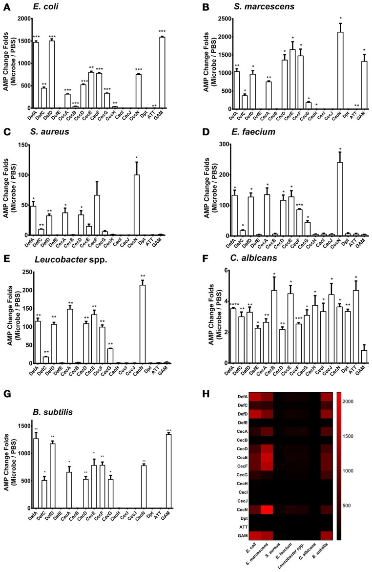
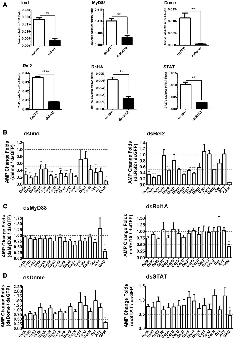
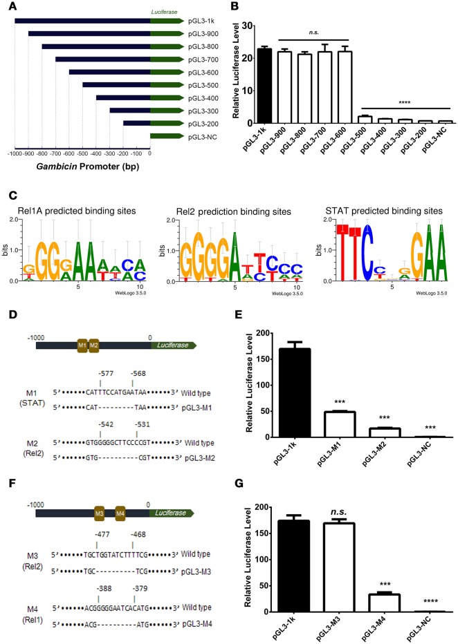
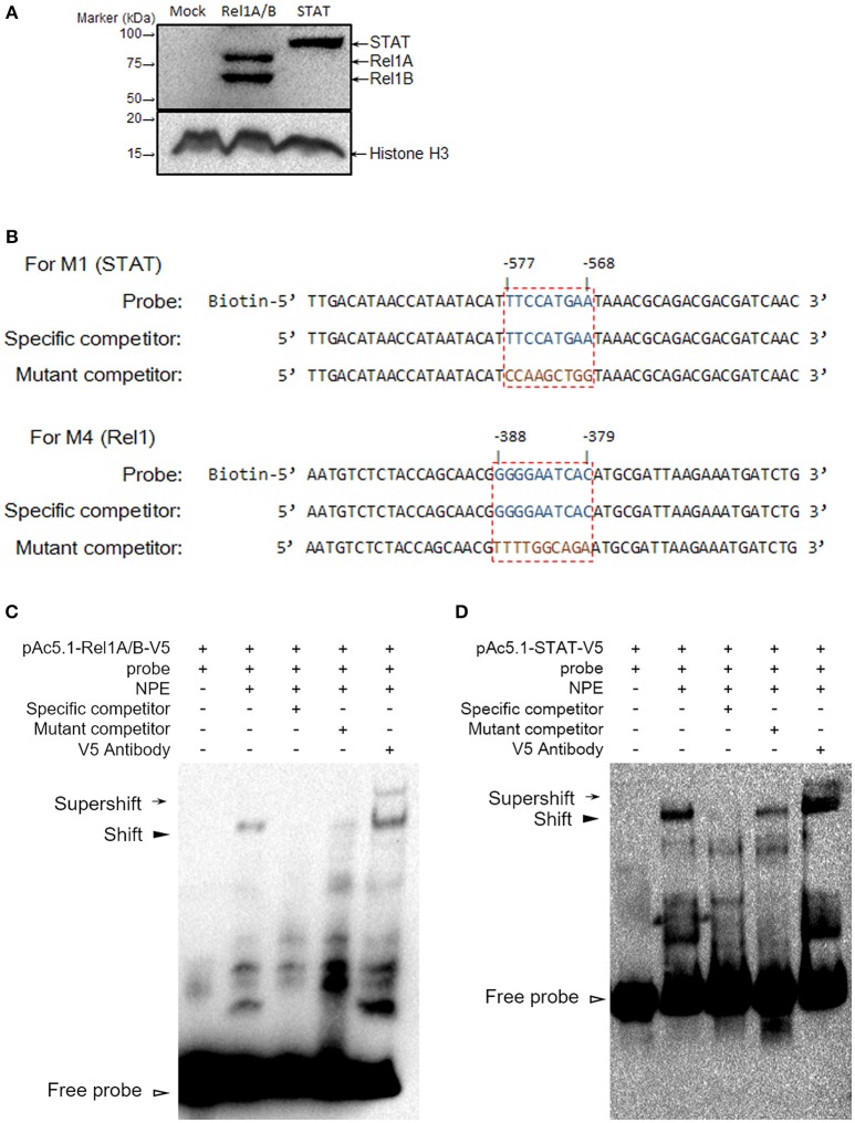
Antimicrobial peptides (AMPs) are an important group of immune effectors that play a role in combating microbial infections in invertebrates. Most of the current information on the regulation of insect AMPs in microbial infection have been gained from Drosophila, and their regulation in other insects are still not completely understood. Here, we generated an AMP induction profile in response to infections with some Gram-negative, -positive bacteria, and fungi in Aedes aegypti embryonic Aag2 cells. Most of the AMP inductions caused by the gram-negative bacteria was controlled by the Immune deficiency (Imd) pathway; nonetheless, Gambicin, an AMP gene discovered only in mosquitoes, was combinatorially regulated by the Imd, Toll and JAK-STAT pathways in the Aag2 cells. Gambicin promoter analyses including specific sequence motif deletions implicated these three pathways in Gambicin activity, as shown by a luciferase assay. Moreover, the recognition between Rel1 (refer to Dif/Dorsal in Drosophila) and STAT and their regulatory sites at the Gambicin promoter site was validated by a super-shift electrophoretic mobility shift assay (EMSA). Our study provides information that increases our understanding of the regulation of AMPs in response to microbial infections in mosquitoes. And it is a new finding that the A. aegypti AMPs are mainly regulated Imd pathway only, which is quite different from the previous understanding obtained from Drosophila.

Keywords: antimicrobial peptides; innate immunity; insect immunity; mosquito; regulation.


Figures

Figure 1
Figure 1
Regulation of AMP genes in microbial infections in the Aag2 cells E. coli (A), S. marcescens (B), S. aureus (C), E. faecium (D), Leucobacter spp. (E) C. albicans (F), and B. subtilis (G) cells at 0.05 OD600 were incubated with the Aag2 cells. Uninfected cells served as controls. 12 h later, the stimulated Aag2 cells were collected to isolate total RNA that was synthesized into cDNA for AMP detection. The qPCR primers for each AMP gene are described in Supplementary Table S2. The AMP stimulation is presented as the fold change relative to that in the control cells without bacterial treatment. The data are presented as the mean ± S.E.M. The differences between microbes treated groups and negative control group were analyzed by using t-test with Welch's correction. (H) The changing folds were presented with a heat map to indicate the AMP induction manner between Gram negative bacteria and B. subtilis infection. Red means high induction as described in the scale bar. The results from 2 independent experiments were combined. *p < 0.05, **p < 0.005, ***p < 0.0005, and ****p < 0.0001. Def, Defensin; Cec, Cecropin; Dpt, Diptericin; ATT, Attacin; GAM, Gambicin.
Figure 2
Figure 2
Role of immune pathways in E. coli-mediated AMP induction in Aag2 cells (A) dsRNA-mediated silencing efficiency of key components of the immune pathways in the Aag2 cells. Expression of the key components in the Imd (Imd and Rel2), Toll (MyD88 and Rel1A), and JAK-STAT (Dome and STAT) pathways were silenced by double-stranded RNA (dsRNA) transfection in the Aag2 cells. GFP dsRNA (dsGFP) served as control. The expression of these genes was determined by qPCR and normalized to the expression of A. aegypti actin. The qPCR primers are shown in Supplementary Table S2. The data were presented as the mean ± S.E.M. The data are analyzed using the non-parametric Mann-Whitney test. (B–D) Expression of the key components in the Imd (Imd and Rel2) (B), Toll (MyD88 and Rel1A) (C), and JAK-STAT (Dome and STAT) (D) pathways were silenced by dsRNA transfection in the Aag2 cells. GFP dsRNA was used as a negative control. Subsequently, E. coli at 0.05 OD600 were incubated with the transfected cells. The AMP expression was then determined by qPCR 12 hrs after the bacterial challenge. The qPCR primers for each AMP genes are described in Supplementary Table S2. The AMP stimulation is presented as the fold change in inducing relative to that in the GFP dsRNA treated (mock control) cells. The data are presented as the mean ± S.E.M. The difference between the AMP induction of gene silenced groups and mock control group were analyzed by using t-test with Welch's correction. The results from 3 independent experiments were combined. *p < 0.05, **p < 0.005, ***p < 0.0005, and ****p < 0.0001. aaActin, A. aegypti Actin.
Figure 3
Figure 3
Gambicin induction is combinatorially regulated by the Imd, Toll and JAK-STAT pathways (A) Schematic representation of the truncation design. The 1000 bp promoter region upstream of the Gambicin gene was cloned into a pGL3-Basic vector (pGL-1k). The truncations of the Gambicin promoter, which were sequentially truncated by the deletion of 100 bp segments from the 5′-end of promoter region, were inserted into the same plasmid. The inserted promoters were followed down-stream by a firefly luciferase gene (the green arrow), thereby enabling the determination of the regulatory activity of the inserted promoters using a luciferase assay. (B) Assessment of promoter activity by 100 bp sequential truncations. The recombinant plasmids with truncated Gambicin promoter were transfected into the Aag2 cells to determine the promoter activity via a luciferase assay. (C) Characterization of the regulatory sites of the Rel1, Rel2 and STAT transcription factors. The regulatory sites were predicted by WebLogo 3.5.0 (http://weblogo.threeplusone.com/create.cgi) and Vector NTI Advanced® 11.5.1 software (Invitrogen, US) with the threshold of 85%. (D) Schematic representation of M1 (STAT) and M2 (Rel2) mutants in the Gambicin promoter. M, Mutation site. (E) Assessment of promoter activity for STAT and Rel2 mutants. The two luciferase plasmids with M1 (STAT) and M2 (Rel2) mutants (please refer to D) were transfected into the Aag2 cells to determine the promoter activity via a luciferase assay. (F) Schematic representation of M3 (Rel2) and M4 (Rel1) mutants in the Gambicin promoter. (G) Assessment of promoter activity for M3 (Rel2) and M4 (Rel1) mutants. The two luciferase plasmids with M3 (Rel2) and M4 (Rel1) mutants (please refer to F) were transfected into the Aag2 cells to determine the promoter activity via a luciferase assay. (B, E, and G) A pAc5.1-Renilla plasmid with constitutive renilla luciferase expression was co-transfected as an internal control. The transfected cells were then challenged by E. coli at 0.05 OD600. The promoter activity in response to the bacterial infection was determined by a luciferase assay. The values of the firefly luciferase were normalized to that of the renilla luciferase. The data were analyzed using the non-parametric Mann-Whitney test. The data are presented as the mean ± S.E.M. Each experiment was biologically reproduced by 3 times. ***p < 0.0005 and ****p < 0.0001. n.s., no significance.
Figure 4
Figure 4
Determine the binding affinity between Rel1 and STAT and their regulatory sites by a super-shift EMSA (A) Ectopic expression of Rel1 and STAT in the Aag2 cells. The A. aegypti Rel1A, Rel1B, and STAT genes were cloned into pAc5.1-V5-His A vectors and designated as pAc-5.1-Rel1A-V5, pAc-5.1-Rel1B-V5, and pAc-5.1-STAT-V5, respectively. Both pAc-5.1-Rel1A-V5 and pAc-5.1-Rel1B-V5 were combined (1:1 w/w) for transfection into the Aag2 cells. The expression of Rel1A/Rel1B and STAT was confirmed by western blot analysis with an anti-V5 antibody. The detection of Histone H3 acts as an internal reference. (B) Design of probes for the EMSA assay. The boxed regions represent the regulatory sites for STAT and Rel1 factors. (C,D) Determine the binding affinity between Rel1 (C) and STAT (D) and their regulatory sites by EMSA. A V5 antibody was used to detect the specific binding between the probes and these ectopically expressed transcription factors (super-shift EMSA). The experiments were repeated 3 times with the similar results. NPE, Nuclear protein extracts.

Similar articles

Cited by

References

    1. Arensburger P., Megy K., Waterhouse R. M., Abrudan J., Amedeo P., Antelo B., et al. . (2010). Sequencing of Culex quinquefasciatus establishes a platform for mosquito comparative genomics. Science 330, 86–88. 10.1126/science.1191864 - DOI - PMC - PubMed
    1. Asling B., Dushay M. S., Hultmark D. (1995). Identification of early genes in the Drosophila immune response by PCR-based differential display: the Attacin A gene and the evolution of attacin-like proteins. Insect Biochem. Mol. Biol. 25, 511–518. 10.1016/0965-1748(94)00091-C - DOI - PubMed
    1. Bangham J., Jiggins F., Lemaitre B. (2006). Insect immunity: the post-genomic era. Immunity 25, 1–5. 10.1016/j.immuni.2006.07.002 - DOI - PubMed
    1. Barletta A. B., Silva M. C., Sorgine M. H. (2012). Validation of Aedes aegypti Aag-2 cells as a model for insect immune studies. Parasit. Vectors 5:148. 10.1186/1756-3305-5-148 - DOI - PMC - PubMed
    1. Bulet P., Dimarcq J. L., Hetru C., Lagueux M., Charlet M., Hegy G., et al. . (1993). A novel inducible antibacterial peptide of Drosophila carries an O-glycosylated substitution. J. Biol. Chem. 268, 14893–14897. - PubMed

Publication types

Substances