Isolated pulmonary hydatid cyst: Misinterpreted as metastatic pulmonary lesion in an operated case of carcinoma breast in young female
- PMID: 28217612
- PMCID: PMC5290789
- DOI: 10.4103/2249-4863.197299
Isolated pulmonary hydatid cyst: Misinterpreted as metastatic pulmonary lesion in an operated case of carcinoma breast in young female
Abstract
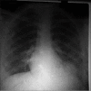
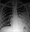
Hydatidosis is caused by Echinococcus granulosus. Humans may be infected incidentally as intermediate host by the accidental consumption of soil, water, or food contaminated by fecal matter of an infected animal. Hydatidosis is one of the most symptomatic parasitic infections in various livestock - raising countries. Lung is the second most commonly affected organ following the liver. The symptoms depend on the size and site of the lesion. It can present as an asymptomatic pulmonary lesion to hemoptysis, chest pain, coughing anaphylaxis, and shock. There are very few reported cases of isolated lung hydatidosis without exposure to animals or nonvegetarian diet. For hydatidosis, serology and imaging are diagnostic tools. Surgical removal and/or chemotherapy are the main-stay of treatment. Here, we discuss a case of persistent left lower lobe cystic lesion in young female with a history of operated left breast carcinoma which was thought to be of metastatic lesion but ultimately confirmed as pulmonary hydatid cyst after unintended aspiration of cystic fluid to rule out malignancy. Pulmonary hydatidosis should always be considered as a differential diagnosis when dealing with a cystic lesion on radiology.
Keywords: Echinococcus granulosus; metastasis; pulmonary hydatidosis.
Conflict of interest statement
There are no conflicts of interest.
Figures


Similar articles
-
Recurrent hydatid cyst of liver with asymptomatic concomitant hydatid cyst of lung: an unusual presentation-case report.Iran J Parasitol. 2015 Jan-Mar;10(1):136-40. Iran J Parasitol. 2015. PMID: 25904958 Free PMC article.
-
Ruptured pulmonary hydatid cyst presenting as hemoptysis in TB endemic country: A case report.Ann Med Surg (Lond). 2022 May 20;78:103836. doi: 10.1016/j.amsu.2022.103836. eCollection 2022 Jun. Ann Med Surg (Lond). 2022. PMID: 35734680 Free PMC article.
-
Bilateral pulmonary hydatidosis associated with uncommon muscular localization.Int J Surg Case Rep. 2020;76:130-133. doi: 10.1016/j.ijscr.2020.09.070. Epub 2020 Sep 16. Int J Surg Case Rep. 2020. PMID: 33035955 Free PMC article.
-
Pulmonary hydatid cyst: Review of literature.J Family Med Prim Care. 2019 Sep 30;8(9):2774-2778. doi: 10.4103/jfmpc.jfmpc_624_19. eCollection 2019 Sep. J Family Med Prim Care. 2019. PMID: 31681642 Free PMC article. Review.
-
Hydatidosis of infratemporal fossa with proptosis - an unusual presentation: a case report and review of the literature.J Med Case Rep. 2018 Oct 17;12(1):309. doi: 10.1186/s13256-018-1812-y. J Med Case Rep. 2018. PMID: 30326941 Free PMC article. Review.
References
-
- McManus DP. Echinococcosis with particular reference to Southeast Asia. Adv Parasitol. 2010;72:267–303. - PubMed
-
- Kulpati DD, Hagroo AA, Talukdar CK, Ray D. Hydatid disease of the lung. Indian J Chest Dis. 1974;16:406–10. - PubMed
-
- Reddy CR, Narasiah IL, Parvathi G, Rao MS. Epidemiology of hydatid disease in Kurnool. Indian J Med Res. 1968;56:1205–20. - PubMed
-
- Morar R, Feldman C. Pulmonary echinococcosis. Eur Respir J. 2003;21:1069–77. - PubMed
Publication types
LinkOut - more resources
Full Text Sources
Other Literature Sources

