

Role of contrast harmonic-endoscopic ultrasound in pancreatic cystic lesions
- PMID: 28218197
- PMCID: PMC5331839
- DOI: 10.4103/2303-9027.190931
Role of contrast harmonic-endoscopic ultrasound in pancreatic cystic lesions
Abstract
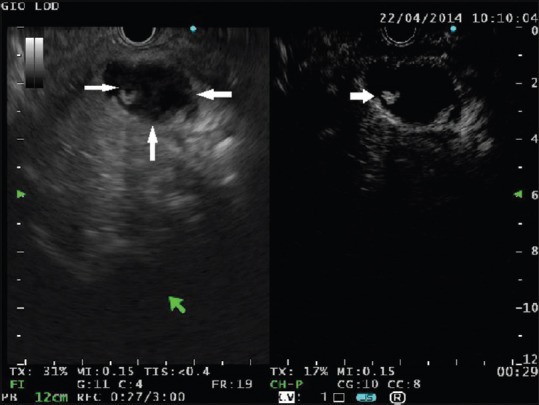
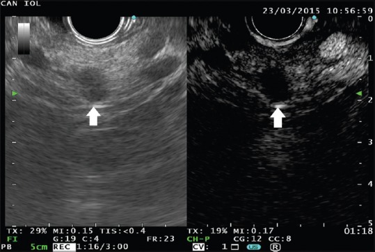
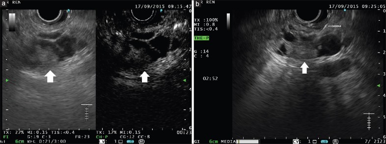
Incidental pancreatic cysts (PCs) are frequently encountered in the general population often in asymptomatic patients who undergo imaging tests to investigate unrelated conditions. The detection of a PC poses a significant clinical dilemma, as the differential diagnosis is quite broad ranging from benign to malignant conditions. Endoscopic ultrasound (EUS) with fine-needle aspiration (FNA) has been reported to be an accurate tool in the differential diagnosis; however, its sensitivity is suboptimal and false negative results do occur. Contrast harmonic EUS (CH-EUS) was demonstrated to be a useful tool to investigate pancreatic solid lesions to differentiate between benign and malignant ones. In the setting of PCs, CH-EUS could help identify areas of malignant growth inside the cystic cavities. Several studies have reported promising results showing malignant areas in PCs as hyperenhanced lesions. Confirmation of malignancy can then be obtained by FNA, which should be precisely targeted according to the findings of the contrast harmonic study.
Conflict of interest statement
There are no conflicts of interest.
Figures
References
-
- Freeny PC, Saunders MD. Moving beyond morphology: New insights into the characterization and management of cystic pancreatic lesions. Radiology. 2014;272:345–63. - PubMed
-
- Zhang XM, Mitchell DG, Dohke M, et al. Pancreatic cysts: Depiction on single-shot fast spin-echo MR images. Radiology. 2002;223:547–53. - PubMed
-
- Lee KS, Sekhar A, Rofsky NM, et al. Prevalence of incidental pancreatic cysts in the adult population on MR imaging. Am J Gastroenterol. 2010;105:2079–84. - PubMed
Publication types
LinkOut - more resources
Full Text Sources
Other Literature Sources
