

STIM1 Phosphorylation at Y361 Recruits Orai1 to STIM1 Puncta and Induces Ca2+ Entry
- PMID: 28218251
- PMCID: PMC5316956
- DOI: 10.1038/srep42758
STIM1 Phosphorylation at Y361 Recruits Orai1 to STIM1 Puncta and Induces Ca2+ Entry
Abstract
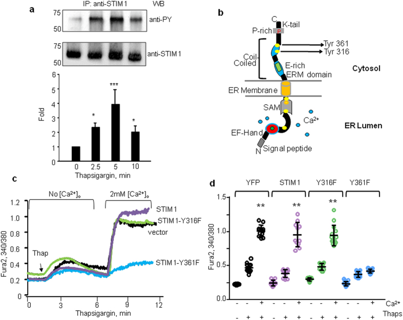
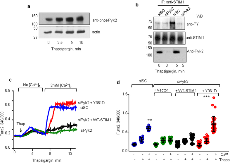
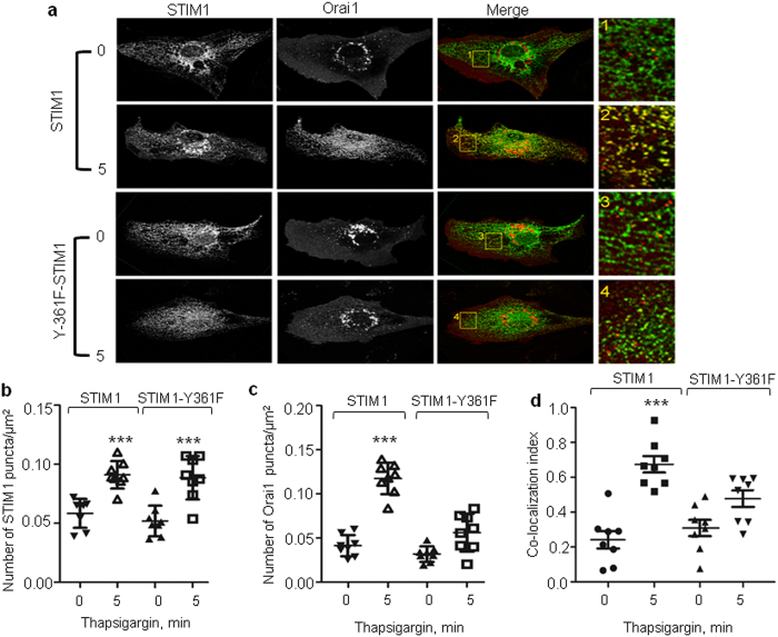
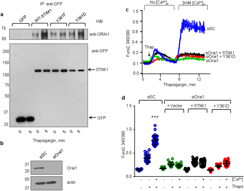
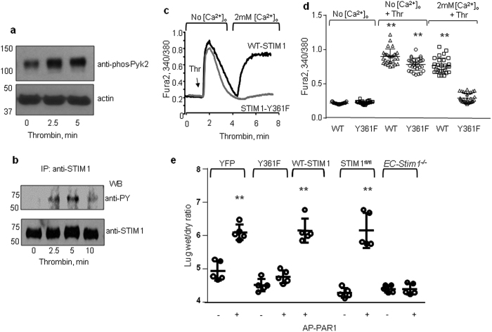
Store-operated Ca2+ entry (SOCE) mediates the increase in intracellular calcium (Ca2+) in endothelial cells (ECs) that regulates several EC functions including tissue-fluid homeostasis. Stromal-interaction molecule 1 (STIM1), upon sensing the depletion of (Ca2+) from the endoplasmic reticulum (ER) store, organizes as puncta that trigger store-operated Ca2+ entry (SOCE) via plasmalemmal Ca2+-selective Orai1 channels. While the STIM1 and Orai1 binding interfaces have been mapped, signaling mechanisms activating STIM1 recruitment of Orai1 and STIM1-Orai1 interaction remains enigmatic. Here, we show that ER Ca2+-store depletion rapidly induces STIM1 phosphorylation at Y361 via proline-rich kinase 2 (Pyk2) in ECs. Surprisingly, the phospho-defective STIM1-Y361F mutant formed puncta but failed to recruit Orai1, thereby preventing. SOCE Furthermore, studies in mouse lungs, expression of phosphodefective STIM1-Y361F mutant in ECs prevented the increase in vascular permeability induced by the thrombin receptor, protease activated receptor 1 (PAR1). Hence, Pyk2-dependent phosphorylation of STIM1 at Y361 is a critical phospho-switch enabling recruitment of Orai1 into STIM1 puncta leading to SOCE. Therefore, Y361 in STIM1 represents a novel target for limiting SOCE-associated vascular leak.
Conflict of interest statement
The authors declare no competing financial interests.
Figures
Similar articles
-
Filamin A Modulates Store-Operated Ca2+ Entry by Regulating STIM1 (Stromal Interaction Molecule 1)-Orai1 Association in Human Platelets.Arterioscler Thromb Vasc Biol. 2018 Feb;38(2):386-397. doi: 10.1161/ATVBAHA.117.310139. Epub 2017 Dec 28. Arterioscler Thromb Vasc Biol. 2018. PMID: 29284605
-
Ca2+/Calmodulin Binding to STIM1 Hydrophobic Residues Facilitates Slow Ca2+-Dependent Inactivation of the Orai1 Channel.Cell Physiol Biochem. 2020 Mar 17;54(2):252-270. doi: 10.33594/000000218. Cell Physiol Biochem. 2020. PMID: 32176842
-
Interplay between ER Ca2+ Binding Proteins, STIM1 and STIM2, Is Required for Store-Operated Ca2+ Entry.Int J Mol Sci. 2018 May 19;19(5):1522. doi: 10.3390/ijms19051522. Int J Mol Sci. 2018. PMID: 29783744 Free PMC article.
-
The Molecular Heterogeneity of Store-Operated Ca2+ Entry in Vascular Endothelial Cells: The Different roles of Orai1 and TRPC1/TRPC4 Channels in the Transition from Ca2+-Selective to Non-Selective Cation Currents.Int J Mol Sci. 2023 Feb 7;24(4):3259. doi: 10.3390/ijms24043259. Int J Mol Sci. 2023. PMID: 36834672 Free PMC article. Review.
-
Key components of store-operated Ca2+ entry in non-excitable cells.J Pharmacol Sci. 2014;125(4):340-6. doi: 10.1254/jphs.14r06cp. Epub 2014 Jul 17. J Pharmacol Sci. 2014. PMID: 25030742 Review.
Cited by
-
Noncanonical function of long myosin light chain kinase in increasing ER-PM junctions and augmentation of SOCE.FASEB J. 2020 Sep;34(9):12805-12819. doi: 10.1096/fj.201902462RR. Epub 2020 Aug 9. FASEB J. 2020. PMID: 32772419 Free PMC article.
-
CLCA2 is a positive regulator of store-operated calcium entry and TMEM16A.PLoS One. 2018 May 14;13(5):e0196512. doi: 10.1371/journal.pone.0196512. eCollection 2018. PLoS One. 2018. PMID: 29758025 Free PMC article.
-
RGS2 is an innate immune checkpoint for suppressing Gαq-mediated IFNγ generation and lung injury.iScience. 2025 Jan 27;28(2):111878. doi: 10.1016/j.isci.2025.111878. eCollection 2025 Feb 21. iScience. 2025. PMID: 40041768 Free PMC article.
-
Tyrosine phosphorylation of S1PR1 leads to chaperone BiP-mediated import to the endoplasmic reticulum.J Cell Biol. 2021 Nov 1;220(12):e202006021. doi: 10.1083/jcb.202006021. Epub 2021 Oct 15. J Cell Biol. 2021. PMID: 34652421 Free PMC article.
-
PAR2-Mediated cAMP Generation Suppresses TRPV4-Dependent Ca2+ Signaling in Alveolar Macrophages to Resolve TLR4-Induced Inflammation.Cell Rep. 2019 Apr 16;27(3):793-805.e4. doi: 10.1016/j.celrep.2019.03.053. Cell Rep. 2019. PMID: 30995477 Free PMC article.
References
-
- Mehta D. et al.. RhoA interaction with inositol 1, 4, 5-trisphosphate receptor and transient receptor potential channel-1 regulates Ca2+ entry. Role in signaling increased endothelial permeability. The Journal of biological chemistry 278, 33492–33500, doi: 10.1074/jbc.M302401200 (2003). - DOI - PubMed
Publication types
MeSH terms
Substances
Grants and funding
LinkOut - more resources
Full Text Sources
Other Literature Sources
Molecular Biology Databases
Miscellaneous
