. 2017 Mar 1;127(3):1090-1098.
doi: 10.1172/JCI90958. Epub 2017 Feb 20.

Endothelial antigen assembly leads to thrombotic complications in heparin-induced thrombocytopenia

Endothelial antigen assembly leads to thrombotic complications in heparin-induced thrombocytopenia

Vincent Hayes et al. J Clin Invest. .

Abstract

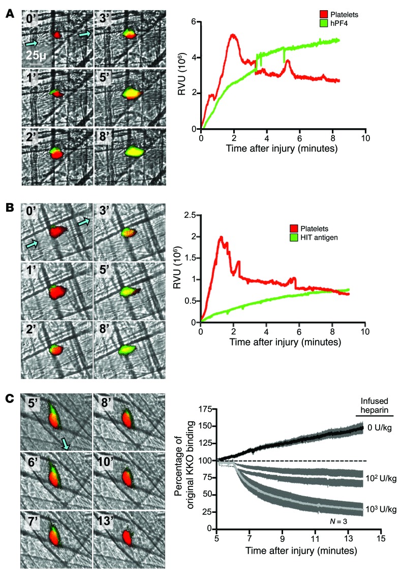
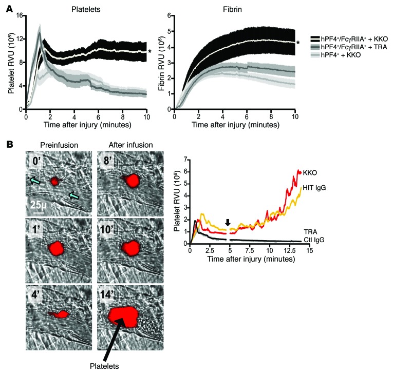
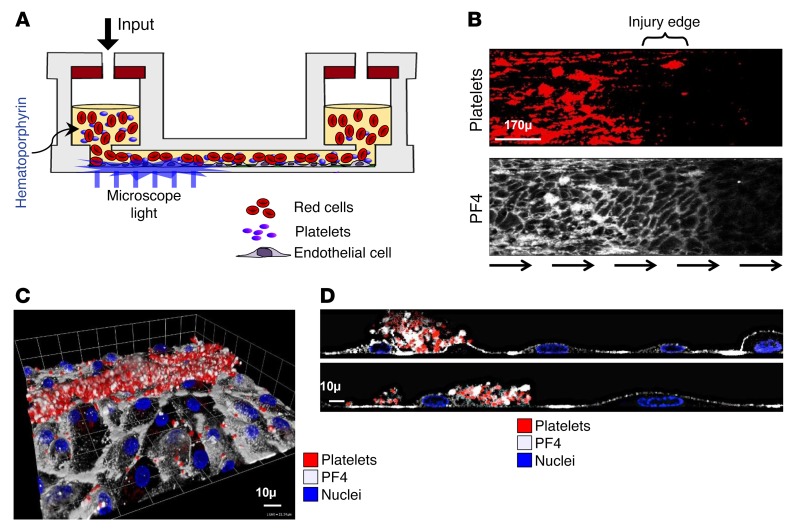
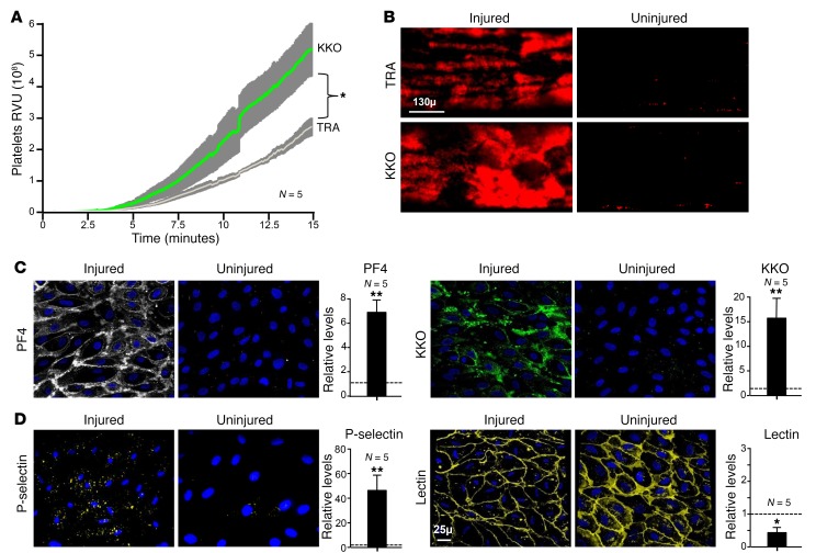
Heparin-induced thrombocytopenia (HIT) is a prothrombotic disorder initiated by antibodies against complexes between human platelet factor 4 (hPF4) and heparin. A better understanding of the events that initiate the prothrombotic state may improve approaches to antithrombotic management. Here, we visualized thrombus formation in an in vivo murine model and an endothelialized microfluidic system that simulate the pathogenesis of HIT. hPF4 released from platelets predominantly bound to peri-injury endothelium and formed HIT antigenic complexes that were dissociated by heparin. In mice expressing both hPF4+ and human platelet IgG Fc receptor IIA (FcγRIIA), infusion of the HIT-like monoclonal antibody KKO increased fibrin and platelet deposition at sites of injury, followed immediately by antigen formation on proximate endothelial cells. After a few minutes, HIT antigen was detected within the thrombus itself at the interface between the platelet core and the surrounding shell. We observed similar results in the humanized, endothelialized microfluidic system. hPF4 and KKO selectively bound to photochemically injured endothelium at sites where surface glycocalyx was reduced. These studies support the concept that the perithrombus endothelium is the predominant site of HIT antigen assembly. This suggests that disrupting antigen formation along the endothelium or protecting the endothelium may provide a therapeutic opportunity to prevent thrombotic complications of HIT, while sparing systemic hemostatic pathways.


Conflict of interest statement

Conflict of interest: The authors have declared that no conflict of interest exists.

Figures

Figure 1
Figure 1. Widefield cremaster laser injury in a non-HIT hPF4+ murine model: In situ studies of hPF4 and HIT antigen distribution in thrombi.
(A) Representative widefield study of more than 10 cremaster laser injuries in hPF4+ mice, with time 0 indicating the onset of injury. Images from a video of a laser injury; platelets are indicated in red, hPF4 is indicated in green, and the direction of blood flow in the vessel is denoted by blue arrows. Graph shows the accumulation of platelets and hPF4 over the study in relative value units (RVU) compared with time 0. (B) Same as in A, but with green showing binding of KKO to indicate the appearance of the HIT antigen. (C) Representative images from Supplemental Video 1 beginning 5 minutes after injury, the point at which 103 U/kg heparin was infused i.v. Platelets are indicated in red and KKO binding in green. The graph indicates that various doses of heparin were infused beginning 5 minutes after the cremaster injury. Percent mean ± 1 SEM for binding of KKO after heparin relative to the 5-minute time point is shown. The dashed line represents no change in KKO binding after heparin infusion compared with the 5-minute heparin time point. Original magnification, ×60.
Figure 2
Figure 2. Widefield in situ studies of the prothrombotic state in a murine HIT model.
(A) Widefield cremaster arteriole laser injuries were performed in hPF4+/FcγRIIA+ and hPF4+ mice infused with KKO or control TRA prior to injury. Platelet accumulation is shown on the left and fibrin on the right. Six mice from each arm were studied, and each mouse had five to six injuries. The AUC was calculated for each injury, then compared between groups by a 2-tailed Mann-Whitney U test. Data represent the mean ± 1 SEM. *P < 0.05 comparing hPF4+/FcγRIIA+ plus KKO with either other group. (B) An experiment similar to the one depicted in A, but the injury was performed at time 0, and KKO (1 μg/g, i.v.) was infused 5 minutes later. Images are from Supplemental Video 2, in which platelets (red) were incorporated into a thrombus 0–4 minutes before or 5–15 minutes after the infusion of KKO. Blue arrows indicate the direction of blood flow in the arteriole; black arrow denotes the growing platelet thrombus. Graph shows the mean values for platelet incorporation into thrombi, with a break in the data just before 5 minutes, when antibodies were infused (vertical arrow). Three injuries were studied in each arm. Original magnification, ×60. Ctl, control.
Figure 3
Figure 3. Confocal microscopic studies of a growing thrombus in HIT showing perithrombotic events.
(A) Confocal microscopic images showing events in a representative cremaster arteriole injury in an hPF4+/FcγRIIA+ mouse infused with KKO. Platelets are shown in red. KKO binding is shown in green. Images on the left are from Supplemental Video 3 and include blue arrows showing the direction of blood flow. The image in the middle is an enlargement of the 5-minute time point, clearly showing a yellow semi-circle at the interface between the core and shell of the growing thrombus just becoming visible at 4 minutes. Images on the right show 3 different views of a 3D reconstruction of a thrombus, with the yellow arrow indicating the direction of blood flow. The dashed white circle indicates the outline of the vessel. (B) Confocal microscopic images, as in A, of another thrombus showing platelet accumulation (red) and binding of KKO (green) as well as staining with annexin V shown in purple as an indication of endothelial injury (50) and overlap with KKO binding shown in white. (C) Mice were preinfused with either KKO or TRA prior to creation of a thrombus. Binding of platelets, KKO, annexin V, and FXa to sites of vascular injury is shown. Data from the individual studies are shown as well as the mean ± 1 SEM (wide and narrow horizontal lines, respectively). The number of injuries studied for each parameter is indicated. *P < 0.001 and **P < 0.0001, by 2-way ANOVA. Original magnification, ×60.
Figure 4
Figure 4. Endothelialized microfluidic channel studies after photochemical injury.
(A) Schematic of microfluidic chamber with whole blood containing hematoporphyrin flowing into an endothelial cell–lined channel, with a light source to injure the endothelium. (B) Adhesion of platelets (red, top) and PF4 (white, bottom) at the junction between the injured and uninjured area from a representative study with infused blood, as in A. (C and D) Confocal images of the injured endothelium. (C) Top-down image of adherent platelets and released PF4 in the photochemically injured area. (D) Same image as in C, in a sagittal view. Original magnification, ×10 (B) and ×40 (C and D).
Figure 5
Figure 5. Studies of HIT in the endothelialized microfluidic channel photochemical injury system.
(A) Platelet accumulation along HUVECs after photochemical injury perfused for 15 minutes with whole human blood containing either KKO or TRA. Data represent the mean ± 1 SEM. *P < 0.0001, for KKO versus TRA exposure by 2-tailed Student t test. (B) Images demonstrate platelets bound to representative fields of photochemically injured and uninjured areas of the channels shown in A from whole blood containing TRA or KKO. (C) Images of representative uninjured and injured areas and graphs of overall comparative measurements showing enhanced binding in injured versus uninjured areas of PF4 (white) and KKO binding (green) after infusion of whole blood, as in A. The mean relative binding for injured versus uninjured endothelium ± 1 SEM is shown. *P < 0.01 and **P < 0.001, by 2-sided Student’s t test for injured versus uninjured areas, with an expectation of 1 and no change in binding (dashed line). (D) Same as in C, except for P-selectin staining (yellow) and lectin binding (yellow) in injured endothelium versus the downstream uninjured area. Lectin staining of the endothelium was decreased after injury. Original magnification, ×20.

References

    1. Amiral J, et al. Platelet factor 4 complexed to heparin is the target for antibodies generated in heparin-induced thrombocytopenia. Thromb Haemost. 1992;68(1):95–96. - PubMed
    1. Greinacher A, Farner B, Kroll H, Kohlmann T, Warkentin TE, Eichler P. Clinical features of heparin-induced thrombocytopenia including risk factors for thrombosis. A retrospective analysis of 408 patients. Thromb Haemost. 2005;94(1):132–135. - PubMed
    1. Hong AP, Cook DJ, Sigouin CS, Warkentin TE. Central venous catheters and upper-extremity deep-vein thrombosis complicating immune heparin-induced thrombocytopenia. Blood. 2003;101(8):3049–3051. doi: 10.1182/blood-2002-05-1448. - DOI - PubMed
    1. Martel N, Lee J, Wells PS. Risk for heparin-induced thrombocytopenia with unfractionated and low-molecular-weight heparin thromboprophylaxis: a meta-analysis. Blood. 2005;106(8):2710–2715. doi: 10.1182/blood-2005-04-1546. - DOI - PubMed
    1. Linkins LA. End of the road for heparin thromboprophylaxis. Blood. 2016;127(16):1945–1946. doi: 10.1182/blood-2016-02-697144. - DOI - PubMed

MeSH terms