. 2017 Mar 1;127(3):1046-1060.
doi: 10.1172/JCI89927. Epub 2017 Feb 20.

Egress of sperm autoantigen from seminiferous tubules maintains systemic tolerance

Egress of sperm autoantigen from seminiferous tubules maintains systemic tolerance

Kenneth S K Tung et al. J Clin Invest. .

Abstract

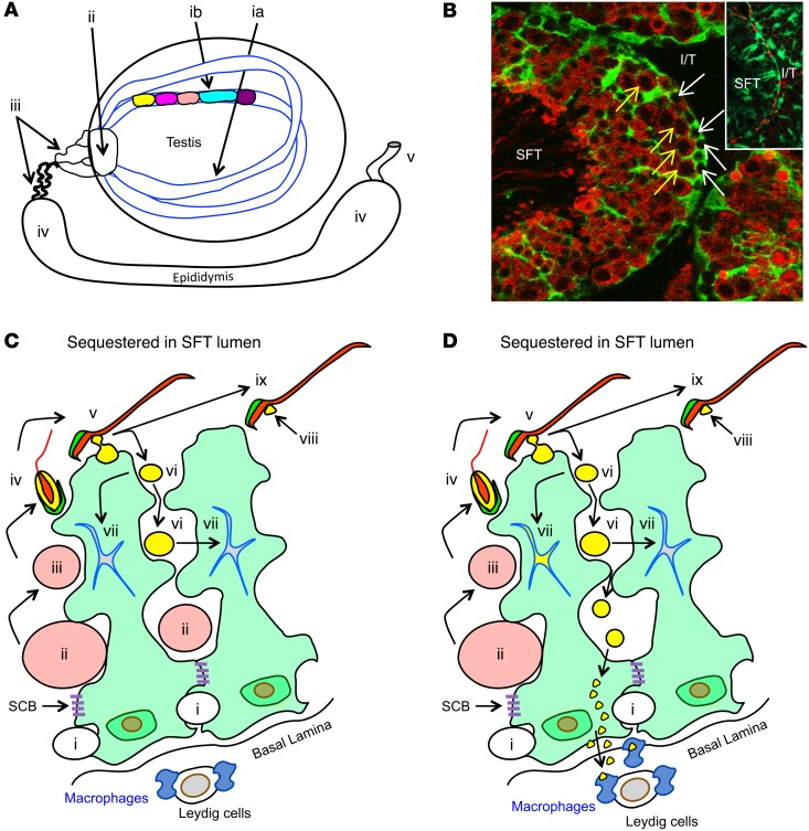
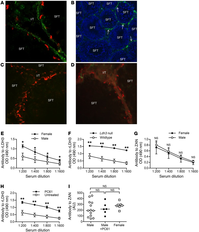
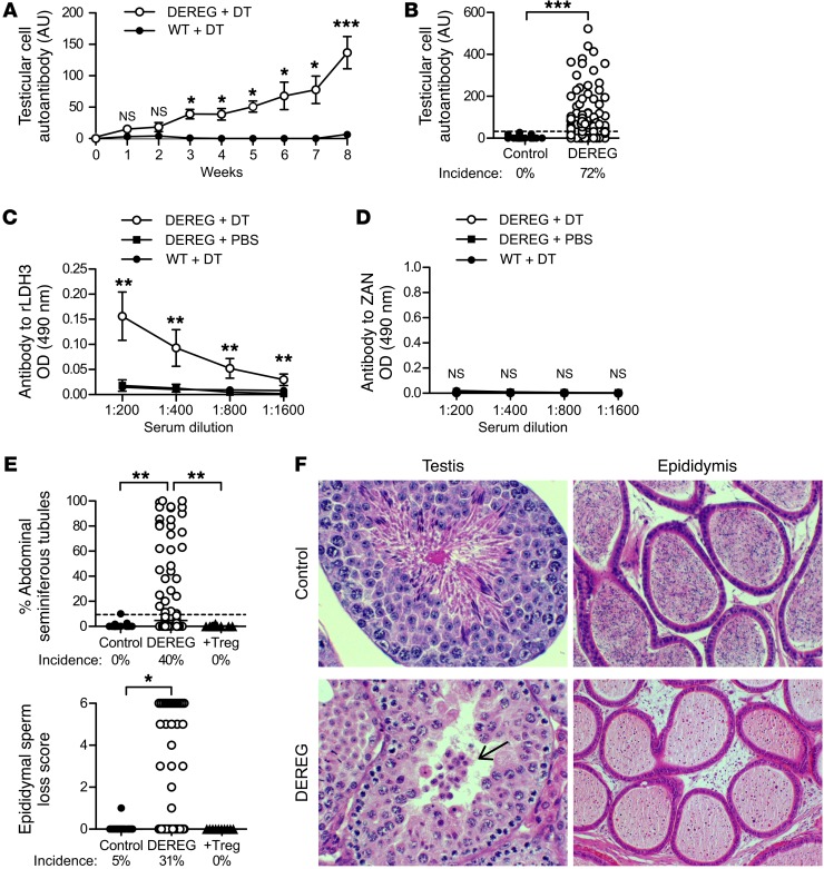
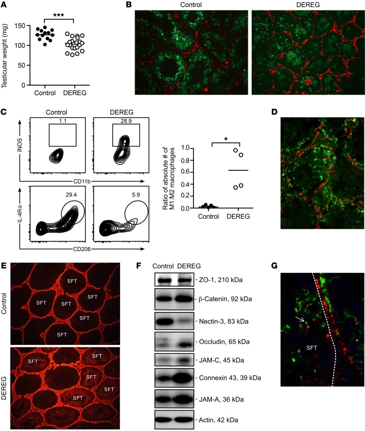
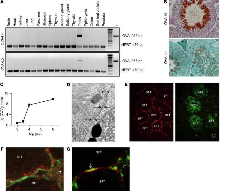
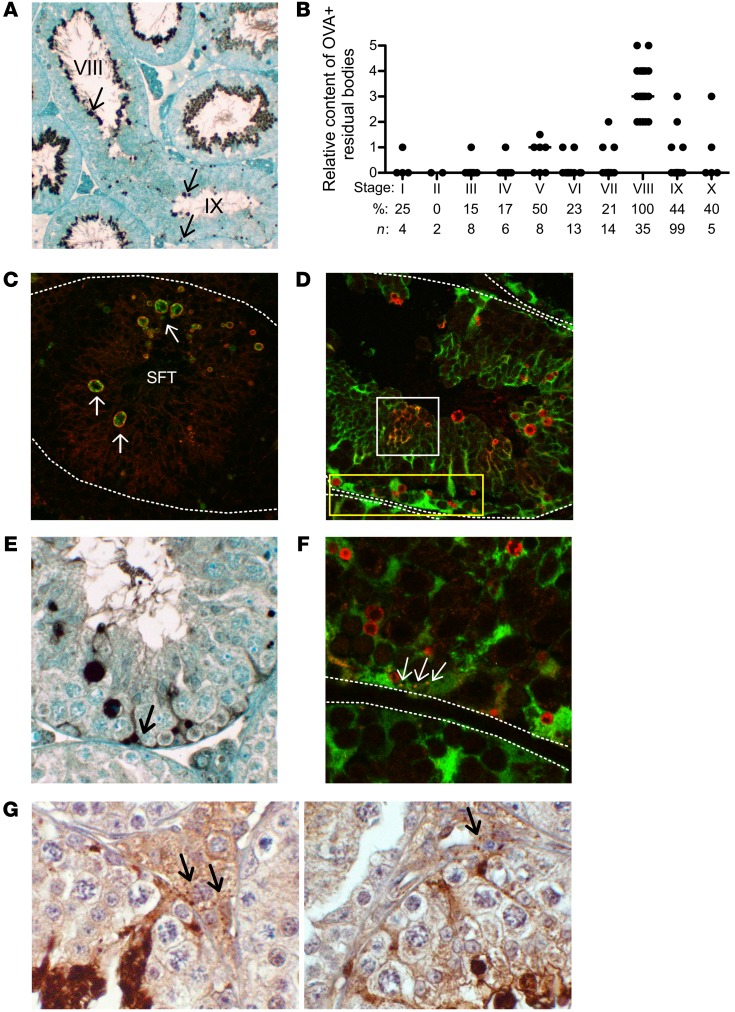
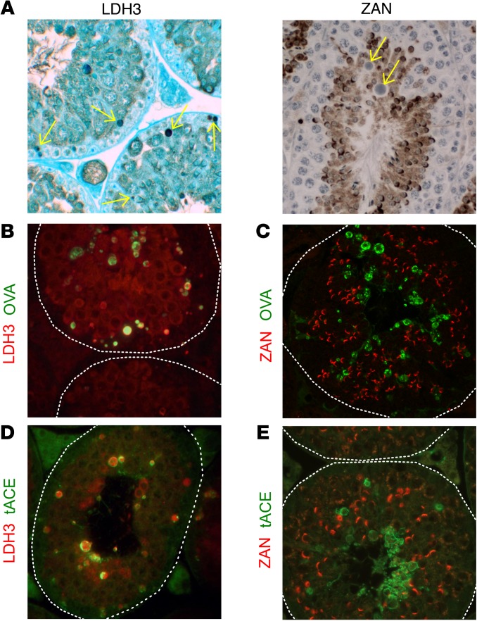
Autoimmune responses to meiotic germ cell antigens (MGCA) that are expressed on sperm and testis occur in human infertility and after vasectomy. Many MGCA are also expressed as cancer/testis antigens (CTA) in human cancers, but the tolerance status of MGCA has not been investigated. MGCA are considered to be uniformly immunogenic and nontolerogenic, and the prevailing view posits that MGCA are sequestered behind the Sertoli cell barrier in seminiferous tubules. Here, we have shown that only some murine MGCA are sequestered. Nonsequestered MCGA (NS-MGCA) egressed from normal tubules, as evidenced by their ability to interact with systemically injected antibodies and form localized immune complexes outside the Sertoli cell barrier. NS-MGCA derived from cell fragments that were discarded by spermatids during spermiation. They egressed as cargo in residual bodies and maintained Treg-dependent physiological tolerance. In contrast, sequestered MGCA (S-MGCA) were undetectable in residual bodies and were nontolerogenic. Unlike postvasectomy autoantibodies, which have been shown to mainly target S-MGCA, autoantibodies produced by normal mice with transient Treg depletion that developed autoimmune orchitis exclusively targeted NS-MGCA. We conclude that spermiation, a physiological checkpoint in spermatogenesis, determines the egress and tolerogenicity of MGCA. Our findings will affect target antigen selection in testis and sperm autoimmunity and the immune responses to CTA in male cancer patients.


Conflict of interest statement

Conflict of interest: The authors have declared that no conflict of interest exists.

Figures

Figure 1
Figure 1. Testis anatomy and selective MGCA sequestration.
(A) Sperm production occurs in cycles (different colors) longitudinally along seminiferous tubules (SFT) (ia, ib). Mature sperm are transported to the vas deferens (v) via the rete testis (ii), ductus efferentes (iii), and epididymis (iv). (B) Seminiferous tubules, visualized by IF on Scx-GFP mouse testes with GFP+ Sertoli cells (green) and LDH3 (red), contain LDH3 spermatogonia (white arrows) and LDH3+ spermatocytes (yellow arrows). Insert: occludin+ Sertoli cell barriers (red) located between adjacent Sertoli cells (green). I/T, interstitial space. Original magnification, ×500; ×800 (insert). (C) A seminiferous tubule segment with 2 Sertoli cells (light green) depicts the complete MGCA sequestration paradigm. Sertoli cells support spermatogenesis (steps i–iv) and spermiation (steps v–ix). Spermatogonia (i) traverse the Sertoli cell barriers to become MGCA+ spermatocytes (ii, pink), then round (iii) and elongating (iv) spermatids. At spermiation, redundant cytoplasm (yellow) and plasma membrane (black) are partially detached from elongated spermatids (v) to form residual bodies (vi) destined for degradation inside Sertoli cells (vii), and retained as cytoplasmic droplets (viii) on mature sperm (ix). The interstitial space contains spermatogonia, basal lamina, peritubular cells (not shown), Leydig cells, macrophages, and afferent lymphatic vessels (not shown). Sertoli cells and Sertoli cell barriers (purple) sequester all MGCA+ meiotic germ cells inside seminiferous tubules. (D) The new selective MGCA sequestration paradigm supported by our study is shown. Tolerogenic NS-MGCA are located in residual bodies and not removed by Sertoli cells. They enter the basal Sertoli cell cytoplasm and egress into the interstitial space. S-MGCA, including those in the sperm acrosome (green crescent), absent from residual bodies, are nontolerogenic. Note that some, but not all, residual bodies are destroyed by the Sertoli cells (vii).
Figure 2
Figure 2. Tolerogenic MGCA egress from seminiferous tubules of normal mouse testes.
(A) After rabbit anti-LDH3 Ab injection, immune complexes appeared as rabbit IgG puncta (green) outside the occludin+ Sertoli cell barrier (red) in adult WT mouse testes (n = 7) surrounding approximately 18% of seminiferous tubules (B arrows, n = 5), but not in (C) testes of adult Ldh3 null mice (n = 7). (D) No immune complexes were detected in the testes of WT mice injected with rabbit anti-ZAN D3p18 Abs. Original magnification, ×800 (A); ×100 (B); ×800 (C); ×800 (D). Tolerance to LDH3 and ZAN were determined by serum IgG Ab responses to rLDH3 (E and F) or ZAN D3p18 (G) at 3 weeks after immunization with testis homogenate in adjuvant. (E) Comparison of rLDH3 responses between WT male mice (n = 5) and WT female mice (n = 5) and (F) between Ldh3 null male mice (n = 6) and WT male mice (n = 6) are shown. (G) Comparison of ZAN D3p18 responses between WT male mice (n = 5) and WT female mice (n = 3) is shown. Ab responses to (H) rLDH3 and (I) ZAN in WT male mice at 3 weeks after immunization with testis homogenate in adjuvants, with (n = 7) and without (n = 13) concomitant CD25 mAb (PC61) injection are shown. Data in EI are from 3 independent experiments. *P < 0.05; **P < 0.01, Mann-Whitney U tests.
Figure 3
Figure 3. Treg-depleted B6AF1-DEREG mice spontaneously develop autoimmune orchitis and produce Abs to LDH3 but not to ZAN.
(A) Kinetics of autoantibody responses to testicular cell antigens (n = 3–43 per time point). (B) Incidence and level of testicular cell autoantibody at 8 weeks between Treg-depleted DEREG mice (n = 106) and WT mice (n = 22) treated with DT. Serum Ab to (C) rLDH3 (n = 5 to n = 10 per group) and (D) ZAN D3p18 (n = 5 to n = 10 per group) in control and Treg-depleted DEREG mice at 8 weeks. (E) Incidences and severity of seminiferous tubules with abnormal spermatogenesis and sperm loss in epididymis at 8 weeks. (F) Representative histopathology of testis and epididymis between DEREG mice treated with DT (bottom) and PBS (top). Arrow points to a cluster of leukocyte-like cells in the tubule lumen (H&E). Original magnification, ×400 (testis); ×200 (epidymis). Data are pooled from 6–12 independent experiments. *P < 0.05; **P < 0.01; ***P < 0.001, Mann-Whitney U tests (AD); Kruskal-Wallis tests with Dunn’s post-tests (E).
Figure 4
Figure 4. EAO of Treg-depleted DEREG mice is characterized by M1-like macrophage infiltration, immune complex deposition, and disrupted Sertoli cell barrier.
(A) Testis weight at 8 weeks after DT treatment. (B) Increased interstitial F4/80+ macrophages (red) and reduced seminiferous tubule diameter and ZAN+ spermatids (green) in DEREG mice with EAO compared with control mice. Original magnification, ×100. (C) Most macrophages of control mice have the characteristic phenotype of M2 macrophages, including the expression of IL-4Rα, CD206, and F4/80 (left), whereas macrophages of Treg-depleted DEREG mice have an M1-like phenotype of high iNOS and MHC class II expression (right); data are expressed as a ratio of absolute numbers (right), where each symbol represents cells from 2 testes of 1 mouse. (D) Mouse IgG immune complexes (green) in the DEREG mouse testes with EAO are located both outside and inside seminiferous tubules relative to the occludin+ Sertoli cell barrier (red). Original magnification, ×300. (E) Biotin (red) exclusion assay in testes of control mice (top) and Treg-depleted DEREG mice (bottom). Original magnification, ×100. (F) Western blot analysis of extractable tight junction proteins of the Sertoli cell barrier from testes of control and Treg-depleted DEREG mice. This is a composite data set of 8 parallel blots wherein 4 control and Treg-depleted DEREG mouse testis samples were analyzed by using corresponding specific Abs in a single experimental session to avoid interexperimental variations. (G) IF staining of murine IgG (green) and rabbit IgG (red) in the testis of a DEREG mouse with EAO at 19 hours after i.p. injection of rabbit Abs to LDH3. Note rabbit (red) and mouse (green) immune complexes were admixed or colocalized inside the seminiferous tubule (arrow). Dotted line, seminiferous tubule boundary. Original magnification, ×800. Data are from 2–12 independent experiments. *P < 0.05; ***P < 0.001, Mann-Whitney U test (A and C).
Figure 5
Figure 5. Testis-specific transgenic OVA expressed in elongated spermatids as a surrogate MGCA also egresses from seminiferous tubules in a concentration-dependent manner.
Comparison between OVA-Hi and OVA-Lo mice: (A) adult OVA expression (reverse transcriptase PCR [RT-PCR]) and (B) OVA localization in elongated spermatids (top, IP with PAS; bottom, IP with hematoxylin). (C) Ontogeny of testicular OVA expression in OVA-Hi mice (ELISA). (D) Tight junction ultrastructure of a Sertoli cell barrier (arrows) in adult OVA-Hi mouse testis. (E) FITC (green) exclusion by the occludin+ Sertoli cell barrier (red) of seminiferous tubules in an OVA-Hi mouse testis (left) compared with testis of a mouse with EAO (right). In OVA-Hi mice injected i.p. with rabbit Abs to OVA, (F) immune complexes detectable as rabbit IgG puncta (green) are detected outside the occludin+ Sertoli cell barrier (red) and (G) inside interstitial MHC class II+ macrophages (green, arrow). Original magnification, ×400 (B); ×10,000 (D); ×800 (E); ×400 (FG).
Figure 6
Figure 6. Stage-specific OVA localization and detection in residual bodies of OVA-Hi mice.
(A) IP staining in a longitudinal seminiferous tubule section of an OVA-Hi mouse testis. Note: OVA+ residual bodies (arrows) are with sperm at stage VIII, but are randomly distributed throughout the seminiferous tubule at stage IX. (B) OVA+ residual bodies with high OVA content are maximally detected at stage IX of the spermatogenic cycle (significance between stage IX and all other stages ranges from significant to highly significant). *P < 0.05; **P < 0.01; ***P < 0.001, Kruskal-Wallis test. (C) Residual bodies (indicated by arrows) in OVA-Hi mouse testis at stage IX: OVA (green) is enclosed by membrane-associated tACE, a residual body marker (red). (D) Most OVA+ residual bodies (red) in OVA-Hi × Scx-GFP F1 testis at stage IX do not costain with Sertoli cell cytoplasm (green), including the very small residual bodies at the base of seminiferous tubules (yellow rectangle). Some OVA is inside the Sertoli cell cytoplasm (yellow) with plexiform distribution (white square). (E) Diffuse OVA IP staining confined to the basal cytoplasm of Sertoli cells in OVA-Hi mouse testis at stage IX (arrow). (F) OVA+ puncta (red) are inside the basal Sertoli cell cytoplasm (yellow, arrows) in OVA-Hi × Scx-GFP F1 mouse testis at stage IX. (G) Small OVA+ speckles detected by IP staining in the interstitial space of OVA-Hi mouse testes (arrows) at stage VIII (left) and stage IX (right). All images are representative of 3–4 experiments. Dotted white lines denote the boundary of seminiferous tubules. Original magnification, ×100 (A); ×400 (CF); ×800 (G).
Figure 7
Figure 7. Detection of LDH3 and ZAN in residual bodies of both OVA-Hi and BALB/c mouse testes.
(A) LDH3 (left, arrows), but not ZAN (right, arrows), is detected in residual bodies of WT mouse testes at stage IX by IP staining (hematoxylin). In OVA-Hi mouse testes at stage IX, OVA (green) in residual bodies is colocalized with (B) LDH3 (red), but not with (C) ZAN (red). In WT testes, tACE (green) in the residual bodies is colocalized with (D) LDH3 (red), but not with (E) ZAN (red). All images are representative of 3–4 independent experiments. Dotted white lines denote the boundary of seminiferous tubules. Original magnification, ×400.

References

    1. Turek PJ, Lipshultz LI. Immunologic infertility. Urol Clin North Am. 1994;21(3):447–468. - PubMed
    1. Duan YG, et al. Immunodeviation towards a Th17 immune response associated with testicular damage in azoospermic men. Int J Androl. 2011;34(6 Pt 2):e536–e545. - PubMed
    1. Simpson AJ, Caballero OL, Jungbluth A, Chen YT, Old LJ. Cancer/testis antigens, gametogenesis and cancer. Nat Rev Cancer. 2005;5(8):615–625. doi: 10.1038/nrc1669. - DOI - PubMed
    1. Whitehurst AW. Cause and consequence of cancer/testis antigen activation in cancer. Annu Rev Pharmacol Toxicol. 2014;54:251–272. doi: 10.1146/annurev-pharmtox-011112-140326. - DOI - PubMed
    1. Xing Y, Hogquist KA. T-cell tolerance: central and peripheral. Cold Spring Harb Perspect Biol. 2012;4(6):doi:10.1101/cshperspect.a006957 - PMC - PubMed