. 2017 Mar 1;127(3):1099-1114.
doi: 10.1172/JCI90086. Epub 2017 Feb 20.

GATA4-dependent organ-specific endothelial differentiation controls liver development and embryonic hematopoiesis

GATA4-dependent organ-specific endothelial differentiation controls liver development and embryonic hematopoiesis

Cyrill Géraud et al. J Clin Invest. .

Abstract

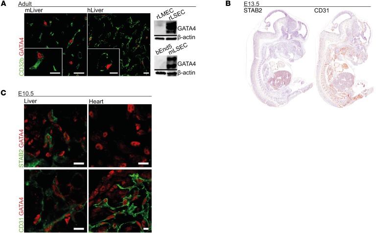
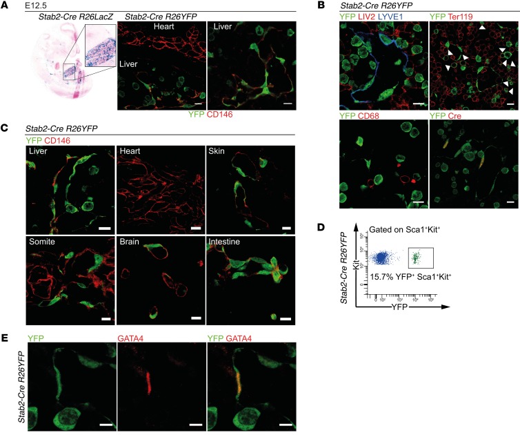
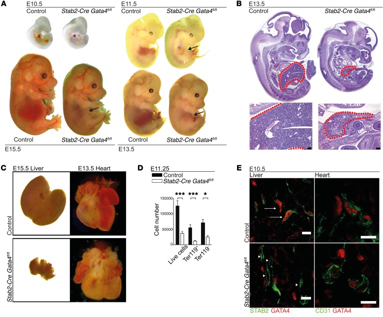
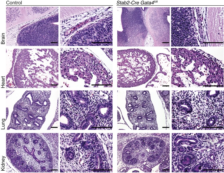
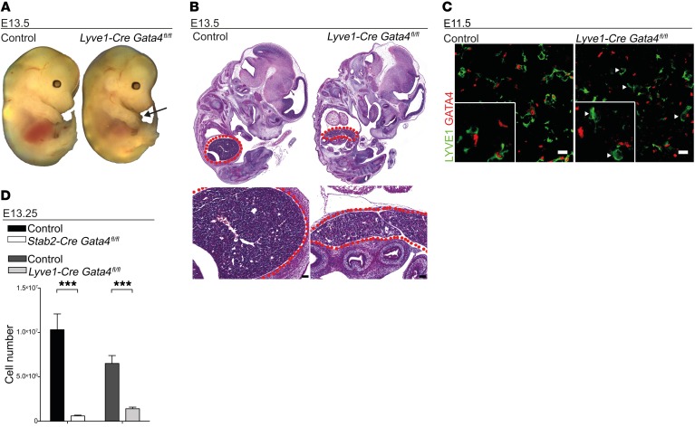
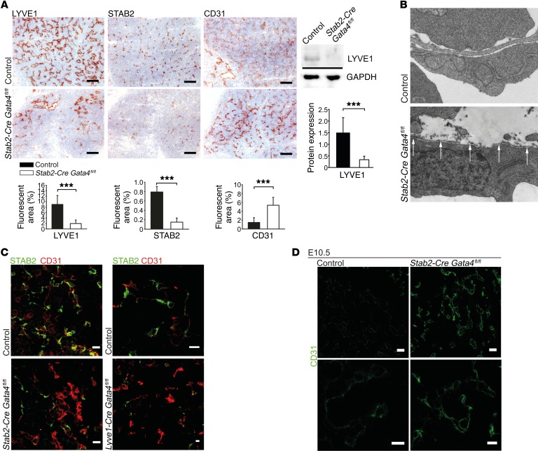
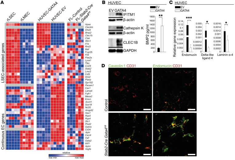
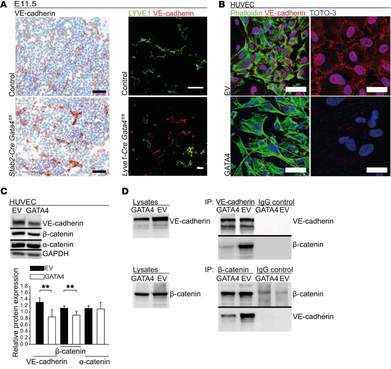
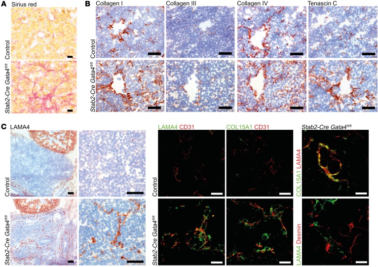
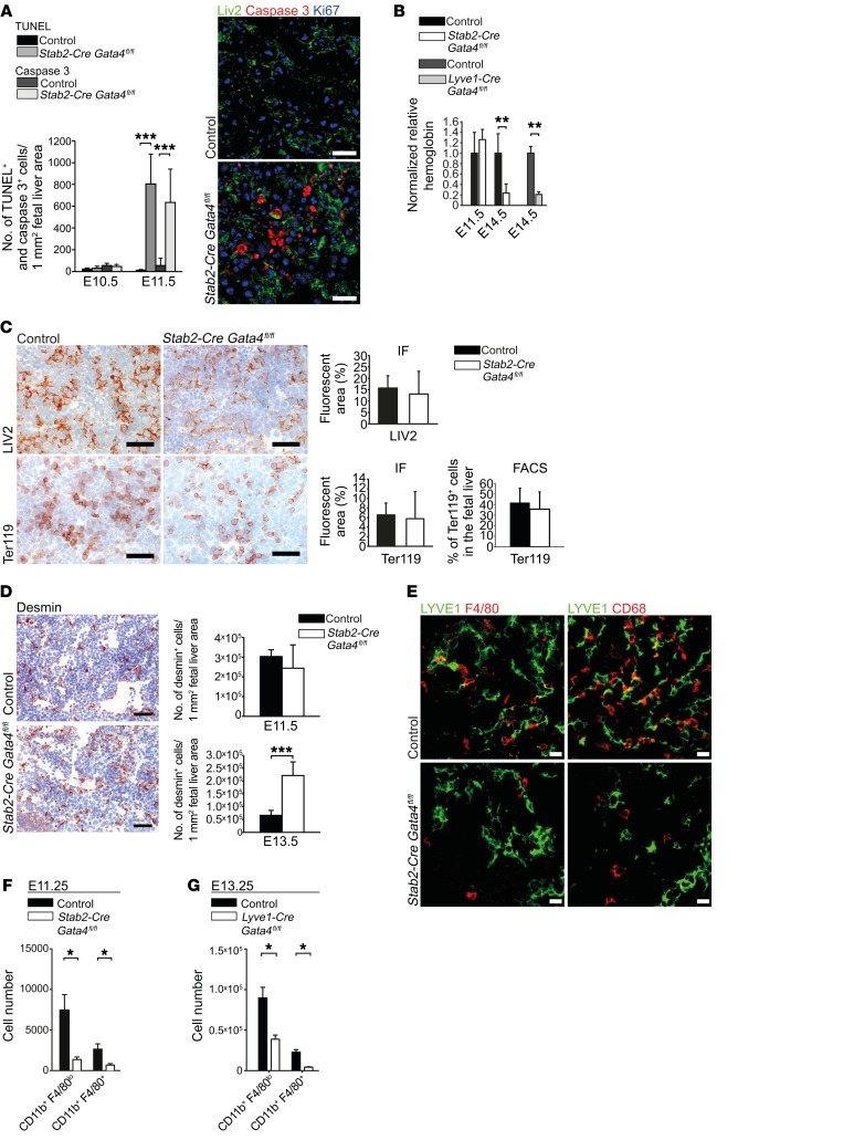
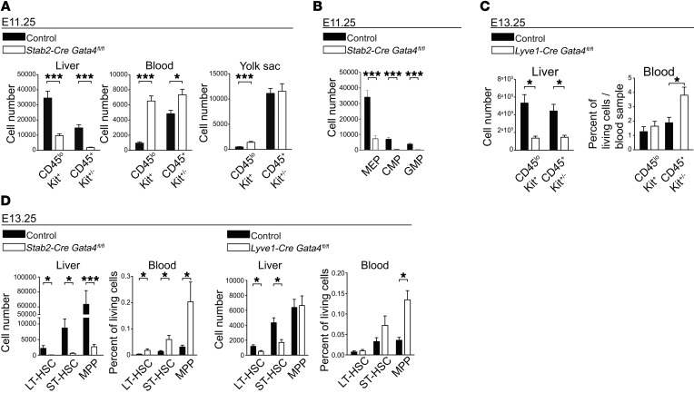
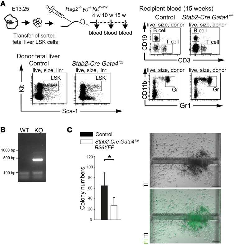
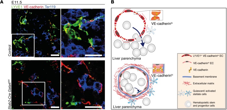
Microvascular endothelial cells (ECs) are increasingly recognized as organ-specific gatekeepers of their microenvironment. Microvascular ECs instruct neighboring cells in their organ-specific vascular niches through angiocrine factors, which include secreted growth factors (angiokines), extracellular matrix molecules, and transmembrane proteins. However, the molecular regulators that drive organ-specific microvascular transcriptional programs and thereby regulate angiodiversity are largely elusive. In contrast to other ECs, which form a continuous cell layer, liver sinusoidal ECs (LSECs) constitute discontinuous, permeable microvessels. Here, we have shown that the transcription factor GATA4 controls murine LSEC specification and function. LSEC-restricted deletion of Gata4 caused transformation of discontinuous liver sinusoids into continuous capillaries. Capillarization was characterized by ectopic basement membrane deposition, formation of a continuous EC layer, and increased expression of VE-cadherin. Correspondingly, ectopic expression of GATA4 in cultured continuous ECs mediated the downregulation of continuous EC-associated transcripts and upregulation of LSEC-associated genes. The switch from discontinuous LSECs to continuous ECs during embryogenesis caused liver hypoplasia, fibrosis, and impaired colonization by hematopoietic progenitor cells, resulting in anemia and embryonic lethality. Thus, GATA4 acts as master regulator of hepatic microvascular specification and acquisition of organ-specific vascular competence, which are indispensable for liver development. The data also establish an essential role of the hepatic microvasculature in embryonic hematopoiesis.


Conflict of interest statement

Conflict of interest: The authors have declared that no conflict of interest exists.

Figures

Figure 1
Figure 1. LSECs represent the only intersection of GATA4 and STAB2 expression during development of the embryo.
(A) Murine liver (mLiver) and human liver (hLiver) show GATA4 in CD32b+ LSECs on co-immunofluorescence (co-IF). Expression of GATA4 detected by Western blot in rat LSECs (rLSEC) and mouse LSECs (mLSEC), but not in rLMECs and bEnd5 murine brain ECs (n = 3). Scale bars: 20 μm and 10 μm (inset). (B) IHC of STAB2 and CD31 at E13.5 (n = 5). (C) Co-IF of GATA4 with STAB2 or CD31 in fetal liver and heart of WT embryos at E10.5 (n = 2). Scale bars: 10 μm.
Figure 2
Figure 2. Characterization of endothelial subtype–specific Stab2-Cre driver mice.
(A) Analysis of Stab2-Cre R26LacZ (β-gal assay) (n = 3) and Stab2-Cre R26YFP (co-IF of YFP and CD146) (n = 3) shows reporter activity in the endothelium of the fetal liver. Scale bars: 10 μm. (BE) Analysis of the Stab2-Cre R26YFP reporter in the embryo. (B) Co-IF of YFP and LYVE1, Cre, LIV2, Ter119, and CD68 in the fetal liver at E12.5. Arrowheads indicate YFP+Ter119+ cells (n = 3). Scale bars: 10 μm. (C) Co-IF of YFP and CD146 at E12.5 (n = 3). Scale bars: 10 μm. (D) FACS analysis of the fetal liver of Stab2-Cre R26YFP reporter mice at E13.25. Representative FACS blot showing YFP reporter activity in Sca1+Kit+ cells (n = 5). (E) Co-IF of YFP and GATA4 in the fetal liver at E12.5 (n = 3). Scale bars: 5 μm.
Figure 3
Figure 3. Deletion of GATA4 in LSECs in Stab2-Cre Gata4fl/fl mice impairs liver development and causes embryonic lethality.
(A) Photomicrographs of Stab2-Cre Gata4fl/fl embryos at different ages. Arrows indicate hypoplastic livers in the mutant embryos from E11.5 to E15.5 (n > 3). (B) H&E staining of Stab2-Cre Gata4fl/fl embryos at E13.5. Red dotted lines indicate the fetal liver (n = 5). Scale bars: 100 μm. (C) Photomicrographs of the fetal liver (E15.5) and fetal heart (E13.5) of Stab2-Cre Gata4fl/fl embryos. (D) FACS analysis of live cells, Ter119+ cells, and Ter119 cells in the liver of Stab2-Cre Gata4fl/fl embryos at E11.25 (n = 14 mutants and 10 controls). Student’s t test; *P < 0.05, ***P < 0.001. (E) Co-IF of GATA4 and STAB2 or CD31 of fetal livers of Stab2-Cre Gata4fl/fl embryos at E10.5. IF shows absence of GATA4 in the mutant liver (arrowheads), but not in controls (arrows) (n = 3). Scale bars: 10 μm.
Figure 4
Figure 4. Deletion of Gata4 in LSECs in Stab2-Cre Gata4fl/fl mice does not affect cardiac, brain, lung, or kidney development.
H&E staining of the brain, heart, lung, and kidney of Stab2-Cre Gata4fl/fl embryos at E13.5 does not show any gross abnormalities (n = 3). Scale bars: 100 μm.
Figure 5
Figure 5. Lyve1-Cre Gata4fl/fl mice show liver hypoplasia and embryonic lethality similar to but less severe than that observed in Stab2-Cre Gata4fl/fl mice.
(A and B) Photomicrographs (A) and H&E staining (B) of Lyve1-Cre Gata4fl/fl embryos at E13.5. Arrow indicates the hypoplastic fetal liver, and red dotted lines indicate the fetal liver (n = 3). Scale bars: 100 μm. (C) Co-IF of GATA4 and LYVE1 of fetal livers of Lyve1-Cre Gata4fl/fl embryos at E11.5. IF shows absence of GATA4 in the mutant liver (arrowheads), but not in controls (n = 3). Scale bars: 10 μm. (D) Total cell numbers of the liver of Stab2-Cre Gata4fl/fl and Lyve1-Cre Gata4fl/fl embryos at E13.25 (Stab2-Cre Gata4fl/fl: n = 13; Lyve1-Cre Gata4fl/fl: n = 6). Student’s t test; ***P < 0.001.
Figure 6
Figure 6. Liver endothelial GATA4 is required for maintenance of discontinuous LSEC differentiation.
(A) Expression of LYVE1, STAB2, and CD31 in the liver of Stab2-Cre Gata4fl/fl embryos at E11.5. IHC (upper panels) and quantification (lower panels) based on IF images of LYVE1, STAB2, and CD31 (n = 4). Scale bars: 100 μm. Western blot of LYVE1 and GAPDH with quantification of densitometric data (n = 5) (right). (B) Transmission electron microscopy (TEM) of the liver of Stab2-Cre Gata4fl/fl embryos at E11.5. Arrows indicate formation of a basement membrane (n = 4). (C) Co-IF of STAB2 and CD31 in the liver of Stab2-Cre Gata4fl/fl (n = 4) (left) and Lyve1-Cre Gata4fl/fl (n = 3) (right) embryos at E11.5. Scale bars: 20 μm (left) and 10 μm (right). (D) IF of CD31 in the fetal liver of Stab2-Cre Gata4fl/fl embryos at E10.5 shows increased expression of CD31 in the endothelium of mutant embryos as an indicator of early hepatic capillarization. Scale bars: 20 μm (upper panels) and 10 μm (lower panels) (n = 2). Student’s t test; ***P < 0.001.
Figure 7
Figure 7. Endothelial GATA4 counterregulates liver sinusoidal and continuous EC differentiation in vivo and in vitro.
(A) Heatmap of LSEC-associated and continuous EC-associated genes comparing primary rLSECs with rLMECs, GATA4-transduced with EV-transduced HUVECs, and liver from Stab2-Cre Gata4fl/fl embryos with control embryos at E11.5. (B) Western blot of IFITM1, cathepsin K, and CLEC1B of GATA4-transduced HUVECs (n = 3) (left). ELISA of BMP2 from supernatants of GATA4-transduced HUVECs (n = 3) (right). (C) qRT-PCR of endomucin, delta-like ligand 4, and laminin α4 in GATA4-transduced HUVECs (n = 3). (D) Co-IF of CD31 with caveolin 1 or endomucin in the liver of Stab2-Cre Gata4fl/fl embryos at E11.5 (n = 4). Scale bars: 20 μm. Student’s t test; *P < 0.05, **P < 0.01,***P < 0.001.
Figure 8
Figure 8. Endothelial GATA4 regulates VE-cadherin expression and junctional stability in liver sinusoidal and continuous ECs in vivo and in vitro.
(A) IHC of VE-cadherin in the liver of Stab2-Cre Gata4fl/fl embryos at E11.5. Scale bars: 50 μm (left). Co-IF of VE-cadherin and LYVE1 in liver of Lyve1-Cre Gata4fl/fl embryos at E11.5 (n = 3). Scale bar: 20 μm (right). (B) Co-IF of VE-cadherin, TOTO-3, and phalloidin of GATA4-transduced HUVECs (n = 3). Scale bars: 60 μm (left) and 30 μm (right). (C) WB and densitometric quantification of VE-cadherin, α-catenin, and β-catenin of GATA4-transduced HUVECs (n = 4). (D) Co-IP of VE-cadherin and β-catenin of GATA4-transduced HUVECs. WB of lysates is shown as control, WB of precipitates (IP) with anti–VE-cadherin, anti–β-catenin, and the respective IgG controls (n = 4). Student’s t test; **P < 0.01.
Figure 9
Figure 9. Liver endothelial GATA4 prevents peri-sinusoidal ECM deposition.
(A) Sirius red staining in the liver of Stab2-Cre Gata4fl/fl embryos at E13.5 (n = 3). Scale bars: 10 μm. (B) IHC of collagen I, collagen III, collagen IV, and tenascin C in the liver of Stab2-Cre Gata4fl/fl embryos at E13.5 (n = 3). Scale bars: 50 μm. (C) Expression of LAMA4 in the liver of Stab2-Cre Gata4fl/fl embryos at E11.5 (n = 3). IHC of LAMA4. Scale bars: 100 μm (overview) and 50 μm (magnification) (left panels). Co-IF of CD31 with LAMA4 or collagen 15a1. Scale bars: 20 μm (middle panels). Co-IF of LAMA4 with COL15A1 or desmin. Scale bars: 10 μm (right panels).
Figure 10
Figure 10. Liver endothelial GATA4 is required for the formation of the fetal hepatic vascular niche.
(A) Quantification of TUNEL assay and IF of cleaved caspase-3 in the fetal liver at E10.5 and E11.5 (n = 3). Co-IF of cleaved caspase-3, Ki67, and LIV2 in the liver of Stab2-Cre Gata4fl/fl embryos (n = 3). Scale bars: 20 μm. (B) Normalized hemoglobin levels from peripheral blood of Stab2-Cre Gata4fl/fl embryos at E11.5 and E14.5 and of Lyve1-Cre Gata4fl/fl embryos at E14.5 (n = 3). (C) Expression of LIV2 and Ter119 in the liver of Stab2-Cre Gata4fl/fl embryos. IHC and quantification based on IF images at E11.5 (n > 3). Scale bars: 50 μm. FACS quantification of Ter119+ cells in relation to total liver cells at E11.25. Data set from Figure 3D. (D) IHC of desmin in the fetal liver of Stab2-Cre Gata4fl/fl embryos at E11.5 (n = 3). Scale bars: 50 μm (left). Quantification based on IF at E11.5 and E13.5 (n = 3). (E) Co-IF of LYVE1, F4/80, and CD68 in the liver of Stab2-Cre Gata4fl/fl embryos at E11.5 (n = 3). Scale bars: 20 μm. (F and G) Quantification upon FACS analysis of CD11b+F4/80lo and CD11b+F4/80+ cells in the liver of Stab2-Cre Gata4fl/fl embryos at E11.25 (F) and in the liver of Lyve1-Cre Gata4fl/fl embryos at E13.25 (G) Stab2-Cre Gata4fl/fl: n = 5 mutants and 6 controls; Lyve1-Cre Gata4fl/fl: n = 6. Student’s t test; *P < 0.05, **P < 0.01,***P < 0.001.
Figure 11
Figure 11. Liver endothelial GATA4 controls hepatic colonization by hematopoietic stem and progenitor cells.
(A) FACS quantification of CD45loKit+ and CD45+Kit+/– hematopoietic progenitors in liver, blood, and YS of Stab2-Cre Gata4fl/fl embryos at E11.25 (liver: n = 14 mutants and 10 controls; blood: n = 15 mutants and 10 controls; YS: n = 8 mutants and 16 controls [YS controls: Stab2-Cretg/WT Gata4fl/WT, Stab2-CreWT/WT Gata4fl/fl, Stab2-CreWT/WT Gata4fl/WT]). (B) FACS quantification of CMPs, GMPs, and myeloid-erythroid progenitors (MEP) in the liver of Stab2-Cre Gata4fl/fl embryos at E11.25 (n = 7). (C) FACS quantification of CD45loKit+ and CD45+Kit+/– hematopoietic progenitors in the liver and blood of Lyve1-Cre Gata4fl/fl embryos at E13.25 (liver: n = 6; blood: n = 6 mutants and 5 controls). (D) FACS quantification of long-term HSCs (LT-HSC), short-term HSCs (ST-HSC), and MPPs in liver and blood of Stab2-Cre Gata4fl/fl (left panels) and Lyve1-Cre Gata4fl/fl (right panels) embryos at E13.25. LT-HSCs (LinSca-1+Kit+CD150+CD48), ST-HSCs (LinSca-1+Kit+CD150CD48), and MPPs (LinSca-1+Kit+CD150CD48+) (Stab2-Cre Gata4fl/fl liver: n = 8; Stab2-Cre Gata4fl/fl peripheral blood: n = 7 mutants and 8 controls; Lyve1-Cre Gata4fl/fl liver: n = 6; Lyve1-Cre Gata4fl/fl peripheral blood: n = 6 mutants and 5 controls). Student’s t test; *P < 0.05, ***P < 0.001.
Figure 12
Figure 12. Analyses of the lineage potential of hematopoietic fetal liver cells from Stab2-Cre Gata4fl/fl embryos.
(A) Fetal liver donor LSK (LinSca-1+Kit+) cells from Stab2-Cre Gata4fl/fl (n = 3) or control (n = 5) embryos at E13.25 were FACS sorted and transferred into Rag2–/– γc–/– KitW/Wv mice. After 4, 10, and 15 weeks, peripheral blood was analyzed for donor-derived lymphoid (CD3+ T cells, CD19+ B cells) and myeloid (CD11b+Gr1+ granulocytes) cells. Flow cytometry blots of donor fetal liver cells from control and Stab2-Cre Gata4fl/fl embryo (left) as well as FACS blots of peripheral blood analysis of the corresponding recipient mouse after 15 weeks (right) are shown. (B) Product from PCR shows the recombination of floxed Gata4 allele in the blood of Stab2-Cre Gata4fl/fl recipient mouse after 15 weeks of transplantation. WT, Stab2-CreWT/WT Gata4fl/WT. (C) Numbers of CFU assay performed with fetal liver cells harvested from Stab2-Cre R26YFP Gata4fl/fl embryos at E12.5 (n = 4 mutants and 7 controls) (left). Representative image shows a fluorescent colony from the mutant embryo (right). Tl, transmission light; Fl, fluorescent light. Scale bars: 200 μm. Student’s t test; *P < 0.05.
Figure 13
Figure 13. Endothelial GATA4 expression preserves liver sinusoidal endothelial identity with high junctional permeability and limited ECM deposition permissive for hepatic stem cell colonization and unperturbed liver development and erythropoiesis.
(A) Co-IF of LYVE1, Ter119, and VE-cadherin in the liver of Stab2-Cre Gata4fl/fl embryos at E11.5 (n = 3). Scale bars: 20 μm. (B) GATA4 serves as the master regulator of the fetal hepatic vascular niche. During normal development, GATA4-dependent gene expression cascades in ECs promote discontinuous sinusoidal endothelial differentiation featuring weak cell-cell contacts, lack of a basement membrane, and low levels of ECM deposition, resulting in an antifibrotic perivascular microenvironment. The hepatic sinusoidal microvasculature supports transmigration of hematopoietic stem and progenitor cells into the liver parenchyma, a process indispensable for liver development. In the absence of GATA4, hepatic sinusoidal microvessels undergo continuous transdifferentiation/capillarization, including basement membrane formation followed by ECM deposition and stellate cell activation, and lethal impairment of hematopoiesis via stabilization of VE-cadherin+ junctions.

Comment in

  • Building discontinuous liver sinusoidal vessels

References

    1. Aird WC. Phenotypic heterogeneity of the endothelium: I. Structure, function, and mechanisms. Circ Res. 2007;100(2):158–173. doi: 10.1161/01.RES.0000255691.76142.4a. - DOI - PubMed
    1. Nolan DJ, et al. Molecular signatures of tissue-specific microvascular endothelial cell heterogeneity in organ maintenance and regeneration. Dev Cell. 2013;26(2):204–219. doi: 10.1016/j.devcel.2013.06.017. - DOI - PMC - PubMed
    1. Aird WC. Phenotypic heterogeneity of the endothelium: II. Representative vascular beds. Circ Res. 2007;100(2):174–190. doi: 10.1161/01.RES.0000255690.03436.ae. - DOI - PubMed
    1. Lawson ND, et al. Notch signaling is required for arterial-venous differentiation during embryonic vascular development. Development. 2001;128(19):3675–3683. - PubMed
    1. You LR, Lin FJ, Lee CT, DeMayo FJ, Tsai MJ, Tsai SY. Suppression of Notch signalling by the COUP-TFII transcription factor regulates vein identity. Nature. 2005;435(7038):98–104. doi: 10.1038/nature03511. - DOI - PubMed

Substances