Review
. 2017 Feb 17;6(1):13.
doi: 10.3390/biology6010013.

Mechanisms of Chromosome Congression during Mitosis

Affiliations
Review

Mechanisms of Chromosome Congression during Mitosis

Helder Maiato et al. Biology (Basel). .

Abstract

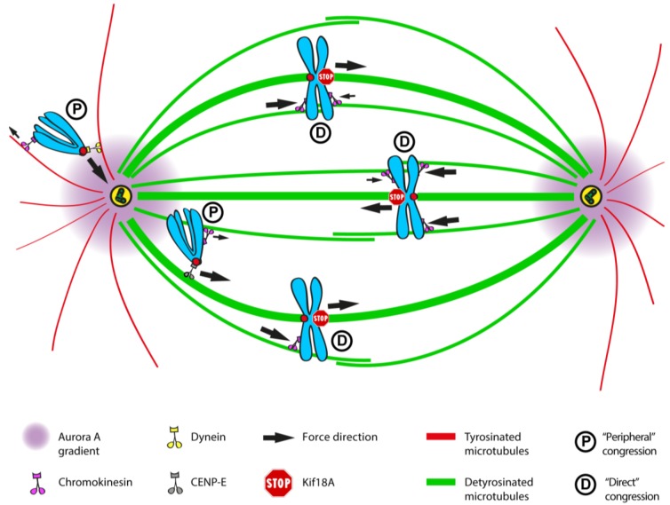
Chromosome congression during prometaphase culminates with the establishment of a metaphase plate, a hallmark of mitosis in metazoans. Classical views resulting from more than 100 years of research on this topic have attempted to explain chromosome congression based on the balance between opposing pulling and/or pushing forces that reach an equilibrium near the spindle equator. However, in mammalian cells, chromosome bi-orientation and force balance at kinetochores are not required for chromosome congression, whereas the mechanisms of chromosome congression are not necessarily involved in the maintenance of chromosome alignment after congression. Thus, chromosome congression and maintenance of alignment are determined by different principles. Moreover, it is now clear that not all chromosomes use the same mechanism for congressing to the spindle equator. Those chromosomes that are favorably positioned between both poles when the nuclear envelope breaks down use the so-called "direct congression" pathway in which chromosomes align after bi-orientation and the establishment of end-on kinetochore-microtubule attachments. This favors the balanced action of kinetochore pulling forces and polar ejection forces along chromosome arms that drive chromosome oscillatory movements during and after congression. The other pathway, which we call "peripheral congression", is independent of end-on kinetochore microtubule-attachments and relies on the dominant and coordinated action of the kinetochore motors Dynein and Centromere Protein E (CENP-E) that mediate the lateral transport of peripheral chromosomes along microtubules, first towards the poles and subsequently towards the equator. How the opposite polarities of kinetochore motors are regulated in space and time to drive congression of peripheral chromosomes only now starts to be understood. This appears to be regulated by position-dependent phosphorylation of both Dynein and CENP-E and by spindle microtubule diversity by means of tubulin post-translational modifications. This so-called "tubulin code" might work as a navigation system that selectively guides kinetochore motors with opposite polarities along specific spindle microtubule populations, ultimately leading to the congression of peripheral chromosomes. We propose an integrated model of chromosome congression in mammalian cells that depends essentially on the following parameters: (1) chromosome position relative to the spindle poles after nuclear envelope breakdown; (2) establishment of stable end-on kinetochore-microtubule attachments and bi-orientation; (3) coordination between kinetochore- and arm-associated motors; and (4) spatial signatures associated with post-translational modifications of specific spindle microtubule populations. The physiological consequences of abnormal chromosome congression, as well as the therapeutic potential of inhibiting chromosome congression are also discussed.

Keywords: CENP-E; Chromokinesin; Dynein; Kinesin; chromosome; kinetochore; microtubule; mitosis; mitotic spindle; polar ejection forces; tubulin code.


Conflict of interest statement

The authors declare no conflict of interest.

Figures

Figure 1
Figure 1
First models of chromosome congression involving either pushing or pulling forces on chromosomes. (a) Model of chromosome congression proposed by Darlington [1] involving a balance of pushing forces on chromosomes. These forces are higher when chromosomes are closer to spindle poles; (b) Model of chromosome congression proposed by Östergren involving pulling forces on chromosomes that are proportional to k-fiber length. Adapted from Östergren, 1950 [13] and displayed under a Creative Commons Attribution-Noncommercial-Share Alike 4.0 International license, as described at https://creativecommons.org/licenses/by-nc-sa/4.0/legalcode.
Figure 2
Figure 2
Evidence that forces on kinetochores are required to position chromosomes at the equator. (a) Original drawings from Izutzu depicting the loss of equatorial position when one of the kinetochore regions from a bivalent chromosome was irradiated with an UV microbeam. Note the displacement of the bivalent from the metaphase plate towards the pole facing the non-irradiated kinetochore after irradiation. Scale bar is 10 μm. Reprinted from Izutsu et al., 1959 [20]; (b) Laser microsurgery of one of the kinetochores from an equatorially-aligned chromosome in a Drosophila S2 cell. Kinetochores were directly labelled with the Centromere Protein A (CENP-A) homologue Cid fused with Green Fluorescent Protein (GFP). Likewise, the chromosome was displaced from the equator after surgery and underwent poleward migration towards the pole facing the undisturbed kinetochore from the pair. Red arrows track the undisturbed kinetochore from the irradiated pair. Green arrows track the congression of an undisturbed chromosome. Laser microsurgery was performed as described in [29]. Scale bar is 2 μm.
Figure 3
Figure 3
Evidence that centrosome-derived microtubules can exert pushing forces. (a) Original Drawings by Drüner depicting the invasion of the chromosomal region by microtubules, which exert a pushing force that assists chromosome alignment at the spindle equator. Reprinted from Drüner, 1895 [12]. Image courtesy of Biodiversity Heritage Library. http://www.biodiversitylibrary.org; (b) Schematic drawing by E. B. Wilson illustrating the pushing action of centrosomal microtubules on the nuclear envelope and subsequent rupture. Reprinted from Wilson, 1925 [10]. Image displayed under a Creative Commons Attribution-Noncommercial-Share Alike 4.0 International license, as described at https://creativecommons.org/licenses/by-nc-sa/4.0/legalcode. Image courtesy of the Wellcome Library. http://wellcomelibrary.org; (c) Schematic drawing by Luykx illustrating the repulsive action of centrosomal microtubules over large organelles (mitochondria). Reprinted from Luykx, 1970 [40]. Courtesy of Elsevier; (d) Original drawings by Darlington illustrating the variability in chromosome positioning in pollen grain cells. Reprinted from Darlington, 1937 [1]. Image courtesy of Biodiversity Heritage Library. http://www.biodiversitylibrary.org; (e,f) Phase contrast image of a newt lung cell undergoing transient monopolar configuration. Kinetochore position was tracked over time, clearly demonstrating the oscillatory behavior of mono-oriented chromosomes in this system. Note that chromosomes do not travel all the way towards the pole. Reprinted from Bajer et al., 1982 [41] and displayed under a Creative Commons Attribution-Noncommercial-Share Alike 4.0 International license, as described at https://creativecommons.org/licenses/by-nc-sa/4.0/legalcode.
Figure 4
Figure 4
Demonstration that polar ejection forces act along the entire chromosome. (a) Phase contrast image of a newt lung cell in which the chromosome arms on one chromosome (arrowheads) were physically separated from the kinetochore region using laser microsurgery. Note the ejection of the acentric chromosome arms away from the polar region. In contrast, the kinetochore-containing region (arrow) moves closer to the polar region. Reprinted from Rieder et al., 1986 [43] and displayed under a Creative Commons Attribution-Noncommercial-Share Alike 4.0 International license, as described at https://creativecommons.org/licenses/by-nc-sa/4.0/legalcode; (b) Schematic representation of the experiment illustrated in (a). Reprinted from Salmon, 1989 [44]. Courtesy of Elsevier.
Figure 5
Figure 5
Demonstration that chromosome congression is independent of bi-orientation. From A-F, the movement of a polar chromosome along a pre-existing k-fiber is illustrated in a PtK1 cell. The leading kinetochore is indicated (yellow arrows). The kinetochore of a neighbor k-fiber on a bi-oriented chromosome is also indicated (yellow arrowheads). Time is in sec. In G, serial sections of a sliding mono-oriented chromosome with the leading kinetochore laterally attached to a neighbor k-fiber. Kinetochores of the congressing chromosome are indicated (white arrows), as well as the kinetochore of a neighbor k-fiber (black arrowheads). Images adapted from Kapoor et al., 2006 [300]. Reprinted with permission from The American Association for the Advancement of Science (AAAS).
Figure 6
Figure 6
Forces at kinetochores are proportional with k-fiber length, but chromosome position at the equator is independent of k-fiber length. (a,b) Laser microsurgery of k-fibers in Drosophila S2 cells stably expressing GFP-α-tubulin to label microtubules (green) and Cid-mCherry to label kinetochores (red). K-fibers were cut (yellow arrowhead) and grew back as described previously [29]. Inverted contrast of GFP-α-tubulin is also shown, as well as the variation of inter-kinetochore distance over time (kymograph; first frame corresponds to pre-surgery distance; second frame onwards are after surgery). Measurement of the inter-kinetochore distance before and after laser surgery ablation of k-fibers (yellow bars) indicates that kinetochores relax after surgery, and this relaxation is more evident the closer the cut is to the kinetochore. Time is in min:sec. White scale bars are 2 μm; (c) Quantification of the percentage of kinetochore relaxation after surgery (determined by the difference between initial inter-kinetochore distance and the minimum observed distance after surgery) indicates a negative correlation (R2 = −0.361; p < 0.001) with the cut distance from the kinetochore (n = 125 cells); (d) Corresponding quantification of the inter-kinetochore distance over time as a function of the cut distance from the kinetochore. Each group was normalized against its initial distance such that one hundred percent corresponds to the average initial distance. The closer the cut is to the kinetochore, the longer the recovery of inter-kinetochore distance and the higher is the relaxation. The inclusion of a kinetochore marker in this study and the observed variability of inter-kinetochore distance after k-fiber cut explains previous observations in which no detectable kinetochore relaxation was observed without the use of a kinetochore marker [29]. Laser microsurgery was performed essentially as described in [373].
Figure 7
Figure 7
Integrated model of chromosome congression in human cells. In this representation, Kif18A is shown to restrict k-fiber length, thereby contributing to a directional switch and regulating chromosome oscillations after bi-orientation. See text for a detailed description.

References

    1. Darlington C.D. Recent Advances in Cytology. 2nd ed. The Blakiston Company; Philadelphia, PA, USA: 1937.
    1. Straight A.F., Marshall W.F., Sedat J.W., Murray A.W. Mitosis in living budding yeast: Anaphase a but no metaphase plate. Science. 1997;277:574–578. doi: 10.1126/science.277.5325.574. - DOI - PubMed
    1. Goshima G., Scholey J.M. Control of mitotic spindle length. Annu. Rev. Cell Dev. Biol. 2010;26:21–57. doi: 10.1146/annurev-cellbio-100109-104006. - DOI - PubMed
    1. Matos I., Pereira A.J., Lince-Faria M., Cameron L.A., Salmon E.D., Maiato H. Synchronizing chromosome segregation by flux-dependent force equalization at kinetochores. J. Cell Biol. 2009;186:11–26. doi: 10.1083/jcb.200904153. - DOI - PMC - PubMed
    1. Joglekar A.P. A cell biological perspective on past, present and future investigations of the spindle assembly checkpoint. Biology. 2016;5:44. doi: 10.3390/biology5040044. - DOI - PMC - PubMed