Review
. 2017 Feb 17;18(2):441.
doi: 10.3390/ijms18020441.

A Review of Gaucher Disease Pathophysiology, Clinical Presentation and Treatments

Affiliations
Review

A Review of Gaucher Disease Pathophysiology, Clinical Presentation and Treatments

Jérôme Stirnemann et al. Int J Mol Sci. .

Abstract

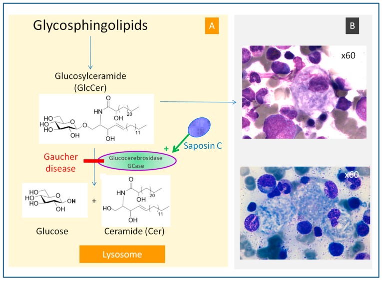
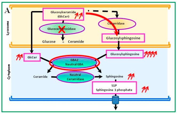
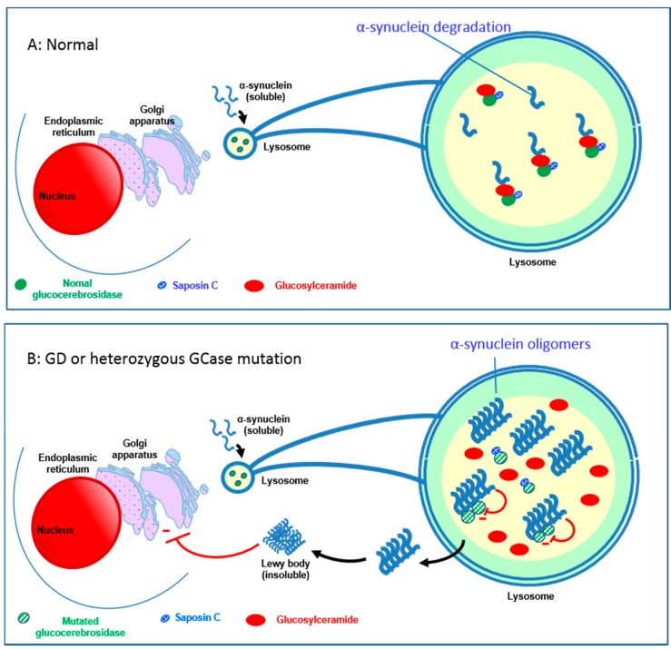
Gaucher disease (GD, ORPHA355) is a rare, autosomal recessive genetic disorder. It is caused by a deficiency of the lysosomal enzyme, glucocerebrosidase, which leads to an accumulation of its substrate, glucosylceramide, in macrophages. In the general population, its incidence is approximately 1/40,000 to 1/60,000 births, rising to 1/800 in Ashkenazi Jews. The main cause of the cytopenia, splenomegaly, hepatomegaly, and bone lesions associated with the disease is considered to be the infiltration of the bone marrow, spleen, and liver by Gaucher cells. Type-1 Gaucher disease, which affects the majority of patients (90% in Europe and USA, but less in other regions), is characterized by effects on the viscera, whereas types 2 and 3 are also associated with neurological impairment, either severe in type 2 or variable in type 3. A diagnosis of GD can be confirmed by demonstrating the deficiency of acid glucocerebrosidase activity in leukocytes. Mutations in the GBA1 gene should be identified as they may be of prognostic value in some cases. Patients with type-1 GD-but also carriers of GBA1 mutation-have been found to be predisposed to developing Parkinson's disease, and the risk of neoplasia associated with the disease is still subject to discussion. Disease-specific treatment consists of intravenous enzyme replacement therapy (ERT) using one of the currently available molecules (imiglucerase, velaglucerase, or taliglucerase). Orally administered inhibitors of glucosylceramide biosynthesis can also be used (miglustat or eliglustat).

Keywords: GBA1 gene; Gaucher disease; biomarkers; enzyme replacement therapy; glucocerebrosidase; lysosomal storage disease; substrate reduction therapy.


Conflict of interest statement

Jérôme Stirnemann has received travel fees from Sanofi-Genzyme. Nadia Belmatoug and Marc G. Berger have received consulting and speaking fees from Sanofi-Genzyme, Shire, Actelion and Pfizer. Roseline Froissart has received travel fees from Sanofi-Genzyme. Grants from these companies were donated to the Department of Clinical Research of the Assistance-Publique Hôpitaux de Paris and of the University Hospital of Clermont-Ferrand. Christine Serratrice and Fabrice Camou have received consultation and speaking fees from Sanofi-Genzyme and Shire. Thierry Levade received support for travel fees from Sanofi-Genzyme and Shire.

Figures

Figure 1
Figure 1
Hydrolysis of glucosylceramide (GlcCer) by glucocerebrosidase (GCase) in the lysosome (A). GCase is activated by saposin C. In lysosomal storage diseases, an enzyme deficiency is responsible for the accumulation of its substrate in the cell lysosome (overload disease). Gaucher disease is caused by a deficiency in glucocerebrosidase (GCase) (or β-glucosidase), which leads to an accumulation of GlcCer. GlcCer forms fibrillar aggregates that accumulate in macrophages and result in the cell cytoplasm presenting a characteristic “crumpled tissue paper” appearance (B), personal pictures, with the courtesy of Fabrice Camou and Rachid Seddik). These cells, known as Gaucher cells, infiltrate various organs (e.g., bone marrow, spleen, and liver) and are responsible for the major signs of the disease.
Figure 2
Figure 2
Alternative metabolic pathway of the glucosylceramide (GlcCer) accumulation due to the glucocerebrosidase (GCase) deficiency. The expression of GCase varies from one cell type to another and depends on the tissue. (A) In a mouse model of GCase deficiency (red cross), GlcCer is transformed via an alternative ceramidase pathway into glucosylsphingosine (red arrow), which is degraded by cytoplasmic GCase2 (GBA2 gene), active at a neutral pH, to S1P, a very active metabolite [20]. (B) Protein maturation takes place in the Golgi apparatus; the transport and delivery of GCase to lysosomes require a particular molecule, LIMP-2, which allows GCase to reach the lysosome where the acidic pH breaks the molecular link [39]. (C) LIMP-2 is a lysosomal membrane protein (LMP) whose highly glycosylated intra-lysosomal part protects the lysosome’s membrane. LIMP-2 anomalies can induce a phenotype rather than GD3 [39].
Figure 2
Figure 2
Alternative metabolic pathway of the glucosylceramide (GlcCer) accumulation due to the glucocerebrosidase (GCase) deficiency. The expression of GCase varies from one cell type to another and depends on the tissue. (A) In a mouse model of GCase deficiency (red cross), GlcCer is transformed via an alternative ceramidase pathway into glucosylsphingosine (red arrow), which is degraded by cytoplasmic GCase2 (GBA2 gene), active at a neutral pH, to S1P, a very active metabolite [20]. (B) Protein maturation takes place in the Golgi apparatus; the transport and delivery of GCase to lysosomes require a particular molecule, LIMP-2, which allows GCase to reach the lysosome where the acidic pH breaks the molecular link [39]. (C) LIMP-2 is a lysosomal membrane protein (LMP) whose highly glycosylated intra-lysosomal part protects the lysosome’s membrane. LIMP-2 anomalies can induce a phenotype rather than GD3 [39].
Figure 3
Figure 3
Relationship between glucocerebrosidase (GCase) and neurological diseases with Lewy bodies. (A) Normally, GCase interacts with its substrate glucosylceramide (GlcCer) as well as monomers of α-synuclein in lysosomes, facilitating the breakdown of both at acidic pH; (B) Mutated GCase or decreased levels of GCase result in a slowdown of α-synuclein degradation and a gradual build up of GlcCer, with the formation of α-synuclein oligomers and fibrils [48,49,54]; GlcCer stabilizes the α-synuclein oligomers [47]. These oligomers are able to bind to the mutated GCase molecules and inhibit the enzymatic activity of GCase, further decreasing the enzyme activity [47,50,55]. These impaired lysosomes show impaired chaperone-mediated autophagy and autophagosome fusion. This results in an increased accumulation of α-synuclein in the cytoplasm, forming insoluble aggregates to form Lewy bodies. These aggregates block trafficking of GCase from the endoplasmic reticulum (ER) to the Golgi [56]. Mutant GCase is retained in the Endoplasmic reticulum, which causes ER stress and evokes the ER stress response (Unfolded Protein Response) [57]. Saposin C can have a modulating effect on this by binding to GCase and thus maintaining its activity [51,58].

Similar articles

Cited by

References

    1. Ginzburg L., Kacher Y., Futerman A.H. The pathogenesis of glycosphingolipid storage disorders. Semin. Cell Dev. Biol. 2004;15:417–431. doi: 10.1016/j.semcdb.2004.03.003. - DOI - PubMed
    1. Hruska K.S., LaMarca M.E., Scott C.R., Sidransky E. Gaucher disease: Mutation and polymorphism spectrum in the glucocerebrosidase gene (GBA) Hum. Mutat. 2008;29:567–583. doi: 10.1002/humu.20676. - DOI - PubMed
    1. Vaccaro A.M., Motta M., Tatti M., Scarpa S., Masuelli L., Bhat M., Vanier M.T., Tylki-Szymanska A., Salvioli R. Saposin C mutations in Gaucher disease patients resulting in lysosomal lipid accumulation, saposin C deficiency, but normal prosaposin processing and sorting. Hum. Mol. Genet. 2010;19:2987–2997. doi: 10.1093/hmg/ddq204. - DOI - PubMed
    1. Sidransky E. Gaucher disease: Insights from a rare Mendelian disorder. Discov. Med. 2012;14:273–281. - PMC - PubMed
    1. Grabowski G.A. Phenotype, diagnosis, and treatment of Gaucher’s disease. Lancet. 2008;372:1263–1271. doi: 10.1016/S0140-6736(08)61522-6. - DOI - PubMed