Review
. 2017 Feb 17;8(2):73.
doi: 10.3390/genes8020073.

Mcm10: A Dynamic Scaffold at Eukaryotic Replication Forks

Affiliations
Review

Mcm10: A Dynamic Scaffold at Eukaryotic Replication Forks

Ryan M Baxley et al. Genes (Basel). .

Abstract

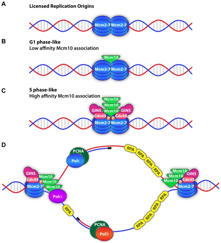
To complete the duplication of large genomes efficiently, mechanisms have evolved that coordinate DNA unwinding with DNA synthesis and provide quality control measures prior to cell division. Minichromosome maintenance protein 10 (Mcm10) is a conserved component of the eukaryotic replisome that contributes to this process in multiple ways. Mcm10 promotes the initiation of DNA replication through direct interactions with the cell division cycle 45 (Cdc45)-minichromosome maintenance complex proteins 2-7 (Mcm2-7)-go-ichi-ni-san GINS complex proteins, as well as single- and double-stranded DNA. After origin firing, Mcm10 controls replication fork stability to support elongation, primarily facilitating Okazaki fragment synthesis through recruitment of DNA polymerase-α and proliferating cell nuclear antigen. Based on its multivalent properties, Mcm10 serves as an essential scaffold to promote DNA replication and guard against replication stress. Under pathological conditions, Mcm10 is often dysregulated. Genetic amplification and/or overexpression of MCM10 are common in cancer, and can serve as a strong prognostic marker of poor survival. These findings are compatible with a heightened requirement for Mcm10 in transformed cells to overcome limitations for DNA replication dictated by altered cell cycle control. In this review, we highlight advances in our understanding of when, where and how Mcm10 functions within the replisome to protect against barriers that cause incomplete replication.

Keywords: CMG helicase; DNA replication; Mcm10; genome stability; origin activation; replication elongation; replication initiation.


Conflict of interest statement

The authors declare no conflict of interest.

Figures

Figure 1
Figure 1
The domain structure of minichromosome maintenance protein 10 (Mcm10). Full-length Mcm10 is depicted for Homo sapiens (875 amino acids (aa)) and Saccharomyces cerevisiae (571 aa). Mcm10 functional domains and the amino acid regions they span depicted. The N-terminal domain (NTD) contains a coiled-coil (CC, orange) motif responsible for Mcm10 self-interaction. The internal domain (ID) mediates Mcm10 interactions with proliferating cell nuclear antigen (PCNA) and DNA polymerase-alpha (Pol-α) through a PCNA-interacting peptide (PIP) box (red) and Hsp10-like domain (purple), respectively. These motifs reside in the oligonucleotide/oligosaccharide binding (OB)-fold (light gray). The OB-fold along with zinc-finger motif 1 (ZnF1, green) serve as a DNA-binding domain. The C-terminal domain (CTD) is specific to metazoa and interacts with DNA primarily through ZnF2 (green). The CTD also includes the zinc ribbon (ZnR, blue) and winged helix motif (WH, dark gray); however their functions are currently unknown. A bipartite nuclear localization sequence (NLS) has been identified in S. cerevisiae.
Figure 2
Figure 2
Evolutionary conservation of functional domains in the Mcm10 ID. (AD) Comparison of the amino acid sequences from Homo sapiens, Mus musculus, Danio rerio, Xenopus laevis, Drosophila melanogaster, Caenorhabditis elegans, Saccharomyces pombe and Saccharomyces cerevisiae of the OB-fold (A), PIP box (B), Hsp10-like (C) and Zinc-Finger 1 (D) domains. The full sequence alignment for the OB-fold is not shown due to size constraints, but can be found in Warren et al., [70]. The percent conservation (% cons.), defined as the percentage of amino acid positions identical (in red) or strongly similar (in blue) to those of human Mcm10, is listed for each domain sequence. The total region aligned for each sequence listed in gray. (E) The crystal structure of the Xenopus Mcm10 (xMcm10) OB-fold (gray), PIP box (red), Hsp10-like (purple) and Zinc-Finger 1 (green) domains is shown. The structure was generated using pdb data file 3EBE and the Chimera program (http://www.cgl.ucsf.edu/chimera) [83].
Figure 3
Figure 3
Evolutionary conservation of functional domains in the Mcm10 NTD. (A) Comparison of the amino acid sequences from H. sapiens, M. musculus, D. rerio, X. laevis, D. melanogaster, C. elegans, S. pombe and S. cerevisiae of the coiled-coil domain. The percent conservation (% cons.), defined as the percentage of amino acid positions identical (in red) or strongly similar (in blue) to those of human Mcm10, is listed for each domain sequence. The total region aligned for each sequence listed in gray. (B) The crystal structure of the Xenopus Mcm10 (xMcm10) coiled-coil domain is shown. The structure was generated using pdb data file 4JBZ and the Chimera program (http://www.cgl.ucsf.edu/chimera) [83].
Figure 4
Figure 4
Evolutionary conservation of functional domains in the Mcm10 CTD. (AC) Comparison of the amino acid sequences from H. sapiens, M. musculus, D. rerio, X. laevis, D. melanogaster and C. elegans of the Winged Helix (A), Zinc-Finger 2 (B) and Zinc-Ribbon (C). The percent conservation (% cons.), defined as the percentage of amino acid positions identical (in red) or strongly similar (in blue) to those of human Mcm10, is listed for each domain sequence. The total region aligned for each sequence listed in gray. (D) The crystal structure of the Xenopus Mcm10 (xMcm10) Zinc-Finger 2 (green) and Zinc-Ribbon (blue) domains is shown. The structure was generated using pdb data file 2KWQ and the Chimera program (http://www.cgl.ucsf.edu/chimera) [83].
Figure 5
Figure 5
Model of the association of Mcm10 with the replisome in initiation and elongation. (A) A Mcm2-7 double hexamer is loaded onto dsDNA and represent a licensed replication origin. (B) Mcm10 directly interacts with the Mcm2-7 with low affinity in G1-phase-like binding prior to CMG assembly. (C) High affinity binding of Mcm10 to the Mcm2-7 complex in S-phase like binding takes place with formation of the CMG complex. (D) Following helicase activation, replication forks progress in opposite directions from each origin. Mcm10 binds and stabilizes ssDNA (right fork) and is later replaced by RPA. Mcm10 loading of DNA polymerase-alpha (Pol-α) (left fork) is repeatedly needed to generate RNA/DNA primers (black DNA regions) for Okazaki fragment synthesis. Processive DNA polymerization is executed by DNA polymerase-epsilon (Pol-ε) (extending the blue leading strand) and DNA polymerase-delta (Pol-δ) (extending the red lagging strand).
Figure 6
Figure 6
MCM10 alterations in human cancer samples and exclusivity with BRCA-associated mutations. (A) Bar graph showing the number and class of alterations including amplifications (red), deletions (blue), mutations (green) or a combination (gray) of MCM10 identified in different cancer types by multiple groups. The tissue/cell type and dataset for each column are listed on the x-axis. Only datasets with 5 or more MCM10 alterations are shown. (B,C) Plots showing the overlap of genetic alterations including amplifications (red), deletions (blue) and mutations (green) in MCM10 or breast cancer (BRCA) associated genes (BRCA1, BRCA2, partner and localizer of BRCA2 (PALB2)) in the Breast Invasive Carcinoma dataset (The Cancer Genome Atlas [TCGA]) (B) or the Cancer Cell Line Encyclopedia (Novartis/Broad) [171]. The data and depictions shown in this figure were accessed via and/or modified from information listed on the cBioPortal for Cancer Genomics (http://www.cbioportal.org/) [168,169].

References

    1. Sun J., Kong D. DNA replication origins, ORC/DNA interaction, and assembly of pre-replication complex in eukaryotes. Acta Biochim. Biophys. Sin. 2010;42:433–439. doi: 10.1093/abbs/gmq048. - DOI - PubMed
    1. Edenberg H.J., Huberman J.A. Eukaryotic chromosome replication. Annu. Rev. Genet. 1975;9:245–284. doi: 10.1146/annurev.ge.09.120175.001333. - DOI - PubMed
    1. Heichinger C., Penkett C.J., Bahler J., Nurse P. Genome-wide characterization of fission yeast DNA replication origins. EMBO J. 2006;25:5171–5179. doi: 10.1038/sj.emboj.7601390. - DOI - PMC - PubMed
    1. Raghuraman M.K., Winzeler E.A., Collingwood D., Hunt S., Wodicka L., Conway A., Lockhart D.J., Davis R.W., Brewer B.J., Fangman W.L. Replication dynamics of the yeast genome. Science. 2001;294:115–121. doi: 10.1126/science.294.5540.115. - DOI - PubMed
    1. Wyrick J.J., Aparicio J.G., Chen T., Barnett J.D., Jennings E.G., Young R.A., Bell S.P., Aparicio O.M. Genome-wide distribution of ORC and Mcm proteins in S. cerevisiae: High-resolution mapping of replication origins. Science. 2001;294:2357–2360. doi: 10.1126/science.1066101. - DOI - PubMed

LinkOut - more resources