. 2017 Feb 18;9(2):68.
doi: 10.3390/toxins9020068.

Bee Venom Phospholipase A2 Ameliorates House Dust Mite Extract Induced Atopic Dermatitis Like Skin Lesions in Mice

Affiliations

Bee Venom Phospholipase A2 Ameliorates House Dust Mite Extract Induced Atopic Dermatitis Like Skin Lesions in Mice

Kyung-Hwa Jung et al. Toxins (Basel). .

Abstract

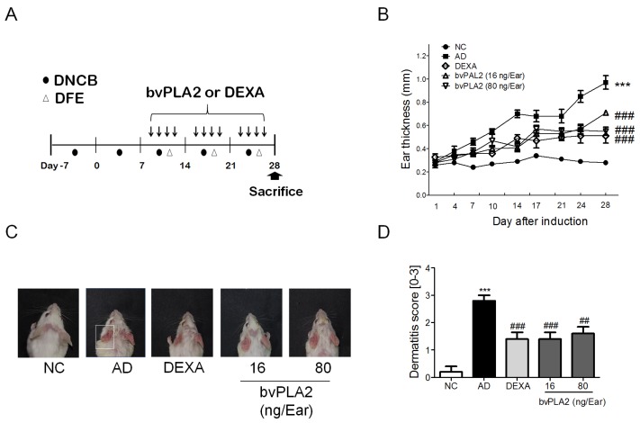
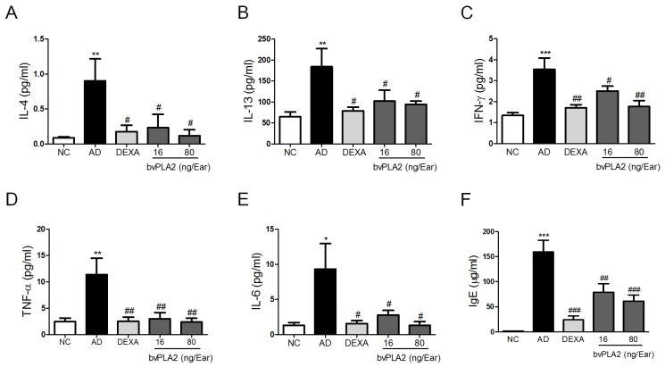
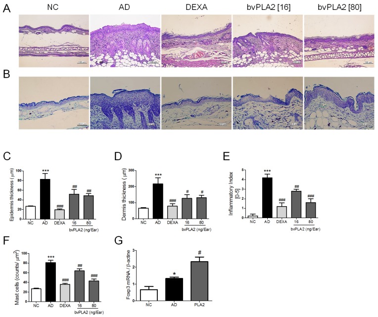
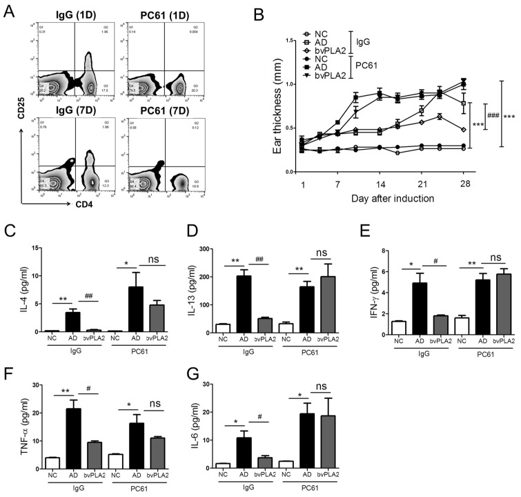
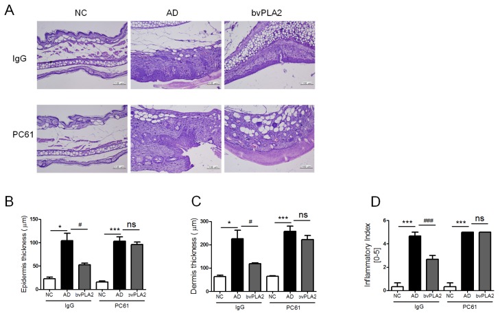
Atopic dermatitis (AD) is a biphasic inflammatory skin disease that is provoked by epidermal barrier defects, immune dysregulation, and increased skin infections. Previously, we have demonstrated that bvPLA2 evoked immune tolerance by inducing regulatory T cells (Treg), and thus alleviated Th2 dominant allergic asthma in mice. Here, we would like to determine whether treatment with bvPLA2 exacerbates the AD-like allergic inflammations induced by house dust mite extract (DFE) in a murine model. Epidermal thickness, immune cell infiltration, serum immunoglobulin, and cytokines were measured. Ear swelling, skin lesions, and the levels of total serum IgE and Th1/Th2 cytokines were elevated in DFE/DNCB-induced AD mice. Topical application of bvPLA2 elicited significant suppression of the increased AD symptoms, including ear thickness, serum IgE concentration, inflammatory cytokines, and histological changes. Furthermore, bvPLA2 treatment inhibited mast cell infiltration into the ear. On the other hand, Treg cell depletion abolished the anti-atopic effects of bvPLA2, suggesting that the effects of bvPLA2 depend on the existence of Tregs. Taken together, the results revealed that topical exposure to bvPLA2 aggravated atopic skin inflammation, suggesting that bvPLA2 might be a candidate for the treatment of AD.

Keywords: IgE; atopic dermatitis; bvPLA2; mast cell; skin lesion.


Conflict of interest statement

The authors declare that they have no competing interests.

Figures

Figure 1
Figure 1
Effects of bvPLA2 treatment on DFE/DNCB-induced AD-like symptoms. (A) Experimental design of the study protocol. To study the effect of bvPLA2 on atopic dermatitis, mice were treated with DFE/DNCB for four weeks and bvPLA2 was injected for three weeks during antigen sensitization; (B) The ear thickness was measured using a dial thickness gauge 24 h after the DFE/DNCB application; (C) Images of the ear skin lesions of the groups of mice taken on the last day of the experiment (day 28); (D) The severities of the symptoms of the ear skin lesions were microscopically indexed with the dermatitis score, which was specified as the sum of the scores (0 = no symptoms; 1 = mild symptoms; 2 = moderate symptoms; and 3 = severe symptoms) for the symptoms of erythema/hemorrhage, edema, excoriation/erosion, and dryness/scaling. NC: normal control group; AD: DFE/DNCB applied group; DEXA: DFE/DNCB applied and dexamethasone-treated group (0.1 mg/Ear, 20 µL) as a positive control; bvPLA2 (16 ng/Ear): DFE/DNC applied and bvPLA2 (16 ng/Ear)-treated group; bvPLA2 (80 ng/Ear): DFE/DNC applied and bvPLA2 (80 ng/Ear)-treated group. The statistical analyses were conducted with one-way ANOVA followed by Newman-Keuls multiple comparison tests (*** p < 0.001 vs. NC and ### p < 0.001, ## p < 0.01 vs. AD; n = 5).
Figure 2
Figure 2
Effects of bvPLA2 on expression of cytokines and serum total IgE. The levels of (A) IL-4; (B) IL-13; (C) IFN-γ; (D) TNF-α; (E) IL-6 in the ear and (F) total IgE in the serum were quantified by mouse CBA (Th1, 2, and 17) kit and ELISA kit. The statistical analyses were conducted with a one-way ANOVA followed by Newman-Keuls multiple comparison tests (*** p < 0.001, ** p < 0.01 and * p < 0.05 vs. NC and ### p < 0.001, ## p < 0.01 and # p < 0.05 vs. AD; n = 5).
Figure 3
Figure 3
Effects of bvPLA2 on histological changes of DNCB/DFE-induced AD-like skin lesions. (A) The ear sections (4 µm thick) were stained with hematoxylin and eosin (H&E) and (B) Toluidine blue (TB) and taken with ×200 and ×400 magnification; (C) The thicknesses of epidermis and (D) dermis were quantified based on the H&E stained sections using Image Pro-Plus 5.1 software as described in the Materials and Methods; (E) Evaluation of ear histological change severity was quantified using a five-point score system; (F) The number of mast cells for five sites selected at random was counted on the ear sections with stained TB. (G) The expression of Foxp3 in the ear tissue was determined by real-time PCR. The statistical analyses were conducted with a one-way ANOVA followed by Newman-Keuls multiple comparisons test (*** p < 0.001 and * p < 0.05 vs. NC and ### p < 0.001, ## p < 0.01 and # p < 0.05 vs. AD; n = 5).
Figure 4
Figure 4
Effects of bvPLA2 on AD-like skin lesions of CD25 depleted mice. The female BALB/C mice received once with a dose of 0.25mg/kg of rat anti-mouse CD25 (PC61) or total rat IgG on days 3, 4, 11, 17, and 25. (A) CD4+CD25+ populations were measured using FACS on day 1 and 7; (B) The ear thickness was measured using a dial thickness gauge 24 h after the DFE/DNCB application. The concentration of (C) IL-4; (D) IL-13; (E) IFN-γ; (F) TNF-α; and (G) IL-6 were measured by mouse CBA (Th1, 2, and 17) kit and ELSA kit. NC: normal control group; AD: DFE/DNCB applied group; bvPLA2: DFE/DNC applied and bvPLA2 (80 ng/Ear)-treated group. IgG: non-CD25 depleted mice; PC61: anti-CD25-depleted mice. The statistical analyses were conducted with a one-way ANOVA followed by Newman-Keuls multiple comparisons test (** p < 0.01 and * p < 0.05, ## p < 0.01 and # p < 0.05; not significant = ns, n = 3).
Figure 5
Figure 5
Effects of bvPLA2 on histological changes in AD-like skin lesions of CD25 depleted mice. (A) Representative ear section images were stained with H&E to assess the histological changes (×200 magnification); (B) the thicknesses of epidermis; and (C) dermis were quantified based on the H&E stained sections using Image Pro-Plus 5.1 software as described in the Materials and Methods; (D) Evaluation of ear histological change severity was quantified using a five-point score system. The statistical analyses were conducted with a one-way ANOVA followed by Newman-Keuls multiple comparisons test (*** p < 0.001 and * p < 0.05 and ## p < 0.01 and # p < 0.05; not significant = ns, n = 3).

References

    1. Berke R., Singh A., Guralnick M. Atopic dermatitis: An overview. Am. Fam. Physician. 2012;86:35–42. - PubMed
    1. Leung D.Y. Atopic dermatitis: New insights and opportunities for therapeutic intervention. J. Allergy Clin. Immunol. 2000;105:860–876. doi: 10.1067/mai.2000.106484. - DOI - PubMed
    1. Leung D.Y., Boguniewicz M., Howell M.D., Nomura I., Hamid Q.A. New insights into atopic dermatitis. J. Clin. Investig. 2004;113:651–657. doi: 10.1172/JCI21060. - DOI - PMC - PubMed
    1. Boniface K., Bernard F.X., Garcia M., Gurney A.L., Lecron J.C., Morel F. IL-22 inhibits epidermal differentiation and induces proinflammatory gene expression and migration of human keratinocytes. J. Immunol. 2005;174:3695–3702. doi: 10.4049/jimmunol.174.6.3695. - DOI - PubMed
    1. Palmer C.N., Irvine A.D., Terron-Kwiatkowski A., Zhao Y., Liao H., Lee S.P., Goudie D.R., Sandilands A., Campbell L.E., Smith F.J., et al. Common loss-of-function variants of the epidermal barrier protein filaggrin are a major predisposing factor for atopic dermatitis. Nat. Genet. 2006;38:441–446. doi: 10.1038/ng1767. - DOI - PubMed

MeSH terms

LinkOut - more resources