. 2017 Apr;24(4):387-394.
doi: 10.1038/nsmb.3381. Epub 2017 Feb 20.

MHC-I peptides get out of the groove and enable a novel mechanism of HIV-1 escape

Affiliations

MHC-I peptides get out of the groove and enable a novel mechanism of HIV-1 escape

Phillip Pymm et al. Nat Struct Mol Biol. 2017 Apr.

Abstract

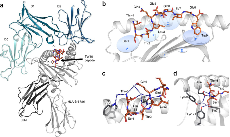
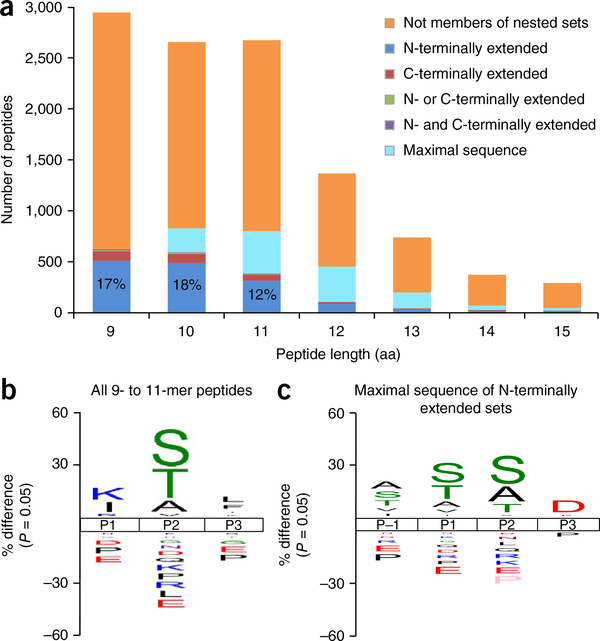
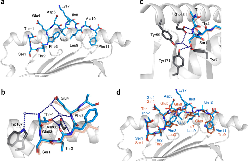
Major histocompatibility complex class I (MHC-I) molecules play a crucial role in immunity by capturing peptides for presentation to T cells and natural killer (NK) cells. The peptide termini are tethered within the MHC-I antigen-binding groove, but it is unknown whether other presentation modes occur. Here we show that 20% of the HLA-B*57:01 peptide repertoire comprises N-terminally extended sets characterized by a common motif at position 1 (P1) to P2. Structures of HLA-B*57:01 presenting N-terminally extended peptides, including the immunodominant HIV-1 Gag epitope TW10 (TSTLQEQIGW), showed that the N terminus protrudes from the peptide-binding groove. The common escape mutant TSNLQEQIGW bound HLA-B*57:01 canonically, adopting a dramatically different conformation than the TW10 peptide. This affected recognition by killer cell immunoglobulin-like receptor (KIR) 3DL1 expressed on NK cells. We thus define a previously uncharacterized feature of the human leukocyte antigen class I (HLA-I) immunopeptidome that has implications for viral immune escape. We further suggest that recognition of the HLA-B*57:01-TW10 epitope is governed by a 'molecular tension' between the adaptive and innate immune systems.


Conflict of interest statement

COMPETING FINANCIAL INTERESTS

The authors declare no competing financial interests.

Figures

Figure 1
Figure 1
HLA-B*57:01 in complex with the TW10 peptide. (a) The overall structure of KIR3DL1 (green) in complex with HLA-B*57:01–TW10 (orange). The structure of KIR3DL1 when bound to HLA B*57:01–LF9 is overlaid (gray) for comparison of the binding modes. (b) Cartoon representation of the crystal structure of HLA-B*57:01 (light gray) in complex with the TW10 peptide TSTLQEQIGW (orange) shown against the α1 helix of HLA and oriented N-C-terminal from left. Anchor pockets of the HLA are indicated along the binding groove at P1 (A) to PΩ (F). (c,d) Orientation of the protruding Thr residue of the peptide at P–1 (orange) and its interaction with residues of the HLA (dark gray sticks) (c), and the conserved hydrogen-bonding network (blue dashed lines) at the N-terminal end of HLA-B*57:01 (dark gray sticks) maintained to Ser at P1 (orange) (d). Hydrogen bonds are shown as blue dashed lines.
Figure 2
Figure 2
Characterization of N-terminally extended peptides. (a) Length distribution of HLA-B*57:01 ligands, showing those classified within nested sets. Definitions of nested-set subcategories can be found in Supplementary Note 1. The values in the first three bars respectively represent the percentage of peptides 9, 10 and 11 amino acids in length that were part of extended sets and extended at the N terminus only. (b,c) Sequence logos showing the percent difference in the abundance of amino acids in comparison to the human proteome at each location in the N-terminal portion of (b) all 9–11-residue HLA-B*57:01 ligands (n = 8268), and in (c) the maximal sequences of N-terminal-extended sets possessing C-terminal aromatic anchors (n = 972). Sequence logos were generated using the iceLogo stand-alone version. Source data are available in Supplementary Tables 1 and 2.
Figure 3
Figure 3
The HIV-1 Gag repertoire of HLA-B*57:01. (a) HIV-1 Gag epitopes presented by HLA-B*57:01. The QW9 and TW10 epitopes form part of N-terminally extended sets. (b) Relative levels of N-terminally extended variants compared with the HIV Gag TW10 epitope. Values are mean and s.d.; n = 3; data represent three independent experiments. Source data are available in Supplementary Table 3.
Figure 4
Figure 4
Cartoon representations of the crystal structure of HLA-B*57:01 (light gray) in complex with the TSTFEDVKILAF peptide (cyan). (a) HLA-B*57:01 in complex with the TSTFEDVKILAF peptide, shown against the α1 helix of HLA and oriented N-C-terminal from the left. (b) The network of direct and water-mediated hydrogen bonds (dark blue dashed lines) around the protruding P–1 residue, showing the interaction of P–1-Thr with Trp167 and Asn66 of HLA. The TW10 peptide is underlaid (orange) for comparison. (c) The network of conserved hydrogen bonds at the N terminus of the HLA-B*57:01 peptide-binding groove, showing the P1-Ser1 side chain replacing contacts normally mediated by the N terminus of the peptide. The TW10 peptide is underlaid (orange) for comparison. (d) Overlay representation of the TSTFEDVKILAF (blue) and TSTLQEQIGW (orange) structures.
Figure 5
Figure 5
Comparison of the HLA-B*57:01–TW10 and HLA-B*57:01–T3N ternary complex structures with KIR3DL1*001. (a) Cartoon representation of the interactions between KIR3DL1 (teal) and the T3N peptide (blue) presented by HLA-B*57:01 (light gray). (b) The interactions between KIR3DL1 (teal) and the TW10 peptide (orange). (c) Cartoon representation of the crystal structure of HLA-B*57:01 (light gray) in complex with the T3N peptide TSNLQEQIGW (blue) shown against the α1 helix of the HLA and oriented N-C-terminal from left. Hydrogen bonds are shown as blue dashed lines. (d) Overlay of the TW10 (orange) and T3N (blue) peptide conformations.
Figure 6
Figure 6
HLA-B*57:01 TW10 and T3N binding to KIR3DL1. (a) (i) Representative surface plasmon resonance (SPR) injection series from two independent experiments for KIR3DL1 binding to the TW10 (top) and T3N (bottom) HLA-B*57:01 complexes. (ii,iii) SPR-based affinity measurements of the interaction between KIR3DL1*001 and B57:01 T3N (ii) and B*57:01 TW10 (iii) complexes; results represent two independent experiments. Data are shown as mean ± range of two values. Concentration values represent the equilibrium binding constants of KIR3DL1 for the HLA-peptide complexes. RU, response units. (b) Staining of KIR3DL1 allotypes with HLA-B*57:01 TW10 (orange) and T3N (blue) tetramers (0.2 μg each), normalized to TW10 binding. Results represent two (015), three (005) and four (001) independent replicates. Data are shown as mean ± range. (c,d) HLA-B*57:01 TW10 and T3N tetramer staining of HEK293 cells transfected with KIR3DL1*001 and a panel of KIR3DL1*001 interface residue mutants, normalized to TW10 tetramer binding to KIR3DL1*001 (c) or to the respective tetramer binding of KIR3DL1*001 transfectants (d). All results for c and d represent three independent experiments and are shown as mean and s.e.m. WT, wild-type.

References

    1. Saper MA, Bjorkman PJ & Wiley DC Refined structure of the human histocompatibility antigen HLA-A2 at 2.6 Å resolution. J. Mol. Biol. 219, 277–319 (1991). - PubMed
    1. Rammensee HG, Friede T & Stevanoviíc S MHC ligands and peptide motifs: first listing. Immunogenetics 41, 178–228 (1995). - PubMed
    1. Deres K, Beck W, Faath S, Jung G & Rammensee HG MHC/peptide binding studies indicate hierarchy of anchor residues. Cell. Immunol. 151, 158–167 (1993). - PubMed
    1. Wilson IA & Fremont DH Structural analysis of MHC class I molecules with bound peptide antigens. Semin. Immunol. 5, 75–80 (1993). - PubMed
    1. Garrett TP, Saper MA, Bjorkman PJ, Strominger JL & Wiley DC Specificity pockets for the side chains of peptide antigens in HLA-Aw68. Nature 342, 692–696 (1989). - PubMed

Publication types

MeSH terms