. 2017 Apr;36(2):111-119.
doi: 10.14366/usg.16054. Epub 2017 Jan 18.

Doppler ultrasonography of the lower extremity arteries: anatomy and scanning guidelines

Affiliations

Doppler ultrasonography of the lower extremity arteries: anatomy and scanning guidelines

Ji Young Hwang. Ultrasonography. 2017 Apr.

Abstract

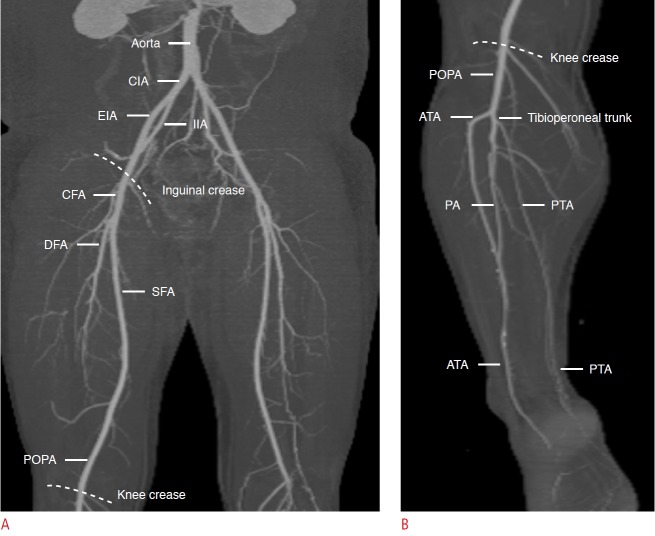
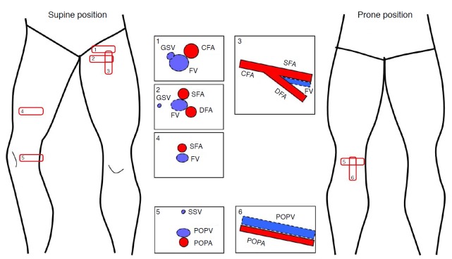
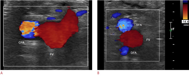
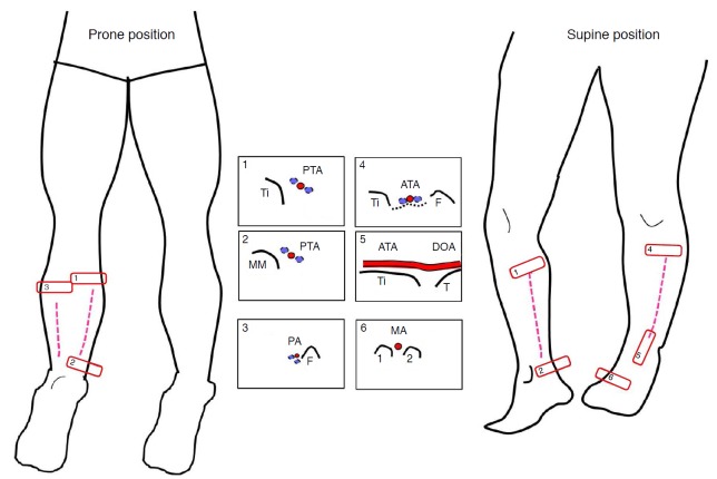
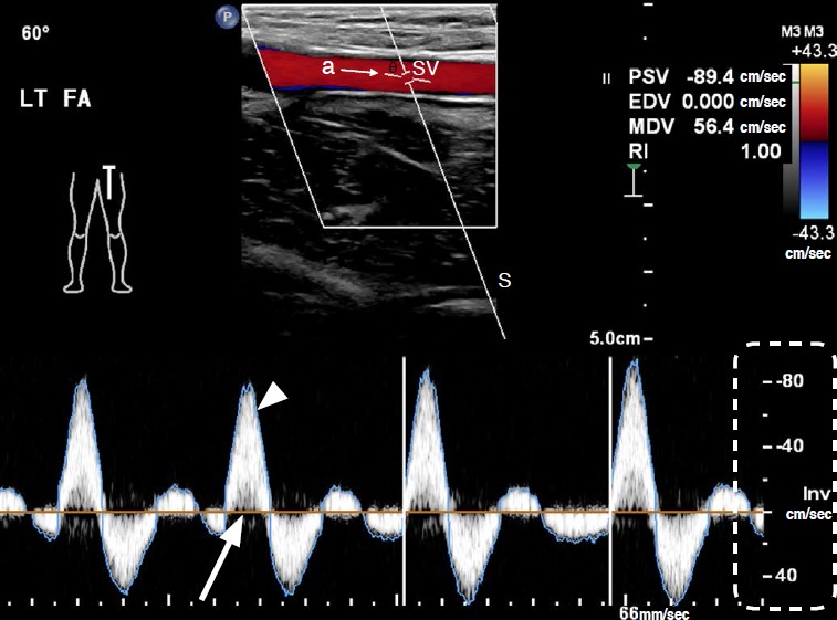
Doppler ultrasonography of the lower extremity arteries is a valuable technique, although it is less frequently indicated for peripheral arterial disease than for deep vein thrombosis or varicose veins. Ultrasonography can diagnose stenosis through the direct visualization of plaques and through the analysis of the Doppler waveforms in stenotic and poststenotic arteries. To perform Doppler ultrasonography of the lower extremity arteries, the operator should be familiar with the arterial anatomy of the lower extremities, basic scanning techniques, and the parameters used in color and pulsed-wave Doppler ultrasonography.

Keywords: Arteries; Lower extremity; Peripheral arterial disease; Ultrasonography, Doppler, color; Ultrasonography, Doppler, pulsed.


Conflict of interest statement

No potential conflict of interest relevant to this article was reported.

Figures

Fig. 1.
Fig. 1.. The anatomy of the lower extremity arteries on computed tomography (CT) angiography.
A. On coronal maximal intensity projection (MIP) CT image above the knee, the external iliac artery (EIA) is continuous with the common femoral artery (CFA) which bifurcates into the superficial femoral artery (SFA) and deep femoral artery (DFA). The SFA is continuous with the popliteal artery (POPA). B. On coronal MIP CT image below the knee, the POPA splits anterior tibial artery (ATA) and tibioperoneal trunk which bifurcates into posterior tibial artery (PTA) and peroneal artery (PA). CIA, common iliac artery; IIA, internal iliac artery.
Fig. 2.
Fig. 2.. The steps of color Doppler ultrasonography (US) for the lower extremities above the knee, with the patient’s position indicated.
The red rectangular boxes are the essential scanning sites and planes for the femoral arteries and the popliteal artery. The numbers within the boxes represent the general steps of scanning. The schema in the box demonstrates the typical US features of arteries and veins at each scanning site. GSV, greater saphenous vein; FV, femoral vein; CFA, common femoral artery; SFA, superficial femoral artery; DFA, deep femoral artery; SSV, small saphenous vein; POPV, popliteal vein; POPA, popliteal artery.
Fig. 3.
Fig. 3.. Normal color Doppler ultrasonography of the femoral arteries in the inguinal area.
A. The common femoral artery (CFA) is lateral to the femoral vein (FV) on a transverse scan at the inguinal crease. Note that the size of the color box is as small as possible. B. The superficial femoral artery (SFA) and the deep femoral artery (DFA) make a shape like Mickey Mouse’s ears, and the FV forms Mickey Mouse’s face.
Fig. 4.
Fig. 4.. The steps of color Doppler ultrasonography for the lower extremities below the knee, with the patient’s position indicated.
The posterior tibial artery (PTA) is seen along the tibia (Ti) at the medial side of the posterior calf (box 1) and behind the medial malleolus (MM) of the ankle (box 2). The peroneal artery (PA) is depicted alongside the fibula (F) on the lateral side of posterior calf in the prone position (box 3). The anterior tibial artery (ATA) is detected over interosseous memberane (black dashed line) between the tibia (Ti) and the fibula (F) at the anterolateral side of the calf (box 4). At the ankle level, the ATA is seen anterior to the tibia plafond (Ti) and the talus (T) (box 5) and continues to the dorsalis pedis artery (DOA) distal to the ankle and metatarsal artery (MA) between the metatarsal bones (box 6).
Fig. 5.
Fig. 5.. Color and pulsed-wave Doppler sonograms of normal lower extremity arteries with parameters.
Top: In a color image of a pulsed-wave Doppler sonogram, the color box is tilted to be parallel to the arterial axis using the “steer” key. Doppler angle (θ) is 60° in this case and is formed by the Doppler line of sight (S) and the axis of arterial flow (a). SV, sample volume; LT FA, left femoral artery; PSV, peak systolic velocity; EDV, end-diastolic velocity; MDV, minimum diastolic velocity; RI, resistivity index. RI=(PSV-EDV)/PSV. Bottom: On the Doppler spectrum, time (seconds) is represented on the x-axis. Blood flow velocity (cm/sec) is shown on the y-axis (dashed line). Flow direction relative to the transducer is shown in relation to the spectrum baseline (arrow). The “high-Q” is the blue outline of the Doppler spectrum (arrowhead).
Fig. 6.
Fig. 6.. Adjustment of pulsed-wave Doppler ultrasonography in a stenotic arterial segment.
The aliasing artifact on the Doppler spectrum can be adjusted by lowering the baseline (arrowhead) and increasing the scale. Note the spectral broadening (arrow) in the Doppler spectrum due to stenosis of the artery. Parameters for color flow (CF) and pulsed-wave (PW) Doppler are peak systolic velocity (PSV) 129 cm/sec, end diastolic velocity (EDV) 15.4 cm/sec, minimum diastolic velocity (MDV) 8.9 cm/sec, resistivity index (RI) 0.88, and wall filter (WF) 120 Hz in CF and 60 Hz in PW. SV, sample volume.
Fig. 7.
Fig. 7.. A 56-year-old man with arterial occlusion.
Color flow is absent in the superficial femoral artery (arrow) on color Doppler sonogram at the inguinal level, representing complete occlusion. The red vessel is the deep femoral artery and the blue vessel is the collapsed femoral vein.

References

    1. Nzeh DA, Allan PL, McBride K, Gillespie I, Ruckley CV. Comparison of colour Doppler ultrasound and digital subtraction angiography in the diagnosis of lower limb arterial disease. Afr J Med Med Sci. 1998;27:177–180. - PubMed
    1. Karacagil S, Lofberg AM, Granbo A, Lorelius LE, Bergqvist D. Value of duplex scanning in evaluation of crural and foot arteries in limbs with severe lower limb ischaemia: a prospective comparison with angiography. Eur J Vasc Endovasc Surg. 1996;12:300–303. - PubMed
    1. Flanigan DP, Ballard JL, Robinson D, Galliano M, Blecker G, Harward TR. Duplex ultrasound of the superficial femoral artery is a better screening tool than ankle-brachial index to identify at risk patients with lower extremity atherosclerosis. J Vasc Surg. 2008;47:789–792. - PubMed
    1. Sensier Y, Bell PR, London NJ. The ability of qualitative assessment of the common femoral Doppler waveform to screen for significant aortoiliac disease. Eur J Vasc Endovasc Surg. 1998;15:357–364. - PubMed
    1. Gooding GA, Perez S, Rapp JH, Krupski WC. Lower-extremity vascular grafts placed for peripheral vascular disease: prospective evaluation with duplex Doppler sonography. Radiology. 1991;180:379–386. - PubMed