. 2017 Aug;140(2):447-453.e3.
doi: 10.1016/j.jaci.2016.12.980. Epub 2017 Feb 20.

Substance P activates Mas-related G protein-coupled receptors to induce itch

Affiliations

Substance P activates Mas-related G protein-coupled receptors to induce itch

Ehsan Azimi et al. J Allergy Clin Immunol. 2017 Aug.

Abstract

Background: Substance P (SP) is linked to itch and inflammation through activation of receptors on mast cells and sensory neurons. There is increasing evidence that SP functions through Mas-related G protein-coupled receptors (Mrgprs) in addition to its conventional receptor, neurokinin-1.

Objective: Because Mrgprs mediate some aspects of inflammation that had been considered mediated by neurokinin-1 receptor (NK-1R), we sought to determine whether itch induced by SP can also be mediated by Mrgprs.

Methods: Genetic and pharmacologic approaches were used to evaluate the contribution of Mrgprs to SP-induced scratching behavior and activation of cultured dorsal root ganglion neurons from mice.

Results: SP-induced scratching behavior and activation of cultured dorsal root ganglion neurons was dependent on Mrgprs rather than NK-1R.

Conclusion: We deduce that SP activates MrgprA1 on sensory neurons rather than NK-1R to induce itch.

Keywords: Mas-related G protein–coupled receptors; Substance P; calcium imaging; dorsal root ganglion neurons; knockout mice; receptor antagonist.


Conflict of interest statement

Disclosure of potential conflict of interest: The authors declare that they have no relevant conflicts of interest.

Figures
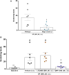
FIG 1
FIG 1
Mrgprs mediate SP-induced itch. A, SP-induced itch (500 µmol/L administered intradermally [i.d.]) is significantly decreased in Mrgpr cluster Δ−/− mice. B, Effects of treatment with the NK-1R antagonist L733060 (500 µmol/L administered intradermally) or the dual NK-1R/Mrgpr antagonist QWF (500 µmol/L administered intradermally) on scratching behavior caused by SP (500 µmol/L administered intradermally). Data are expressed as means ± SEMs. The unpaired Student t test (n = 7 mice per group in Fig 1, A) and 1-way ANOVA followed by the Tukey test (n = 6 mice per group in Fig 1, B) were used. Significant differences from wild-type mice (†P < .05), baseline (#P < .05), SP plus vehicle (*P < .05), or SP plus L733060 ($$P < .01) are indicated.
FIG 2
FIG 2
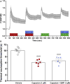
Effect of QWF on capsaicin-induced Ca2+ flux and thermal hyperalgesia. A, QWF (1 µmol/L) does not affect Ca2+ flux induced by capsaicin (1 µmol/L) and KCl (40 mmol/L) in cultured wild-type DRG neurons. B, QWF (1 µmol/L, intraplantar) has no effect on capsaicin-induced thermal hyperalgesia in wild-type mice. Data are expressed as means ± SEMs. One-way ANOVA followed by the Tukey test (n = 6 mice per group). Statistical differences from vehicle are indicated as follows: ###P < .001. Total number of cells analyzed in Fig 2, A, was 267.
FIG 3
FIG 3
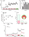
SP activates NK-1R−/− nociceptors. A, SP (10–1000 nmol/L) induces Ca2+ flux in wild-type (gray) and NK-1R−/− (black) cultured DRG neurons. B, Percentage of SP-responding cells from wild-type or NK-1R−/− mice. C, Maximal Ca2+ flux in DRG neurons from wild-type (gray) and NK-1R−/− (black) mice. D, Size of SP-responsive cells from NK-1R−/− mice. Inset, Venn diagram showing overlapping populations of KCl-positive, capsaicin-positive, and SP-responsive neurons. E, Ca2+ flux induced by SP (1 µmol/L), capsaicin (1 µmol/L), and KCl (40 mmol/L) in DRG neurons from wild-type (gray) and TRPV1-Cre+/−DTA+/−(red) mice. The unpaired Student t test was used. Significant differences from NK-1R−/− were indicated as follows: **P < .01 and ***P < .001. Total number of cells analyzed was as follows: Fig 3, A–C, 328 (wild-type) and 1463 (NK-1R−/−); Fig 3, D, 1463; Fig 3, D, inset, 314; and Fig 3, E, 787 (wild-type) and 246 (TRPV1-Cre+/−DTA+/−).
FIG 4
FIG 4
Effect of QWF on SP-induced Ca2+ flux in cultured DRG neurons from NK-1R−/− mice. A, QWF (1 µmol/L) blocks SP-induced (300 nmol/L) Ca2+ flux in DRG neurons from NK-1R−/− mice. Cells blocked by QWF also respond to allyl isothiocyanate (1 µmol/L), capsaicin (1 µmol/L), and KCl (40 mmol/L). B, Amplitude of SP-induced Ca2+ flux in NK-1R−/− neurons before and after treatment with QWF (1 µmol/L). Data are expressed as means ± SEMs. The paired Student t test was used. Statistical difference from SP is indicated as follows: ***P < .001. Total number of cells analyzed was as follows: Fig 4, A, 314; Fig 4, B, 492.

References

    1. Duval A, Dubertret L. Aprepitant as an antipruritic agent? N Engl J Med. 2009;361:1415–6. - PubMed
    1. Stander S, Siepmann D, Herrgott I, Sunderkotter C, Luger TA. Targeting the neurokinin receptor 1 with aprepitant: a novel antipruritic strategy. PLoS One. 2010;5:e10968. - PMC - PubMed
    1. Santini D, Vincenzi B, Guida FM, Imperatori M, Schiavon G, Venditti O, et al. Aprepitant for management of severe pruritus related to biological cancer treatments: a pilot study. Lancet Oncol. 2012;13:1020–4. - PubMed
    1. Wallengren J. Topical aprepitant in clinical and experimental pruritus. Arch Dermatol. 2012;148:957–9. - PubMed
    1. Wallengren J, Edvinsson L. Topical non-peptide antagonists of sensory neurotransmitters substance P and CGRP do not modify patch test and prick test reactions: a vehicle-controlled, double-blind pilot study. Arch Dermatol Res. 2014;306:505–9. - PubMed