

Comparison of Carotid-Femoral and Brachial-Ankle Pulse-Wave Velocity in Association With Target Organ Damage in the Community-Dwelling Elderly Chinese: The Northern Shanghai Study
- PMID: 28219916
- PMCID: PMC5523744
- DOI: 10.1161/JAHA.116.004168
Comparison of Carotid-Femoral and Brachial-Ankle Pulse-Wave Velocity in Association With Target Organ Damage in the Community-Dwelling Elderly Chinese: The Northern Shanghai Study
Abstract
Background: Carotid-femoral pulse-wave velocity (cf-PWV) and brachial-ankle PWV (ba-PWV) are the 2 most frequently applied PWV measurements. However, little is known about the comparison of hypertensive target organ damage (TOD) with cf-PWV and ba-PWV.
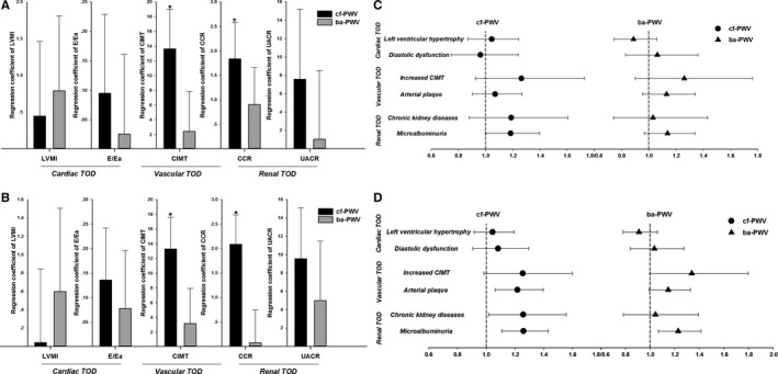
Methods and results: A total of 1599 community-dwelling elderly subjects (age >65 years) in northern Shanghai were recruited from June 2014 to August 2015. Both cf-PWV and ba-PWV were measured using SphygmoCor and VP1000 systems, respectively. Within the framework of comprehensive cardiovascular examinations, risk factors were assessed, and asymptomatic TOD, including left ventricular mass index, peak transmitral pulsed Doppler velocity/early diastolic tissue Doppler velocity (E/Ea), carotid intima-media thickness, arterial plaque, creatinine clearance rate, and urinary albumin-creatinine ratio were all evaluated. Both PWVs were significantly associated with male sex, age, waist/hip circumference, fasting plasma glucose, and systolic blood pressure, and ba-PWV was also significantly related to body mass index. Both PWVs were significantly correlated with most TOD. When cf-PWV and ba-PWV were both or separately put into the stepwise linear regression model together with cardiovascular risk factors and treatment, only cf-PWV, but not ba-PWV, was significantly associated with carotid intima-media thickness and creatinine clearance rate (P<0.05). When cf-PWV and ba-PWV were both or separately put into the same full-mode model after adjustment for confounders, only cf-PWV, but not ba-PWV, showed significant association with carotid intima-media thickness and creatinine clearance rate (P<0.05). Similar results were observed in logistic regression analysis.
Conclusions: Taken together, in the community-dwelling elderly Chinese, cf-PWV seems to be more closely associated with hypertensive TOD, especially vascular and renal TOD, as compared with ba-PWV.
Clinical trial registration: URL: http://www.clinicaltrials.gov. Unique identifier: NCT02368938.
Keywords: brachial‐ankle pulse‐wave velocity; cardiovascular disease risk factors; carotid‐femoral pulse‐wave velocity; target organ damage.
© 2017 The Authors. Published on behalf of the American Heart Association, Inc., by Wiley Blackwell.
Figures
References
-
- Zhang Y, Agnoletti D, Protogerou AD, Topouchian J, Wang JG, Xu Y, Blacher J, Safar ME. Characteristics of pulse wave velocity in elastic and muscular arteries: a mismatch beyond age. J Hypertens. 2013;31:554–559; discussion 559. - PubMed
-
- Yu WC, Chuang SY, Lin YP, Chen CH. Brachial‐ankle vs carotid‐femoral pulse wave velocity as a determinant of cardiovascular structure and function. J Hum Hypertens. 2008;22:24–31. - PubMed
-
- Kass DA. Ventricular arterial stiffening: integrating the pathophysiology. Hypertension. 2005;46:185–193. - PubMed
-
- Mancia G, Fagard R, Narkiewicz K, Redon J, Zanchetti A, Bohm M, Christiaens T, Cifkova R, De Backer G, Dominiczak A, Galderisi M, Grobbee DE, Jaarsma T, Kirchhof P, Kjeldsen SE, Laurent S, Manolis AJ, Nilsson PM, Ruilope LM, Schmieder RE, Sirnes PA, Sleight P, Viigimaa M, Waeber B, Zannad F; Task Force Members . 2013 ESH/ESC guidelines for the management of arterial hypertension: the Task Force for the management of arterial hypertension of the European Society of Hypertension (ESH) and of the European Society of Cardiology (ESC). J Hypertens. 2013;31:1281–1357. - PubMed
-
- Zhang Y, Agnoletti D, Xu Y, Wang JG, Blacher J, Safar ME. Carotid‐femoral pulse wave velocity in the elderly. J Hypertens. 2014;32:1572–1576; discussion 1576. - PubMed
Publication types
MeSH terms
Associated data
LinkOut - more resources
Full Text Sources
Other Literature Sources
Medical
