Comparative Study
. 2017 Apr 4;474(8):1373-1394.
doi: 10.1042/BCJ20160851.

Peroxiredoxin 1 (Prx1) is a dual-function enzyme by possessing Cys-independent catalase-like activity

Affiliations
Comparative Study

Peroxiredoxin 1 (Prx1) is a dual-function enzyme by possessing Cys-independent catalase-like activity

Cen-Cen Sun et al. Biochem J. .

Abstract

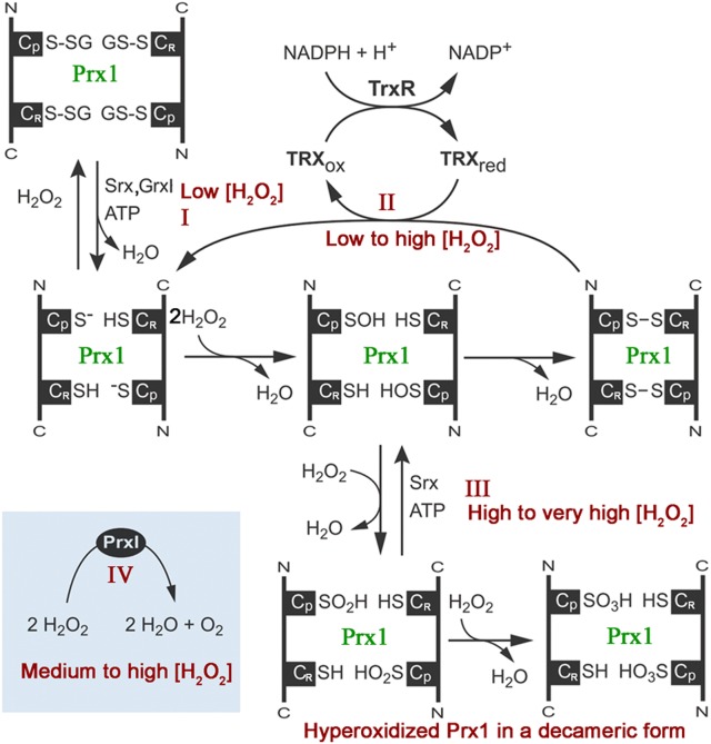
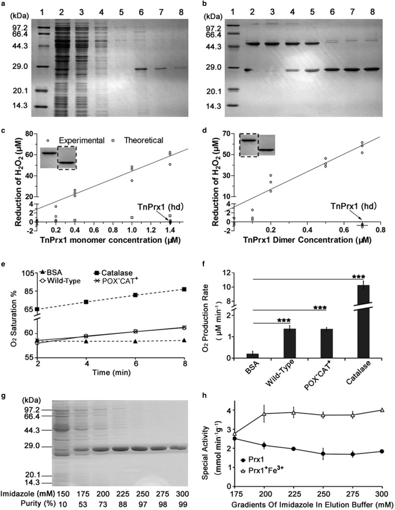
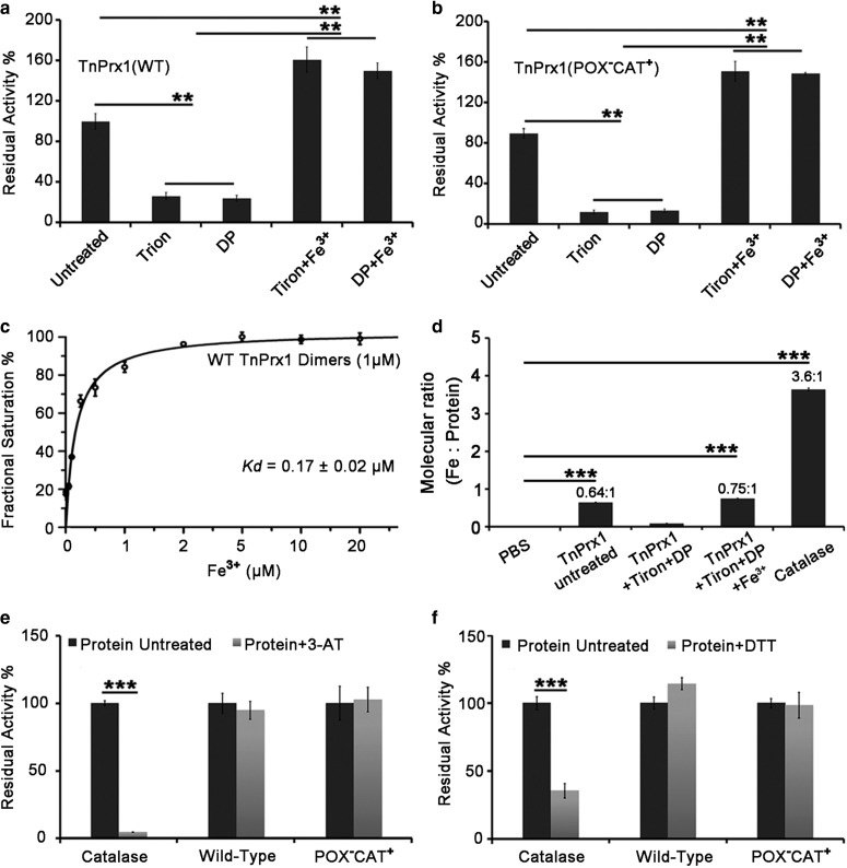
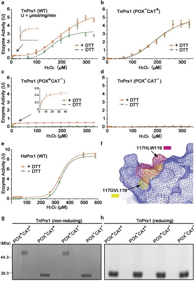
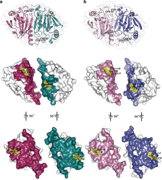
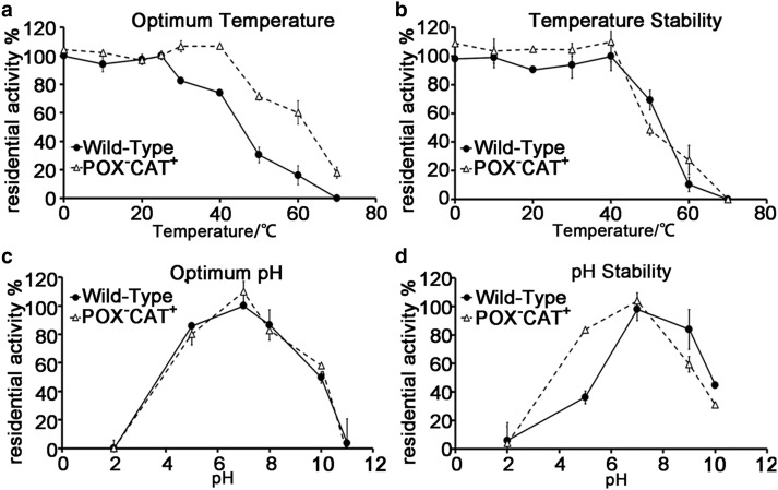
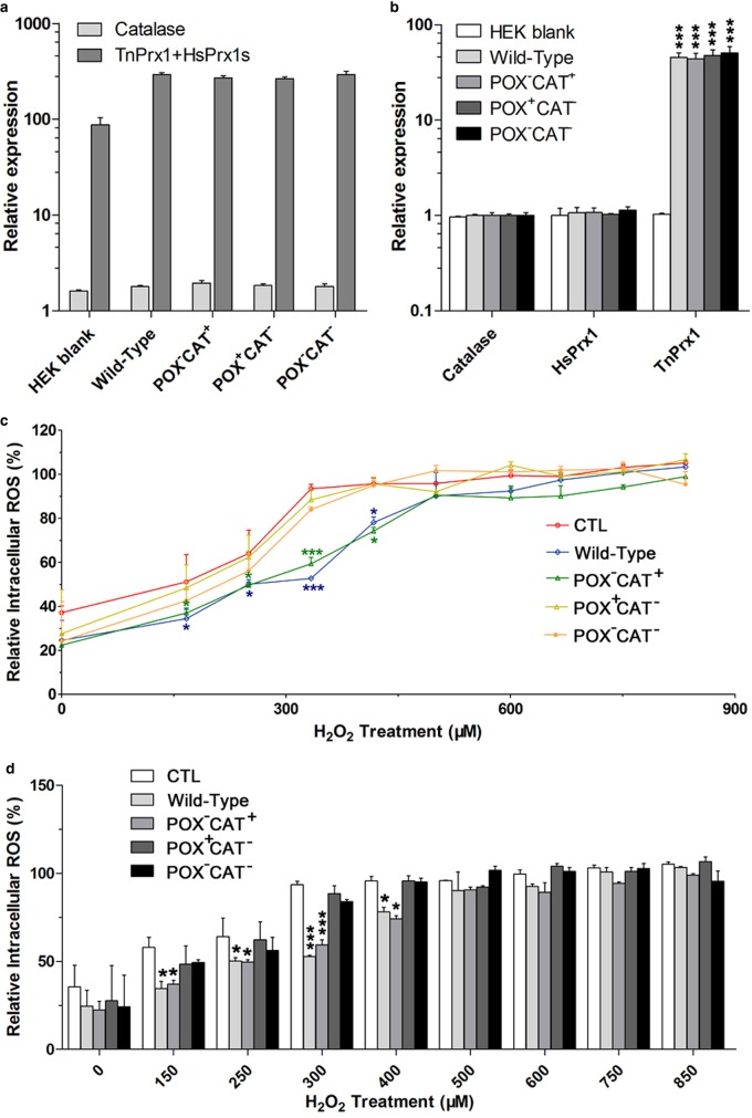
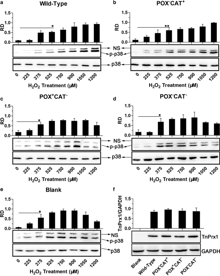
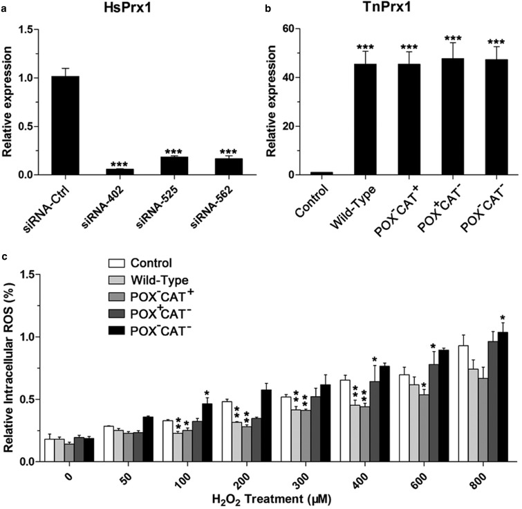
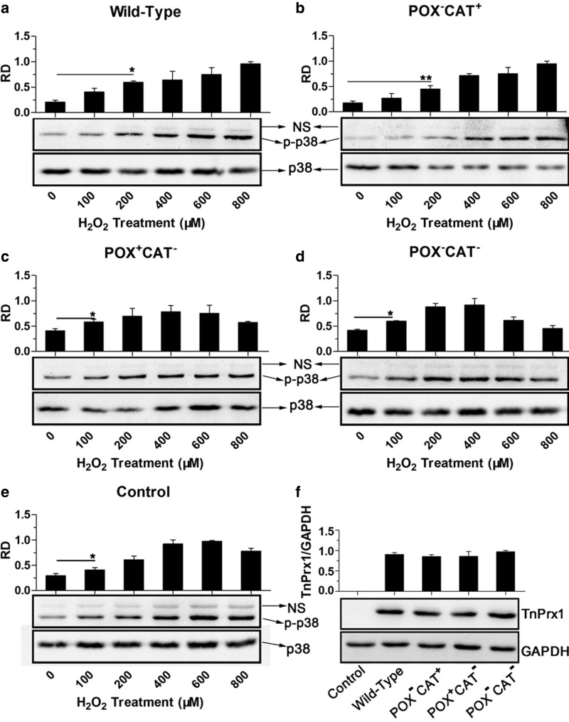
Peroxiredoxin (Prx) was previously known as a Cys-dependent thioredoxin. However, we unexpectedly observed that Prx1 from the green spotted puffer fish Tetraodon nigroviridis (TnPrx1) was able to reduce H2O2 in a manner independent of Cys peroxidation and reductants. This study aimed to validate a novel function for Prx1, delineate the biochemical features and explore its antioxidant role in cells. We have confirmed that Prx1 from the puffer fish and humans truly possesses a catalase (CAT)-like activity that is independent of Cys residues and reductants, but dependent on iron. We have identified that the GVL motif was essential to the CAT-like activity of Prx1, but not to the Cys-dependent thioredoxin peroxidase (POX) activity, and generated mutants lacking POX and/or CAT-like activities for individual functional validation. We discovered that the TnPrx1 POX and CAT-like activities possessed different kinetic features in the reduction of H2O2 The overexpression of wild-type TnPrx1 and mutants differentially regulated the intracellular levels of reactive oxygen species (ROS) and the phosphorylation of p38 in HEK-293T cells treated with H2O2 Prx1 is a dual-function enzyme by acting as POX and CAT with varied affinities towards ROS. This study extends our knowledge on Prx1 and provides new opportunities to further study the biological roles of this family of antioxidants.

Keywords: ROS; catalase-like activity; peroxiredoxin 1 (Prx1); puffer fish.


Conflict of interest statement

The Authors declare that there are no competing interests associated with the manuscript.

Figures

Figure 1.
Figure 1.. Illustration of pathways regulating vertebrate peroxiredoxin 1 (Prx1) to scavenge hydrogen peroxide.
Pathway I, the peroxidatic cysteine (CP-SH) and resolving cysteine (CR-SH) of Homo sapiens Prx1 (HsPrx1) can be glutathionylated in the presence of a small amount of H2O2, and deglutathianylated by sulfiredoxin (Srx) or glutaredoxin I (GrxI). Pathway II, the CP-SH of Prx1 is oxidized by H2O2 to cysteine-sulfenic acid (CP-SOH), which then reacts with CR-SH of the other subunit to produce an intermolecular disulfide, and the reducing equivalents for such typical peroxidase activity of Prx1 are ultimately derived from NADPH (reduced form of nicotinamideadenine dinucleotide phosphate) via thioredoxin reductase (TrxR) and Trx. Pathway III, the CP-SH is selectively oxidized by H2O2 to CP-SO2H or even CP-SO3H, which can be reversed by Srx and ATP (adenosine triphosphate). Pathway IV, the newly discovered catalase-like activity of Prx1, which directly converts H2O2 to O2.
Figure 2.
Figure 2.. Verification of the catalase (CAT)-like activity of TnPrx1 and mutants.
(a) Expression and purification of soluble TnPrx1 protein. Lanes: 1, protein markers; 2, crude cell lysate; 3, flow-through; 4–5, 40 mM imidazole wash; 6–8, eluted recombinant protein (250 mM imidazole); (b) Reductive dissociation of TnPrx1 dimer induced by DTT. Lanes: 1, protein markers; 2–8, proteins treated with different concentrations of DTT (dithiothreitol) (0, 0.1, 0.5, 1, 5, 10 and 50 mM); (c,d) Activities of TnPrx1 in monomers (c) and dimers (d) in the absence of a reducing agent by detecting the reduction of H2O2 using a luminol chemiluminescence assay after incubation with 300 µM H2O2 for 10 min at 25°C. The bands in the dashed box denote the TnPrx1 monomers and dimers used in the corresponding assays. Theoretical values represented the maximal reduction of H2O2 possibly achieved by the oxidation of three TnPrx1 Cys residues in a given amount of TnPrx1 protein in the absence of reductants or redox recycling. hd = heated denatured TnPrx1 protein (at 100°C for 10 min); (e,f) Detection of O2 production in reactions containing H2O2 and various protein constructs (i.e. 0.32 µM wild-type dimers, 0.64 µM POXCAT+ monomers, 8 nM catalase and 6 µM BSA) with oxygen electrode technique; POX = Cys-dependent thioredoxin peroxidase; (g,h) Gradient elution of TnPrx1 protein with varied concentrations of imidazole in elution buffer (g), and their corresponding activity by measuring the reduction of H2O2 with or without the addition of extra iron (200 µM) by luminol chemiluminescence (h). TnPrx1 concentration was determined by thin-layer gel optical scanning. Activity was normalized to mmol of H2O2 reduced per min per gram of TnPrx1 protein. Data are representative of at least three independent experiments. The error bars represent standard deviations (SDs), and statistical significances between experimental and control groups were determined by Student's t-test. ***P < 0.001.
Figure 3.
Figure 3.. Iron-dependency and inhibition of TnPrx1 and mutants determined by measuring the reduction of H2O2.
(a,b) Effects of iron chelators (25 mM 4,5-dihydroxy-1,3-benzene disulfonic acid [tiron] and 50 mM 2,2′-dipyridyl [DP]) on the CAT activity of wild-type (WT) TnPrx1 and mutants, and restoration of the activity by the addition of Fe3+ (200 µM). Residual activities were expressed as the percentage activity (vs. untreated WT TnPrx1); (c) Dose-dependent WT TnPrx1 activity on Fe3+. Residual activities were expressed as the percentage activity (vs. WT TnPrx1 treated with 200 µM Fe3+); (d) The molar ratio between TnPrx1 protein and bound iron determined by ICP-OES (inductively coupled plasma optical emission spectroscopy). TnPrx1 was treated as specified, followed by extensive washes with water by ultrafiltration prior to ICP. Bovine catalase and PBS were used as controls; (e,f) Effects of catalase inhibitors 3-Amino-1,2,4-triazole (3-AT) and dithiothreitol (DTT) on the CAT activity of WT TnPrx1 and mutants. CAT was used as positive control. Protein samples (50 µl, 50 µg ml−1, pretreated with 3-AT or DTT) were reacted with 50 µl of H2O2 solutions (300 µM) for 1 min. The residual activities were measured by luminol chemiluminescence assay and expressed as the percent activity (vs. untreated WT TnPrx1). Data are representative of at least three independent experiments. The error bars represent standard deviations (SDs), and statistical significances between experimental and control groups were determined by Student's t-test. **P < 0.01, ***P < 0.001. CAT = Catalase-like activity of peroxiredoxin 1.
Figure 4.
Figure 4.. Kinetic features of Prx1 proteins.
(a–e) Enzyme kinetics curves for wild-type (WT) and mutated pufferfish Prx1 (TnPrx1) and WT human Prx1 (HsPrx1) with or without DTT (dithiothreitol). (f) Structural comparison of the potential cavity of WT Prx1 protein (yellow) and its mutant (warm pink) in mesh form. The image is a merged model of the two Prx1 proteins; (g,h) The dimeric versus monomeric status of TnPrx1 proteins in non-reducing or reducing SDS-PAGE (Sodium dodecyl sulfate-Polyacrylamide Gel Electrophoresis), respectively. All TnPrx1 proteins were treated with monomer-to-dimer transition protocol prior to the assays.
Figure 5.
Figure 5.. Structural comparison between rat Prx1 (PDB ID:1QQ2) (a) and TnPrx1 determined by homology-modelling (b).
Structural models were represented in surface forms prepared using PyMOL software (www.pymol.org). The amino acids located at the dimer interface are shown in colors. The pockets containing the 117GVL119 motif in rat Prx1 and TnPrx1 are highlighted in yellow.
Figure 6.
Figure 6.. The effect of pH and temperature on catalase (CAT)-like activity and the stability of TnPrx1 proteins.
(a) The effect of temperature on residual Prx1-CAT activity. The activity assay was performed at pH 7.0 and at various temperatures. (b) The effect of temperature on Prx1-CAT stability. All the proteins were incubated at pH 7.0 and at various temperatures for 1 h and then the residual activity was estimated. (c) The effect of pH on residual Prx1-CAT activity. The activity assay was performed at room temperature and at various pH values. (d) The effect of pH on Prx1-CAT stability. The proteins were incubated with various pH at 4°C for 6 h and then the residual activity was measured.
Figure 7.
Figure 7.. Involvement of TnPrx1 constructs in regulating intracellular ROS (iROS) in HEK-293T cells.
(a,b) The expression of various TnPrx1 constructs in transfected cells were confirmed by qRT-PCR (real-time quantitative polymerase chain reaction) compared with those of endogenous HsPrx1 and catalase (CAT) genes. The relative levels of Prx1 transcripts (HsPrx1) only in blank control, or HsPrx1 + TnPrx1 in transfected cells were determined using a pair of primers derived from regions conserved between fish and mammalian Prx1 genes (Table 2). (a) Fold changes of Prx1 and catalase transcripts were expressed relative to the catalase transcripts in the blank control. (b) Fold changes of HsPrx1 and TnPrx1 transcripts were expressed relative to the transcripts of their own genes in the blank control. (c,d) Effects of TnPrx1 constructs on intracellular ROS in transfected cells treated with exogenous H2O2 as determined by DCFH (dichlorodihydrofluorescein diacetate) fluorescence assay. Cells transfected with blank vector were used as the negative control. The error bars represent standard deviations (SDs), and statistical significances between experimental and control groups were determined by Student's t-test. *P < 0.05, ***P < 0.001.
Figure 8.
Figure 8.. Effects of various TnPrx1 constructs on ROS-mediated phosphorylation of p38 MAPK in HEK-293T cells.
(a–e) Western blot analysis was conducted on total protein extracts from cells transfected with various TnPrx1 constructs and treated with various concentrations of exogenous H2O2 using antibodies specific to p38 and phosphorylated p38 (p-p38), respectively. Blank control cells received no transfection. Representative data from one of the three or more independent experiments were shown. Note: Antibody to p38 recognized a single band at 43 kDa, while antibody to p-p38 also produced a nonspecific band (NS) above the major band at 43 kDa for each sample. The NS bands were much weaker than—but proportional to—the major bands, a finding that was also observed by other investigators (e.g. [–57]). Bar charts showed relative density (RD) of p-p38 major bands (vs. p38). The error bars represented standard deviations (SDs) from three replicated blots. An asterisk in each bar chart indicates the lowest concentration of exogenous H2O2 starting to give statistical significance between H2O2-treated and untreated controls (*P < 0.05 by Student's t-test); (f) Confirmation on the protein expression from TnPrx1 constructs by Western blot analysis using antibodies specific to His-tag and to human GAPDH control, respectively. RD = relative density; POX = Cys-dependent thioredoxin peroxidase; CAT = Catalase-like activity of peroxiredoxin 1.
Figure 9.
Figure 9.. Involvement of TnPrx1 constructs in regulating intracellular ROS (iROS) in wild-type and HsPrx1-silienced HEK-293T cells.
(a) Interference of HsPrx1 expression in HEK-293T cells by three siRNA species targeting HsPrx1. Negative control used siRNA-Ctrl. The interference efficiency was shown as relative levels of HsPrx1 transcripts determined by qRT-PCR (real-time quantitative polymerase chain reaction). (b) Expression of various TnPrx1 constructs in cells co-transfected with siRNA-402. (c) Effects of TnPrx1 constructs on iROS (intracellular reactive oxygen species) levels in siRNA-402-transfected cells treated with exogenous H2O2 as determined by DCFH Dichlorodihydrofluorescein Diacetate fluorescence assay. Cells transfected with siRNA-402 only were used as control. Error bars represent standard deviations (SDs). Asterisks show statistical significances between the experimental and control groups by Student's t-test (*P < 0.05, **P < 0.01 and ***P < 0.001). POX = Cys-dependent thioredoxin peroxidase; CAT = Catalase-like activity of peroxiredoxin 1.
Figure 10.
Figure 10.. Effects of various TnPrx1 constructs on the ROS-mediated phosphorylation of p38 MAPK in HsPrx1-silienced HEK-293T cells.
(a–e) Western blot analysis of total protein extracts from cells co-transfected with HsPrx1-siRNA-402 and various TnPrx1 constructs after the treatment of various concentrations of exogenous H2O2 using antibodies specific to p38 and phosphorylated p38 (p-p38) proteins. Control cells were transfected with siRNA-402 only. Representative data from one of the three independent experiments are shown. (f) Protein expression of TnPrx1 constructs by Western blot analysis using antibodies specific to His-tag and to human GAPDH (glyceraldehyde-3-phosphate dehydrogenase) (control). An asterisk in each bar chart indicates the lowest concentration of exogenous H2O2 yielding statistical significance between H2O2-treated cells and untreated controls (*P < 0.05, **P < 0.01 by Student's t-test). RD = relative density.

References

    1. Rhee S. G. (2016) Overview on peroxiredoxin. Mol. Cells 39, 1–5 - PMC - PubMed
    1. Netto L. E. and Antunes F. (2016) The roles of peroxiredoxin and thioredoxin in hydrogen peroxide sensing and in signal transduction. Mol. Cells 39, 65–71 - PMC - PubMed
    1. Perkins A., Nelson K. J., Parsonage D., Poole L. B. and Karplus P. A. (2015) Peroxiredoxins: guardians against oxidative stress and modulators of peroxide signaling. Trends Biochem. Sci. 40, 435–445 - PMC - PubMed
    1. Rhee S. G., Kang S. W., Jeong W., Chang T.-S., Yang K.-S. and Woo H. A. (2005) Intracellular messenger function of hydrogen peroxide and its regulation by peroxiredoxins. Curr. Opin. Cell Biol. 17, 183–189 - PubMed
    1. Rhee S. G., Woo H. A., Kil I. S. and Bae S. H. (2012) Peroxiredoxin functions as a peroxidase and a regulator and sensor of local peroxides . J. Biol. Chem. 287, 4403–4410 - PMC - PubMed

Publication types

MeSH terms