. 2017 Mar 22;37(12):3246-3263.
doi: 10.1523/JNEUROSCI.3128-16.2017. Epub 2017 Feb 20.

Activity-Induced Synaptic Structural Modifications by an Activator of Integrin Signaling at the Drosophila Neuromuscular Junction

Affiliations

Activity-Induced Synaptic Structural Modifications by an Activator of Integrin Signaling at the Drosophila Neuromuscular Junction

Joo Yeun Lee et al. J Neurosci. .

Abstract

Activity-induced synaptic structural modification is crucial for neural development and synaptic plasticity, but the molecular players involved in this process are not well defined. Here, we report that a protein named Shriveled (Shv) regulates synaptic growth and activity-dependent synaptic remodeling at the Drosophila neuromuscular junction. Depletion of Shv causes synaptic overgrowth and an accumulation of immature boutons. We find that Shv physically and genetically interacts with βPS integrin. Furthermore, Shv is secreted during intense, but not mild, neuronal activity to acutely activate integrin signaling, induce synaptic bouton enlargement, and increase postsynaptic glutamate receptor abundance. Consequently, loss of Shv prevents activity-induced synapse maturation and abolishes post-tetanic potentiation, a form of synaptic plasticity. Our data identify Shv as a novel trans-synaptic signal secreted upon intense neuronal activity to promote synapse remodeling through integrin receptor signaling.SIGNIFICANCE STATEMENT The ability of neurons to rapidly modify synaptic structure in response to neuronal activity, a process called activity-induced structural remodeling, is crucial for neuronal development and complex brain functions. The molecular players that are important for this fundamental biological process are not well understood. Here we show that the Shriveled (Shv) protein is required during development to maintain normal synaptic growth. We further demonstrate that Shv is selectively released during intense neuronal activity, but not mild neuronal activity, to acutely activate integrin signaling and trigger structural modifications at the Drosophila neuromuscular junction. This work identifies Shv as a key modulator of activity-induced structural remodeling and suggests that neurons use distinct molecular cues to differentially modulate synaptic growth and remodeling to meet synaptic demand.

Keywords: Drosophila; Shriveled; activity-dependent structural remodeling; integrin; neuromuscular junction; synaptic boutons.


Figures

Figure 1.
Figure 1.
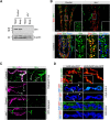
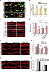
Shriveled is a presynaptically secreted protein. A, Representative Western blot showing that Shv protein is expressed in the Drosophila nervous system and shv1 is virtually a null allele. B, Immunostaining of Drosophila larval brain of control and shv1 with indicated antibodies. Elav is a marker for neurons, and Repo is a marker for glial cells. Bottom panels show that a majority of Shv-positive cells are also Elav positive, and a minor population of Repo-positive cells is positive for Shv staining (arrow). Boxed region is enlarged, representing Shv expression in the ventral ganglion and motor neurons. Scale bar, 20 μm. C, Representative images of the NMJs stained with Shv antibody with or without detergent. *Background due to autofluorescence coming from tracheal branch. Scale bar, 5 μm. D, Images of synaptic terminal highlighting the presence of Shv-GFP outside of the boutons but not when the signal peptide has been deleted. Anti-GFP antibody (red) stained using detergent-free condition colocalizes with extracellular Shv-GFP (green) driven by pan-neuronal driver (arrowhead highlights colocalization), but not when the signal peptide was truncated. Despite the presence of Shv-GFP in muscles when Shv-GFP was expressed using 24B-GAL4 (green signal), anti-GFP antibody (red) did not detect extracellular Shv-GFP signal in full-length or truncated form. Scale bar, 5 μm.
Figure 2.
Figure 2.
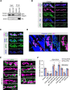
Shriveled modulates synaptic growth. A, Muscle area 6/7 NMJ at A2 stained by HRP for the indicated genotypes. Scale bar, 10 μm. B, Number of boutons normalized to muscle surface area (MSA). C, Representative Western blot showing relative levels of Shv protein in protein extracts of dissected larval brains. D, Relative ghost bouton index across genotypes. E, Images of NMJs labeled with the indicated antibodies. Arrowheads point to ghost boutons that are recognized by HRP labeling while lacking Dlg immunostaining. Scale bar, 5 μm. *p < 0.05 compared with control. All values represent the mean ± SEM.
Figure 3.
Figure 3.
Shv activates integrin receptor at NMJ. A, Immunoprecipitation (IP) assay followed by Western blots with the indicated antibodies demonstrate interaction between Shv and βPS integrin. The control used is transgenic flies without the nSyb-GAL4 driver; thus, it does not express Shv-HA. B, The presynaptic pFAK signal was calculated by outlining the presynaptic bouton using the HRP signal, whereas total pFAK signal was determined by outlining the postsynaptic area using Dlg staining. C, Wild-type NMJs stained with the indicated antibodies. Presynaptic membrane is demarcated by HRP staining (white outline). The pFAK signal is not enriched at the active zone as detected by NC82. D, Phalloidin staining highlights the postsynaptic muscle of a wild-type NMJ. pFAK signal can be seen throughout the muscle but is enriched at the synapse and is present both presynaptically and postsynaptically. HRP highlights the presynaptic bouton, and some phalloidin staining, which detects F-actin, is also enriched at the postsynaptic bouton. E, Representative images of NMJs stained with HRP and pFAK across genotypes. F, Quantification of presynaptic pFAK and total pFAK levels at synaptic terminal by outlining HRP or Dlg, respectively. Scale bars, 5 μm. *p < 0.05 compared with control. All values represent the mean ± SEM.
Figure 4.
Figure 4.
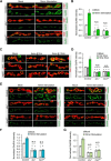
Shv genetically interacts with integrin. A, Representative images of NMJs stained with HRP for the indicated genotypes. Scale bar, 10 μm. B, Quantification of bouton numbers normalized to muscle surface area (MSA). C, Images of NMJs labeled with HRP and Dlg. Arrowheads point to ghost boutons that are HRP positive but Dlg negative. Scale bar, 5 μm. D, Quantification of relative ghost bouton index. E, Quantification of presynaptic pFAK and total pFAK levels at synaptic terminal by outlining HRP or Dlg, respectively. F, Representative images of NMJs stained with HRP and pFAK across genotypes. Scale bar, 5 μm. *p < 0.05 compared with control; **p < 0.05 compared with indicated genotypes; ***p < 0.05 when comparing the value of total pFAK between the indicated genotypes. All values represent the mean ± SEM.
Figure 5.
Figure 5.
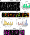
Pulsed high K+ stimulation did not trigger Shv release or change in pFAK levels. A, F, Representative images and quantification of ghost boutons after 3× spaced high K+ stimulation (A) or 5× spaced high K+ stimulation (F). Arrowheads highlight the ghost boutons. B, G, Representative images of Shv at the NMJs following 3× spaced high K+ stimulation (B) or 5× spaced high K+ stimulation (G), which were stained using detergent-free condition. C, H, Quantification of relative Shv intensity. D, I, Images of NMJ stained with pFAK and HRP antibodies following stimulation with the indicated conditions. E, J, Quantification of pFAK levels upon 3× spaced K+ stimulation (E) or 5× spaced K+ stimulation (J) compared with mock treatment (without high K+). *p < 0.05 when compared with mock-treated control; **p < 0.05 when comparing mock treated or 3× stimulated. Scale bars, 5 μm. All values represent the mean ± SEM.
Figure 6.
Figure 6.
Shv is secreted during intense synaptic stimulation to acutely activate integrin signaling. A, Representative images of NMJ stimulated with high K+ for 10 min followed by detergent-free Shv antibody labeling. B, Images representing Shv release after electrical stimulation at 10 Hz for 5 min but not by stimulation at 1 Hz for 5 min. Extracellular Shv was stained using detergent-free condition. C, Quantification of total and presynaptic pFAK levels before and after 10 min high K+ stimulation. D, Levels of pFAK immunostaining within NMJs treated with a persistent pulse of high K+ stimulation across genotypes. E, Images representing the elevated pFAK level following high-frequency electrical stimulation but not in low-frequency or shv1 and mysts1. F, Quantification of normalized total pFAK level upon high-frequency electrical stimulation. G, Representative images showing that presynaptic knockdown of βν integrin results in a synaptic overgrowth phenotype. H, Representative images of UASνRNAi/nSyb-GAL4 showing elevated pFAK staining after high K+ stimulation for 10 min. I, Quantification of presynaptic pFAK levels upon high K+ stimulation. Scale bars: A, B, E, H, 5 μm; D, G, 10 μm. *p < 0.05 compared with control. All values represent the mean ± SEM.
Figure 7.
Figure 7.
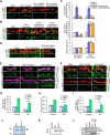
Shv release during intense activity induces local bouton enlargement. A, Representative images of ghost bouton before and after intense stimulation with high K+ application (highlighted with arrowhead). B, Normalized ghost bouton index present upon 10 min high K+ depolarization. C, Images representing an increase in individual synaptic bouton size after high K+ stimulation. D, Quantification of the average size of type 1b bouton following 10 min high K+ application. E, G, Representative images of NMJ stained with HRP staining showing size of synaptic boutons following nerve-evoked stimulation (E) and with and without pulses of high K+ incubation (G), as marked. F, H, Quantification of bouton size. Scale bars: A, C, E, G, 5 μm. *p < 0.05 compared with control; **p < 0.05 when comparing the indicated conditions. All values represent the mean ± SEM.
Figure 8.
Figure 8.
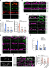
Release of Shv during neuronal stimulation induces synapse maturation. A, Images representing staining of GluRIII at the NMJ with or without 10 min high K+ stimulation. B, Quantification of relative GluRIII staining intensity at the NMJ with and without 10 min high K+ depolarization. C, Representative images of GluRIII staining upon low-frequency (1 Hz) or high-frequency (10 Hz) electrical stimulation. D, Quantification of relative GluRIII intensity at synaptic terminals. E, Images of GluRIIA and GluRIIB intensity at the NMJ with and without 10 min high K+ stimulation. F, G, Quantification of GluRIIA (F) and GluRIIB (G) staining intensities at the NMJ. *p < 0.05 compared with control. All values represent the mean ± SEM. Scale bars: A, C, E, 5 μm.
Figure 9.
Figure 9.
Presynaptic and postsynaptic βPS integrin activation differentially affects bouton size and glutamate receptor abundance. A, Representative images of third-instar NMJ stained with GluRIII antibody and HRP with and without 10 min high K+ depolarization. For genotypes containing tubulin-GAL80ts (GAL80ts), embryos were grown at 18°C until the L1 stage and then were shifted to 29°C to inactivate tubulin-GAL80ts. Scale bar, 5 μm. B, C, Quantification of relative GluRIII staining intensity (B) and bouton size (C). *p < 0.05 compared with control; **p < 0.05 when comparing the indicated conditions. All values represent the mean ± SEM.
Figure 10.
Figure 10.
Extracellular incubation of Shv is sufficient to trigger synapse maturation. A, B, Representative images of HRP and GluRIII immunoreactivity at synaptic boutons of control NMJ subjected to 5×spaced depolarization (A) and 10 min high K+ depolarization (B) in the presence of transcriptional inhibitor actinomycin or translational inhibitor cycloheximide. The ghost bouton is highlighted by a white arrowhead. C, Quantifications of normalized ghost bouton index, GluRIII levels, and bouton size upon 5× spaced or 10 min high K+ stimulation. *p < 0.05 when compared with respective mock-treated control (5× spaced stimulation); **p < 0.05 when compared with respective mock-treated control (10 min stimulation). D, Representative images of Shv release at the synapse upon prolonged high K+ stimulation with and without translational inhibitor cycloheximide. Detergent-free staining condition was used. E, Representative images of pFAK level changes upon incubation with purified Shv or mutated Shv (ShvLNV) protein. F, Images representing the elevation of GluRIII level upon Shv treatment. G, Quantification of total pFAK levels at the NMJs after Shv or ShvLNV application normalized to mock-treated control. H, Enlargement of individual bouton size upon the addition of purified Shv but not ShvLNV. I, Quantification of GluRIII intensity at the NMJ following Shv or ShvLNV protein incubation normalized to mock-treated control. *p < 0.05 compared with mock-treated control; **p < 0.05 when comparing the indicated conditions. All values represent the mean ± SEM. J, Coomassie gel showing relative amounts of Shv or ShvLNV following purification. The arrowhead points to the position of the expected protein size. K, Western blot confirming the expression and isolation of Shv and ShvLNV proteins. Western blot detected with HA antibody. L, Pull-down experiment using HA fusion proteins and fly protein lysates reveals that ShvLNV does not interact with integrin. Representative blots are shown, and results were confirmed in three independent experiments. Scale bar, 5 μm in all images.
Figure 11.
Figure 11.
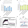
Shv regulates functional plasticity. A, Representative mEPSP and average mEPSP amplitude recorded using an HL-3 solution containing 0.5 mm Ca2+. B, Representative evoked EPSP recording in HL3 containing 0.5 mm Ca2+. Average EPSP was corrected using nonlinear summation. C, Normalized EPSP amplitude upon tetanus stimulation shows synaptic augmentation and post-tetanic potentiation in control NMJ, whereas shv1 failed to respond to the activity. Upregulation of Shv in shv1 mutant restored synaptic augmentation and PTP but overexpression of shv alone inhibited augmentation and PTP. Control, n = 8; shv1, n = 11; shv1;UAS-shv/nSyb-GAL4, n = 10; UAS-shv/nSyb-GAL4, n = 4. *p < 0.05 compared with control. All values represent the mean ± SEM. D, Model demonstrating an Shv-dependent synaptic remodeling event upon intense neuronal activity. Presynaptically released Shv activates integrin signaling pathways bidirectionally and influences local synapse growth and glutamate receptor levels at the NMJ.

Similar articles

Cited by

References

    1. Albin SD, Davis GW (2004) Coordinating structural and functional synapse development: postsynaptic p21-activated kinase independently specifies glutamate receptor abundance and postsynaptic morphology. J Neurosci 24:6871–6879. 10.1523/JNEUROSCI.1538-04.2004 - DOI - PMC - PubMed
    1. Arikkath J. (2010) N-cadherin: stabilizing synapses. J Cell Biol 189:397–398. 10.1083/jcb.201004022 - DOI - PMC - PubMed
    1. Ataman B, Ashley J, Gorczyca M, Ramachandran P, Fouquet W, Sigrist SJ, Budnik V (2008) Rapid activity-dependent modifications in synaptic structure and function require bidirectional Wnt signaling. Neuron 57:705–718. 10.1016/j.neuron.2008.01.026 - DOI - PMC - PubMed
    1. Bahr BA, Staubli U, Xiao P, Chun D, Ji ZX, Esteban ET, Lynch G (1997) Arg-Gly-Asp-Ser-selective adhesion and the stabilization of long-term potentiation: pharmacological studies and the characterization of a candidate matrix receptor. J Neurosci 17:1320–1329. - PMC - PubMed
    1. Berninger B, Bi GQ (2002) Synaptic modification in neural circuits: a timely action. Bioessays 24:212–222. 10.1002/bies.10060 - DOI - PubMed

Publication types

MeSH terms

Substances

LinkOut - more resources