. 2017 Feb 21:7:43010.
doi: 10.1038/srep43010.

A novel role of the organizer gene Goosecoid as an inhibitor of Wnt/PCP-mediated convergent extension in Xenopus and mouse

Affiliations

A novel role of the organizer gene Goosecoid as an inhibitor of Wnt/PCP-mediated convergent extension in Xenopus and mouse

Bärbel Ulmer et al. Sci Rep. .

Abstract

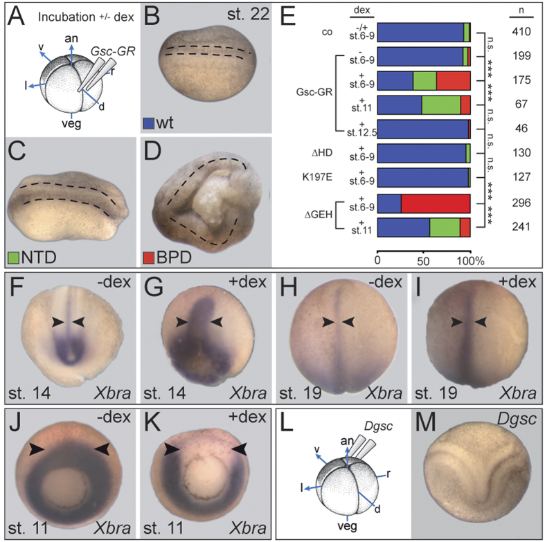
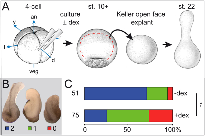
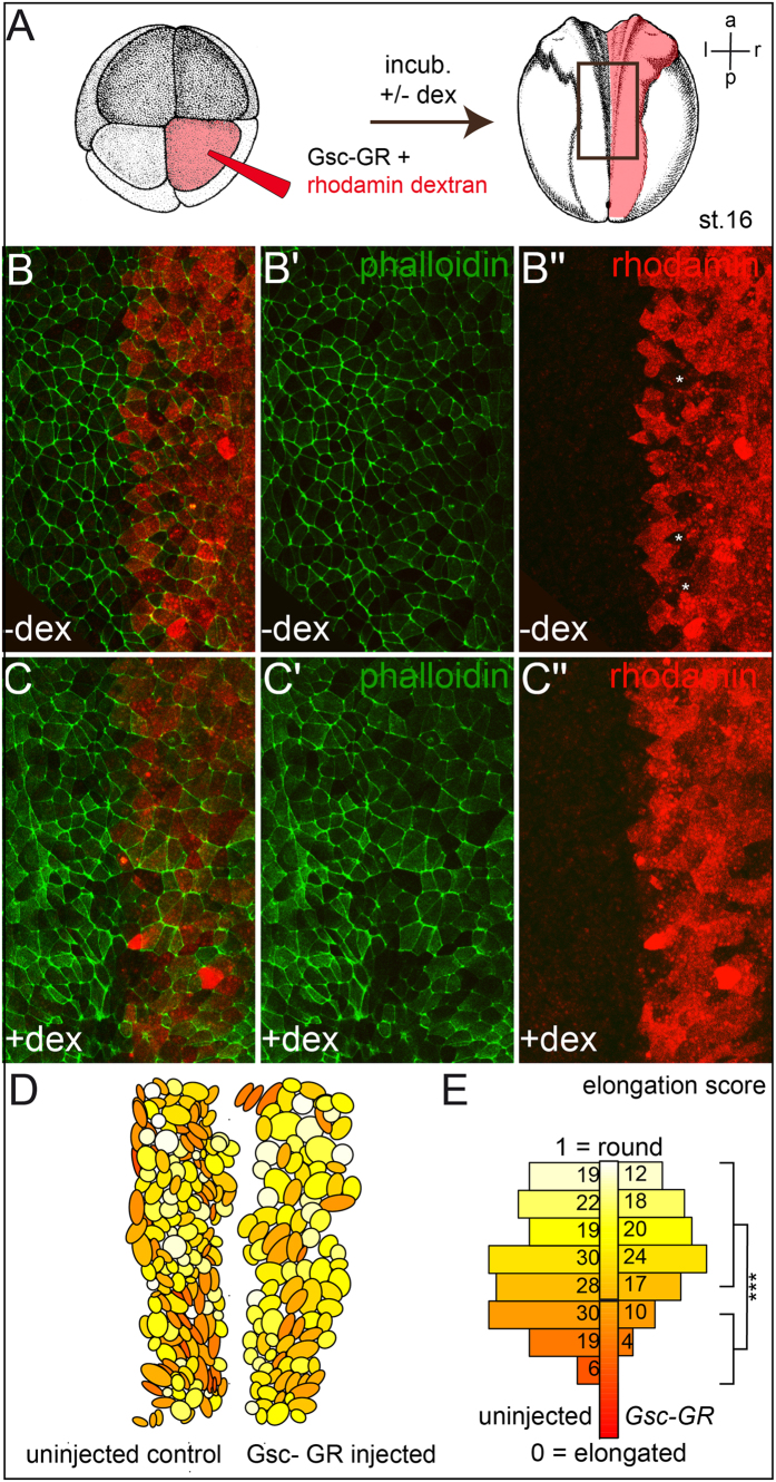
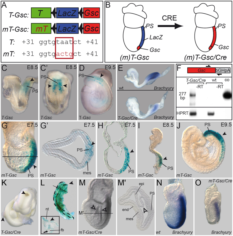
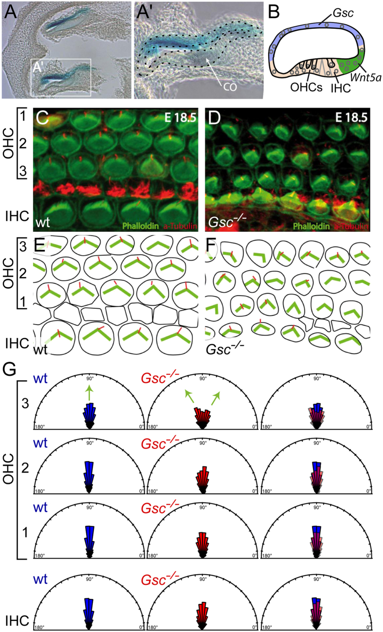
Goosecoid (Gsc) expression marks the primary embryonic organizer in vertebrates and beyond. While functions have been assigned during later embryogenesis, the role of Gsc in the organizer has remained enigmatic. Using conditional gain-of-function approaches in Xenopus and mouse to maintain Gsc expression in the organizer and along the axial midline, neural tube closure defects (NTDs) arose and dorsal extension was compromised. Both phenotypes represent convergent extension (CE) defects, arising from impaired Wnt/planar cell polarity (PCP) signaling. Dvl2 recruitment to the cell membrane was inhibited by Gsc in Xenopus animal cap assays and key Wnt/PCP factors (RhoA, Vangl2, Prickle, Wnt11) rescued Gsc-mediated NTDs. Re-evaluation of endogenous Gsc functions in MO-mediated gene knockdown frog and knockout mouse embryos unearthed PCP/CE-related phenotypes as well, including cartilage defects in Xenopus and misalignment of inner ear hair cells in mouse. Our results assign a novel function to Gsc as an inhibitor of Wnt/PCP-mediated CE. We propose that in the organizer Gsc represses CE as well: Gsc-expressing prechordal cells, which leave the organizer first, migrate and do not undergo CE like the Gsc-negative notochordal cells, which subsequently emerge from the organizer. In this model, Gsc provides a switch between cell migration and CE, i.e. cell intercalation.


Conflict of interest statement

The authors declare no competing financial interests.

Figures

Figure 1
Figure 1. Gsc-mediated CE phenotypes in Xenopus.
(A) Experimental design. Specimens were injected with Gsc-GR into the dorsal marginal region of the 4-cell embryo and cultured to the stages indicated, with or without addition of dex. (B–E) Gsc-GR induced NTD and BPD in whole embryos. Specimens were scored for wt appearance (blue; B), NTD (green; C) and BPD (red; D). Anterior is to the left in (BD). (E) Compilation of results. Note that Gsc-GR caused CE phenotypes in a highly significant proportion of embryos, but only when activated before and during gastrulation. Note also that deletion of the homeodomain (∆HD) or altering the DNA-binding specificity (K197E) prevented BPD/NTD-induction, while the repression domain GEH was not required for BPD/NTD. (F–I) Impaired CE of the notochord upon sustained dorsal Gsc-GR expression. Note that the notochord was wider and shorter in dex-treated (G,I) as opposed to untreated (F,H) specimens, both at stage 14 (F,G) and stage 19 (H,I). (J,K) Repression of Xbra transcription on the dorsal side upon Gsc-GR activation. (L,M) Double axis formation (M) following ventral injections of Dgsc mRNA into 4-cell Xenopus embryos (L).
Figure 2
Figure 2. Gsc inhibits CE in Keller open face explants.
(A–C) CE defects in Keller open face explants (schematically depicted in (A) upon activation of Gsc-GR. (B) Explants were classified as class 2 (blue) when extensions showed a constriction (left), as class 1 (green) when elongation occurred without constriction (middle), and as class 0 (red) when no elongation ensued (right). an, animal; uninj., uninjected control; d, dorsal; l, left; r, right; v, ventral; veg, vegetal. (C) Summary of results.
Figure 3
Figure 3. Gsc compromises bipolar elongation of neural plate cells.
(A) Targeted injection scheme of Gsc-GR and linage tracer (rhodamine red) into the right side of the neural plate (B,C). Drawings taken from Xenbase (www.xenbase.org/anatomy/alldevo.do). (D,E) Analysis of cell elongation. The color gradient ranging from pale yellow (round, width = length, 1) to dark red (elongated, 0) exemplifies the change from bipolar cells on the un-injected (right) side towards rounded cells upon activation of Gsc-GR (D). (E) Significant decrease of percentage of elongated cells (elongation score <1/2) after Gsc-GR missexpression. a, anterior; l, left; p, posterior; r, right.
Figure 4
Figure 4. Gsc-mediated CE phenotypes in the mouse.
Conditional misexpression of Gsc in the entire primitive streak of the mouse. (A) Constructs used to generate transgenic mouse lines. T, wt Brachyury streak enhancer; mT, mutant enhancer not repressed by Gsc; triangles, loxP sites. (B) Schematic depiction of Gsc (red) and LacZ (blue) expression at E7.5 before (left) and after (right) Cre-mediated recombination. (C,D) LacZ expression (arrowheads) in the primitive streak (PS) mesoderm of E8.5 (lateral view in C, posterior view in C’) and E9.5 (D) T-Gsc embryos. (E) Reduced Brachyury mRNA expression upon transgene activation (T-Gsc/Cre, lower panel) compared to wt embryo (upper panel). (F) Detection of transgenic Gsc mRNA by RT-PCR from T-Gsc/Cre and wt E8.5 embryos. A 277 bp fragment specific for transgenic Gsc mRNA was amplified using a Gsc primer and a primer derived from the bovine growth hormone polyadenylation (bGHpA) signal present in the construct. Note that no signal was detected in wt embryos, and that a band identical in size to one amplified from the T-Gsc control plasmid was seen in T-Gsc/Cre embryos. (G–J) LacZ expression (arrowheads) in the PS mesoderm of E7.5 (G,H) plane of histological section G’ indicated in (G), E8.5 (I) and E9.5 (J) mT-Gsc embryos. (K) Cranial and caudal NTD (arrowheads) in E10.5 T-Gsc/Cre embryo. (L) Craniorachischisis in chimeric E10.5 embryo generated from ES cells expressing LacZ and Gsc. Note that, except for the forebrain region (arrow; cross section shown in inset), the entire neural tube stayed open (arrowheads). (M) Malformation of mt-Gsc/Cre gastrula embryo. Note irregular folding of epiblast (open arrowheads). (M’) Histological section at level indicated in (M). (N,O) Repression of Brachyury transcription in mT-Gsc/Cre (O) compared to wt (N) E7.5 embryos. end, endoderm; epi, epiblast; fb, forebrain; mes, mesoderm; nt, neural tube; PS, primitive streak.
Figure 5
Figure 5. Gsc-GR inhibits membrane recruitment of Dvl2.
(A) Co-injection of mRNAs as indicated into the animal region of all cells at the 4-cell stage or of selected cells at the 8-cell stage. Embryos were cultured ± dex (added at st. 6/7), animal cap tissues were excised at stage 10 and subjected to live imaging. (B–E) Membrane localization of Dvl2-GFP was significantly impaired upon Gsc-GR activation. (B–D) Examples of specimens from the same batch of embryos and photographed with the same exposure times showing lack of localization (B; red), good (C; green) and attenuated localization (D; blue). (E) Quantification of results (p = 0.002). (F,G) Cell-autonomous effect of Gsc-GR. Injection of Gsc-GR in 1/4 animal cap cells at the 8-cell stage (cf. A) resulted in attenuation of Dvl2-GFP membrane recruitment upon dex treatment (cf. F’ and G’). *mark Gsc-GR-injected cells, as revealed by fluorescence of lineage tracer mRFP.
Figure 6
Figure 6. Rescue of Gsc-GR mediated NTD/BPD by Wnt/PCP pathway components.
Xenopus embryos were injected with the indicated mRNAs into the dorsal marginal region of all cells at the 4-cell and cultured to stage 22. Dex was added when Gsc-GR was used. Specimens were scored for normal appearance (blue bars), NTD (green) and BPD (red). (A) constitutively active RhoA; (ca; A) dominant-negative (dn) RhoA; (C) Prickle; (D) Vangl2; (E) Brachyury; (F) Wnt11. Uninjected embryos (uninj.) served as controls. Note that rescue was observed upon co-injection of Gsc-GR with ca-RhoA, Prickle, Vangl2, Brachyury and Wnt11, while enhanced phenotypes were seen with co-injected dn-RhoA. As embryos in the latter combination showed high rates of lethality, the dose of injected Gsc-GR was reduced from 400 pg to 160 pg. Cf. Table S1 for numbers and statistics.
Figure 7
Figure 7. Prechordal plate and cartilage defects in Gsc morphant Xenopus tadpoles.
(A–E) Prechordal plate defects. (AC) Close-set eyes in Gsc morphants. Distance between left and right eye (red lines) was reduced in morphants. Arithmetic mean of control specimens was set to 1.0 in (C). Note that this phenotype was rescued by co-injection of a mouse Gsc cDNA construct. (D,E) Shh mRNA expression in control (D) and high dose Gsc morphant (E). Note that the prechordal plate (arrowheads) was severely reduced in morphants. (F–I) Cartilage phenotypes in Gsc morphant frog tadpoles. Cartilage was stained with alcian blue in wt (F,H) or Gsc morphant (G,I) tadpoles at stage 45. Shape of cartilage cells of was analyzed in frontal sections of embryos (F,G). (H,I) Cells were outlined with ImageJ and aspect ratios were calculated and visualized. Cell shapes are indicated by a color gradient from yellow to red, with round cells depicted in light yellow and elongated bipolar cells in deep red. Note that the majority of cartilage cells in Gsc morphants had lost their bipolar appearance.
Figure 8
Figure 8. Disrupted alignment of outer hair cells (OHCs) in the cortical organ of Gsc knockout embryos.
(A) Gsc transcription (blue) locates opposite of the cortical organ (CO). (B) Schematic depiction of Gsc expression in blue and Wnt5a expression in green. OHCs and inner hair cells (IHC) are highlighted by arrows. (C–G) Confocal imaging of kinocilia (red, tubulin) and stereocilia (green, phalloidin) in the cortical organ of Gsc knockout mouse embryos (D,F), compared to wt littermates (C,E) schematically depicted in (E,F). (G) Quantification of alignments, depicted as rose plots. According to the angle of deviations from the normal perpendicular orientation (90°), vectors were plotted in 11.25° sectors. The area of a sector represents the number of cells with this directionality. Note that significantly higher deviations from the normal perpendicular orientation (90°) were observed in OHC3 of Gsc knockout specimens (middle, red, n = 390) compared to wildtype littermates (left, blue, n = 308, p = 0.03).
Figure 9
Figure 9. Vertebrate-specific Gsc functions: a model.
(A) Domain structure of invertebrate and vertebrate Gsc proteins; numbers indicate ranges of amino acids. The engrailed homology (eh1/GEF) repression domain and the homeodomain (HD) are common to all Gsc proteins. Two highly conserved domains (X, Y) flanking the HD emerged at the base of the vertebrates. Note that invertebrates, besides lacking X/Y, possess variable length N- and C-terminal sequences and that the linker region between eh1/GEF and HD also varies greatly in length. (B) All Gsc proteins have the potential to act as transcriptional repressors through HD-binding to DNA and Groucho-recruitment to eh1/GEF. When assayed in Xenopus, Drosophila and vertebrate Gsc proteins act in organizer patterning and axis development through their conserved anti-BMP function. (C) Vertebrate Gsc proteins in addition affect cell shape and behavior through their anti PCP/CE function. We propose that X- and Y-domain interacting factors XIF and YIF function in recruiting novel transcriptional target genes under Gsc control. We further propose that this novel function of Gsc co-evolved with the vertebrate-specific novelties of an enlarged brain, skull and placodes, as vertebrate Gsc is expressed in the prechordal plate/floor plate of the diencephalon, neural crest mesenchyme and derivatives as well as otic vesicle/nasal cavity.

Similar articles

Cited by

References

    1. Paré A. C. et al. A positional Toll receptor code directs convergent extension in Drosophila. Nature 515, 523–527 (2014). - PMC - PubMed
    1. Ninomiya H., Elinson R. P. & Winklbauer R. Antero-posterior tissue polarity links mesoderm convergent extension to axial patterning. Nature 430, 364–367 (2004). - PubMed
    1. Blum M. et al. Ciliation and gene expression distinguish between node and posterior notochord in the mammalian embryo. Differentiation 75, 133–146 (2007). - PubMed
    1. Spemann H. & Mangold H. Über Induktion von Embryonalanlagen durch Implantation artfremder Organisatoren. Archiv für Entwicklungsmechanik der Organismen 100, 599–638 (1924).
    1. Blum M. et al. Gastrulation in the mouse: the role of the homeobox gene goosecoid. Cell 69, 1097–1106 (1992). - PubMed

Publication types

MeSH terms