. 2017 Feb 21:7:43256.
doi: 10.1038/srep43256.

Clinical Investigation of the Posterior scleral contraction to Treat Macular Traction Maculopathy in Highly Myopic Eyes

Affiliations

Clinical Investigation of the Posterior scleral contraction to Treat Macular Traction Maculopathy in Highly Myopic Eyes

An-Peng Pan et al. Sci Rep. .

Abstract

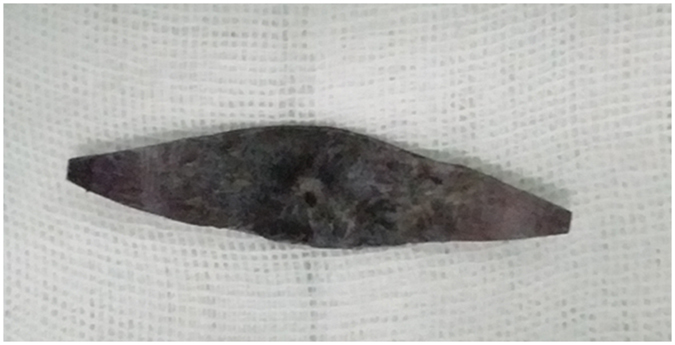
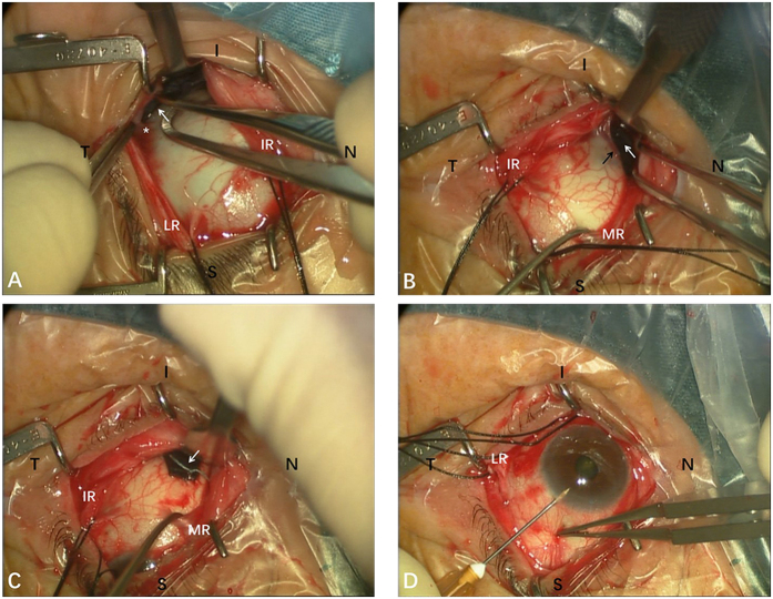
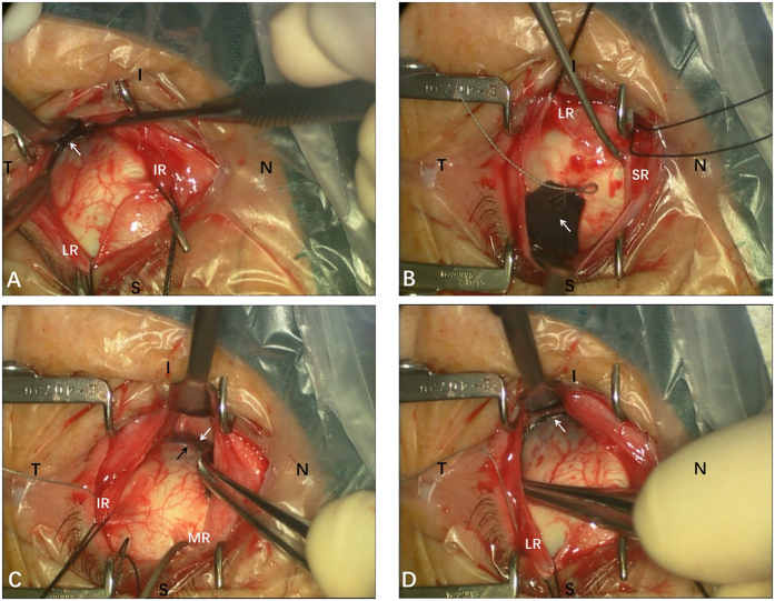
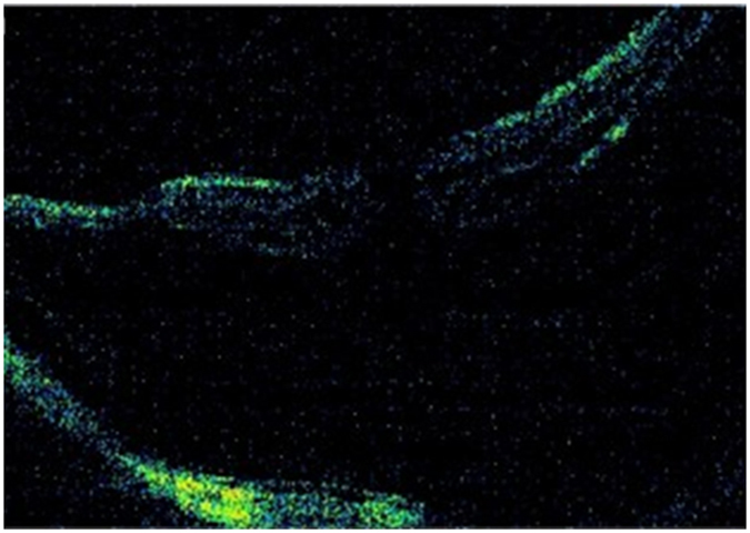
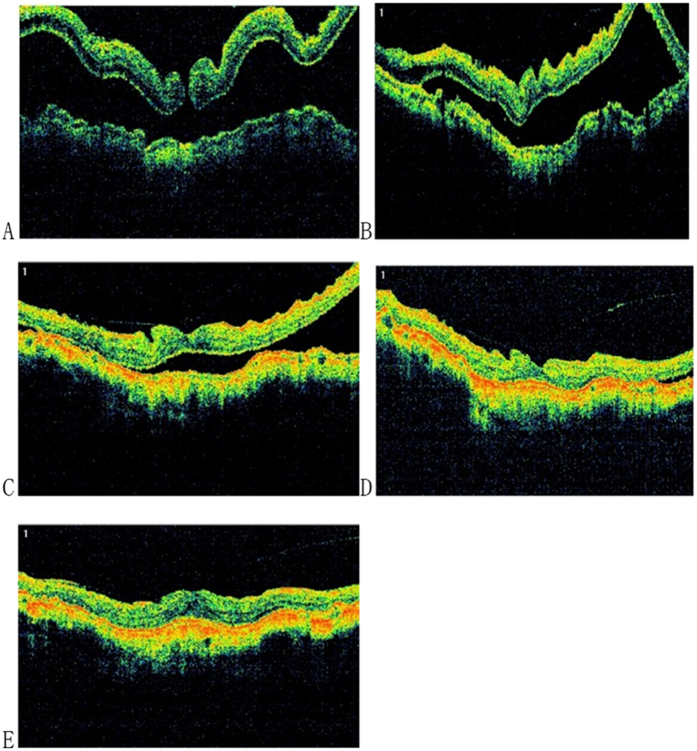
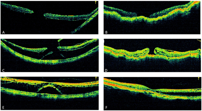
Myopic traction maculopathy (MTM) can cause vision disabilities in highly myopic eyes. This retrospective case series investigated the clinical outcomes of posterior scleral contraction (PSC) using genipin-cross-linked sclera as the material to treat MTM in highly myopic eyes. In total, 32 eyes from 29 highly myopic patients who underwent PSC for MTM were recruited. The changes in best-corrected visual acuity (BCVA) and axial length were evaluated, macular reattachment and macular hole (MH) closure was assessed by optical coherence tomography, and complications were evaluated. At the final follow-up, the retina was completely reattached in 25 eyes (78.1%), essentially reattached in 4 eyes (12.5%), and partially reattached in 3 eyes (9.4%). The logMAR BCVA improved significantly from 1.18 ± 0.45 preoperatively to 0.87 ± 0.45 postoperatively (P < 0.001). The 32 eyes were further divided into the MH group (16 eyes) and the non-MH group (16 eyes) for comparison. The MH was closed in 9 eyes (56.3%). The retinal reattachment rate was 75.0% in the MH group and 81.25% in the non-MH group, and the logMAR BCVA improved significantly in both groups. The PSC using genipin-cross-linked sclera as the material can effectively treat MTM in highly myopic eyes, and significant visual improvement can be achieved with minimal complications.


Conflict of interest statement

The authors declare no competing financial interests.

Figures

Figure 1
Figure 1. The genipin-cross-linked sclera used in posterior scleral contraction (PSC) surgery.
The donor sclera was cut into spindle strips.
Figure 2
Figure 2. Intraoperative photographs of the posterior scleral contraction (PSC) surgery in one case (Left eye).
(A) With the assistance of the traction sutures, the sclera strip (white arrow) was passed underneath the inferior oblique, next to the insertion point of the inferior oblique (asterisk). (B) The sclera strip (white arrow) was passed underneath the inferior rectus with caution, efforts were made to protect vortex vein (black arrow) from damage. (C) The inferior nasal end of sclera strip (white arrow) was fixed to the pre-equatorial sclera, 2 mm behind and outside the insertion point of the medial rectus (MR) muscle. (D) A 25-gauge syringe needle (1 mL) was inserted into the superior anterior chamber to release the aqueous humor. S: superior; I: inferior; N: nasal; T: temporal; LR: lateral rectus; IR: inferior rectus; MR: medial rectus.
Figure 3
Figure 3. Intraoperative photographs of the posterior scleral contraction (PSC) surgery, the same case as in Fig. 2.
(A) Ensured that the middle part of the sclera strip (white arrow) was placed at the posterior pole. (B) The superior temporal end of sclera strip (white arrow) was fixed to the pre-equatorial sclera, 2 mm behind and outside the insertion point of the superior rectus (SR) muscle. (C) The scleral strip (white arrow) was checked to assure that it was correctly positioned without damaging the vortex vein (black arrow). (D) Ensured that the sclera strip (white arrow) was stretched into a U shape to surround the posterior pole. S: superior; I: inferior; N: nasal; T: temporal; LR: lateral rectus; IR: inferior rectus; MR: medial rectus; SR: superior rectus.
Figure 4
Figure 4. The patient was 59 years old, female, with a logMAR BCVA of 1.30, and the axial length was 29.99 mm.
The preoperative optical coherence tomography (OCT) image of the left eye revealed a significant macular hole retinal detachment (MHRD).
Figure 5
Figure 5. The patient’s left eye received posterior scleral contraction (PSC) in June 2013, the genipin-cross-linked sclera was spindle shaped, and the length of the sclera strip was 45 mm with a width of 12 mm in the middle.
(A) One week after the surgery, significant retinal folds were noted, and the macular hole was still present. The axial length (AL) was 27.09 mm with a reduction of 2.90 mm compared to the preoperative AL (29.99 mm), and the LogMAR BCVA was 1.30. (B) One month after the surgery, the retinal detachment area was reduced, the AL was 27.47 mm, and the logMAR BCVA was 1.0. (C) Six months after the surgery, the retinal detachment area continued to reduce, retinal folds were relieved, the AL was 27.78 mm, and the logMAR BCVA was 1.0. (D) Twelve months after the surgery, the retina was essentially reattached, the AL was 27.83 mm, and the logMAR BCVA was 1.0. (D) Twenty-four months after surgery, the retina was completely reattached, the AL was 27.78 mm with a reduction of 2.21 mm compared to the preoperative AL (29.99 mm), and the logMAR BCVA was 0.7.
Figure 6
Figure 6
(A) For eye No. 8, preoperatively, OCT images showed a huge macular hole and associated retinal detachment. The logMAR best-corrected visual acuity was 2.0. The axial length was 30.0 mm. (B) After posterior scleral contraction (PSC), there was a complete retinal reattachment, but the macular hole was not closed. The logMAR best-corrected visual acuity was 1.4. The axial length was 26.63 mm. (C) For eye No. 26, preoperatively, OCT images showed a macular hole and associated retinal detachment, and the epiretinal membrane was present as well. The logMAR best-corrected visual acuity was 0.52. The axial length was 30.49 mm. (D) After PSC, the macular hole was not closed, and the retinal detachment area was reduced by more than 80% but not completely. The retinal detachment was still present around the macular hole edge, which was defined as essential reattachment. The logMAR best-corrected visual acuity was 0.4. The axial length was 27.88 mm. (E) For eye No. 31, preoperatively, OCT images showed foveal detachment and myopic foveoschisis. The logMAR best-corrected visual acuity was 0.52. The axial length was 27.98 mm. (F) After PSR, the retinal detachment area was reduced by 40%~79%, and retinal detachment was still present, which was defined as partial reattachment. The logMAR best-corrected visual acuity was 0.22. The axial length was 26.87 mm.

Similar articles

Cited by

References

    1. Buch H., Vinding T. & Nielsen N. V. Prevalence and causes of visual impairment according to World Health Organization and United States criteria in an aged, urban Scandinavian population: the Copenhagen City Eye Study. Ophthalmology 108, 2347–2357 (2001). - PubMed
    1. Iwase A. et al.. Prevalence and causes of low vision and blindness in a Japanese adult population: the Tajimi Study. Ophthalmology 113, 1354–1362, doi: 10.1016/j.ophtha.2006.04.022 (2006). - DOI - PubMed
    1. Wang Y., Xu L. & Jonas J. B. Prevalence and causes of visual field loss as determined by frequency doubling perimetry in urban and rural adult Chinese. American journal of ophthalmology 141, 1078–1086, doi: 10.1016/j.ajo.2006.01.023 (2006). - DOI - PubMed
    1. Yamada M. et al.. Prevalence of visual impairment in the adult Japanese population by cause and severity and future projections. Ophthalmic epidemiology 17, 50–57, doi: 10.3109/09286580903450346 (2010). - DOI - PubMed
    1. Wong T. Y., Ferreira A., Hughes R., Carter G. & Mitchell P. Epidemiology and disease burden of pathologic myopia and myopic choroidal neovascularization: an evidence-based systematic review. Am J Ophthalmol 157, 9–25 e12, doi: 10.1016/j.ajo.2013.08.010 (2014). - DOI - PubMed