. 2017 Feb 21:7:42885.
doi: 10.1038/srep42885.

Epigenetic determinants of space radiation-induced cognitive dysfunction

Affiliations

Epigenetic determinants of space radiation-induced cognitive dysfunction

Munjal M Acharya et al. Sci Rep. .

Abstract

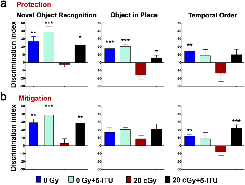
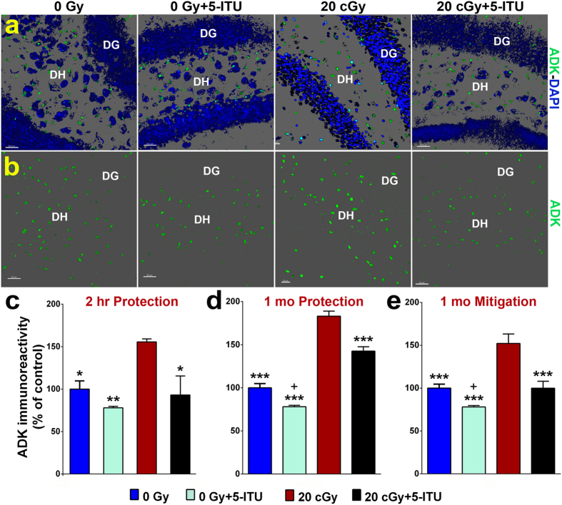
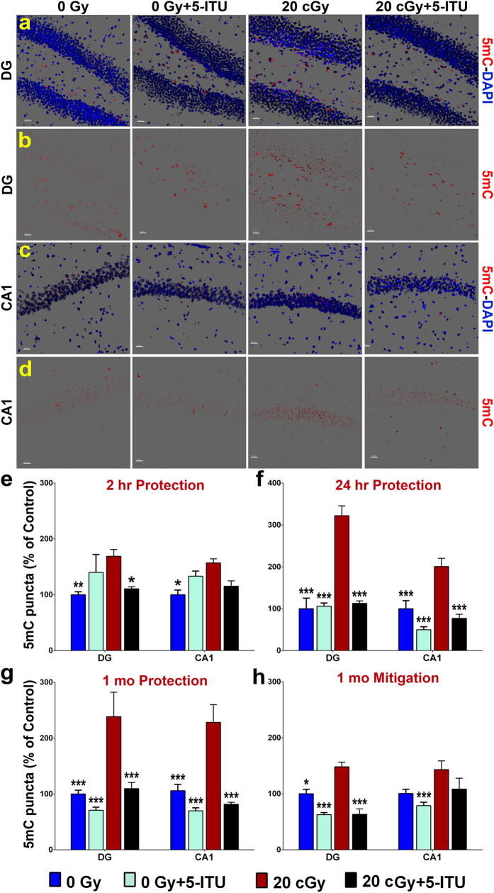
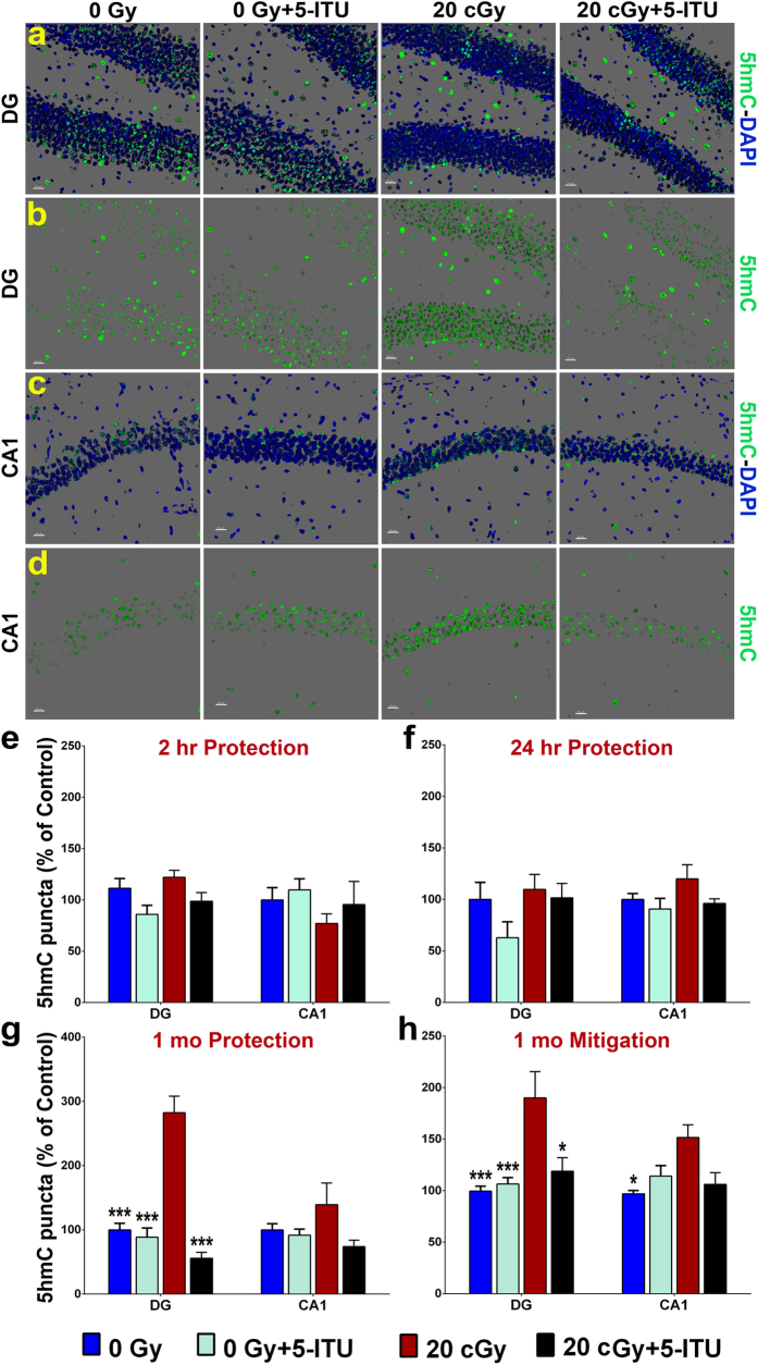
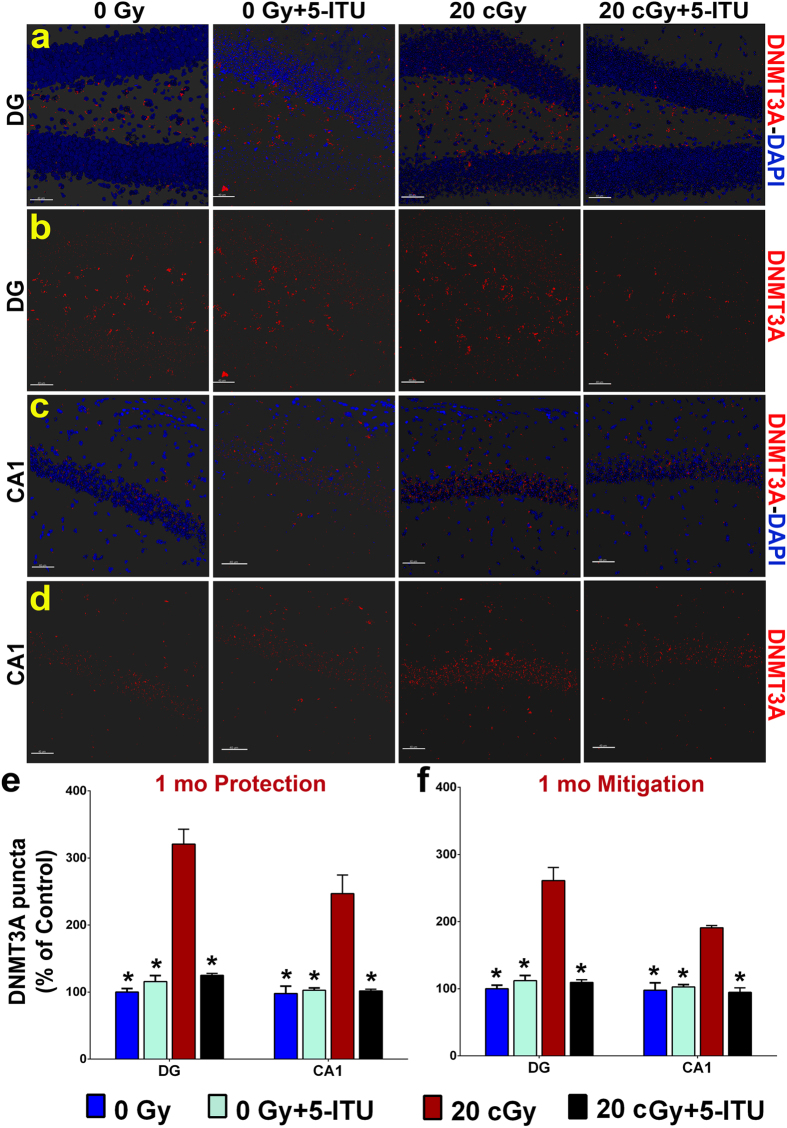
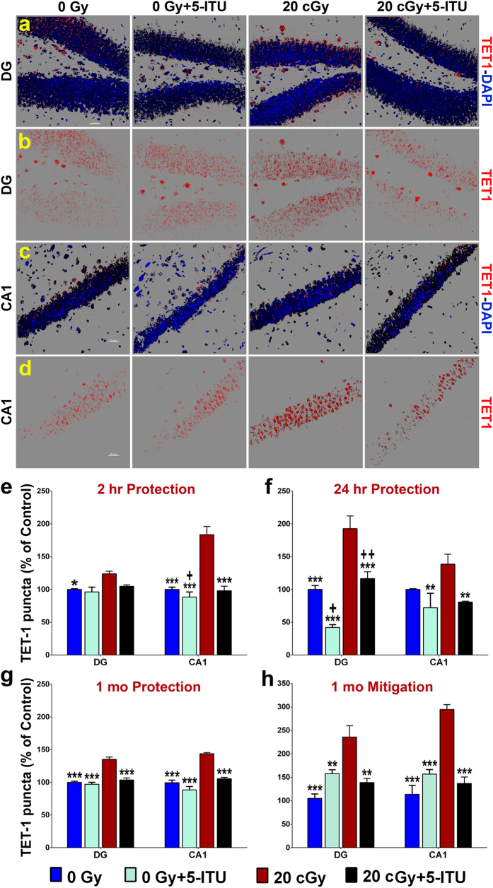
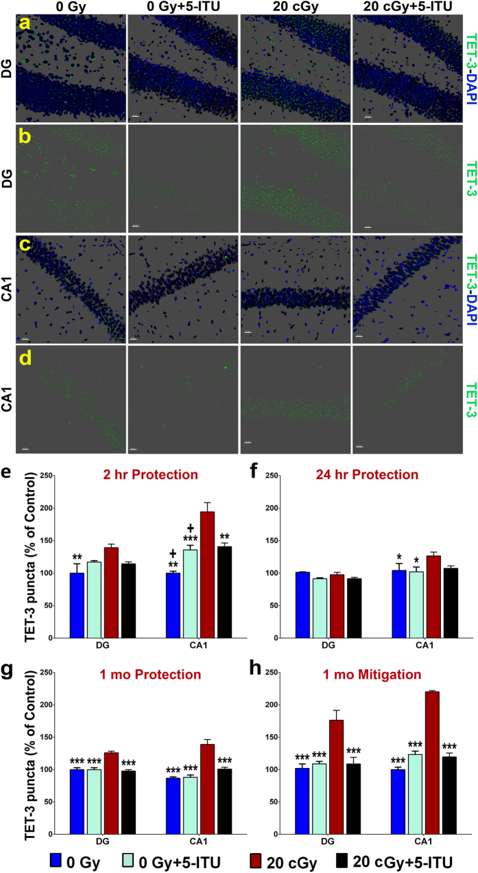
Among the dangers to astronauts engaging in deep space missions such as a Mars expedition is exposure to radiations that put them at risk for severe cognitive dysfunction. These radiation-induced cognitive impairments are accompanied by functional and structural changes including oxidative stress, neuroinflammation, and degradation of neuronal architecture. The molecular mechanisms that dictate CNS function are multifaceted and it is unclear how irradiation induces persistent alterations in the brain. Among those determinants of cognitive function are neuroepigenetic mechanisms that translate radiation responses into altered gene expression and cellular phenotype. In this study, we have demonstrated a correlation between epigenetic aberrations and adverse effects of space relevant irradiation on cognition. In cognitively impaired irradiated mice we observed increased 5-methylcytosine and 5-hydroxymethylcytosine levels in the hippocampus that coincided with increased levels of the DNA methylating enzymes DNMT3a, TET1 and TET3. By inhibiting methylation using 5-iodotubercidin, we demonstrated amelioration of the epigenetic effects of irradiation. In addition to protecting against those molecular effects of irradiation, 5-iodotubercidin restored behavioral performance to that of unirradiated animals. The findings of this study establish the possibility that neuroepigenetic mechanisms significantly contribute to the functional and structural changes that affect the irradiated brain and cognition.


Conflict of interest statement

The authors declare no competing financial interests.

Figures

Figure 1
Figure 1. Experimental Design.
(a) DNA methyltransferases (DNMT) convert cytosine to 5-methylcytosine (5mC) and ten-eleven translocation (TET) enzymes convert 5mC to 5-hydroxymethylcytosine (5hmC). (b) DNA methylation is linked to the S-adenosylmethionine (SAM) dependent transmethylation pathway that is regulated by adenosine under the control of adenosine kinase (ADK). DNMT uses SAM as a methyl donor, reducing it to S-adenosylhomocysteine (SAH) and then to adenosine and homocysteine (HCY) by SAH hydrolase. This process is dependent on removal of adenosine and HCY. Blockage of this pathway by 5-Iodotubercidin (5-ITU) and buildup of adenosine inhibits DNMT activity and reduces DNA methylation. (c) Schematic of the experimental time line. Mice in the protection study received 5-ITU treatments on 6 consecutive days pre-irradiation with the last injection 30 minutes prior to irradiation (28Si particles, 600 MeV/n, 20cGy at the Brookhaven National Laboratory, BNL). Mice in the mitigation study received 5-ITU treatments on 6 consecutive days post-irradiation with the first injection 30 minutes after irradiation. One month post-irradiation mice were administered behavioral testing (weeks 4–6) on the novel object recognition (NOR), object in place (OiP) and temporal order tasks (TO), after which brains were harvested for tissue analyses.
Figure 2
Figure 2. Protection and mitigation of radiation induced cognitive dysfunction by 5-ITU.
(a) During the protection study, irradiation using 20cGy of 28Si particles impaired exploration on the novel object recognition (NOR), object in place (OiP) and temporal order (TO) tasks 1 month post exposure, but 5-ITU treatment protected against these adverse effects in the NOR and OiP tasks (20cGy + 5-ITU). (b) During the mitigation study, irradiation using 20cGy of 28Si particles impaired exploration on the NOR and TO tasks, but 5-ITU mitigated these adverse effects. All data are presented as mean ± SEM (N = 8–10 animals per group). P values are derived from ANOVA and Bonferroni’s multiple comparisons test. *P < 0.05, **P < 0.01, ***P < 0.001 for 0Gy, 0Gy + 5-ITU and 20cGy + 5-ITU as compared to 20cGy.
Figure 3
Figure 3. 5-ITU treatment protects against 28Si particle irradiation induced increased ADK immunoreactivity.
(a,b) Representative images illustrate that ADK protein levels are elevated 1 month post-irradiation (20cGy) that is reduced by 5-ITU treatment (ADK, green; DAPI nuclear counterstain, blue) in the hippocampal dentate hilus (DH) and dentate gyrus (DG). Deconvolution of images and quantification of ADK protein demonstrate that irradiation increased ADK at (c) 2 hrs post-irradiation and at (d,e) 1 month post-irradiation and that 5-ITU treatment protects against and mitigates this increase. Data are presented as mean ± SEM (N = 3–4 mice/group). P values are derived from ANOVA and Bonferroni’s multiple comparisons test (*P < 0.05, **P < 0.01, ***P < 0.001 for 0Gy, 0Gy + 5-ITU and 20cGy + 5-ITU as compared to 20cGy). +P < 0.01 for 0Gy as compared to 0Gy + 5-ITU. (a,b) Scale bars 20 μm.
Figure 4
Figure 4. 28Si particle irradiation increases levels of 5-methylcytosine in the hippocampus.
Representative images show that radiation exposure causes increased 5mC levels in (a,b) DG and (c,d) CA1 regions of the brain and that is reduced by 5-ITU treatment (5mC, red; DAPI nuclear counterstain, blue). Deconvolution of images and quantification of 5mC demonstrate that irradiation induces increased 5mC at (e) 2 hrs, (f) 24 hrs and (g) 1 month post-irradiation and that 5-ITU treatment prior to irradiation protects against this increase. (h) Post-irradiation 5-ITU treatment also mitigates the radiation induced increase in 5mC. Data are presented as mean ± SEM (N = 3–4 mice/group). P values are derived from ANOVA and Bonferroni’s multiple comparisons test. *P < 0.05, **P < 0.01, ***P < 0.001 for 0Gy, 0Gy + 5-ITU and 20cGy + 5-ITU as compared to 20cGy. (a–d) Scale bars 20 μm.
Figure 5
Figure 5. 28Si particle irradiation causes increased levels of 5-hydroxymethylcytosine in the hippocampus 1 month post-irradiation.
Representative images show that irradiation causes increased 5mC levels in the (a,b) DG and (c,d) CA1 regions of the brain and that is reduced by 5-ITU treatment (5hmC, green; DAPI nuclear counterstain, blue). Deconvolution of images and quantification of 5hmC demonstrate that 5hmC marks are not increased at (e) 2 hrs or (f) 24 hrs post-irradiation. Radiation exposure does cause elevated 5hmC at (g) 1 month post-irradiation and that 5-ITU treatment prior to irradiation provides some protection against this increase. (h) Post-irradiation 5-ITU treatment also provides some mitigation of this radiation effect. Data are presented as mean ± SEM (N = 3–4 mice/group). P values are derived from ANOVA and Bonferroni’s multiple comparisons test. *P < 0.05, **P < 0.01, ***P < 0.001 for 0Gy, 0Gy + 5-ITU and 20cGy + 5-ITU as compared to 20cGy. (ad) Scale bars 20 μm.
Figure 6
Figure 6. 28Si particle irradiation increases levels of DNMT3a protein in the hippocampus.
Representative images show that irradiation causes increased levels of DNMT3a in (a,b) DG and (c,d) CA1 regions of the brain and that is reduced by 5-ITU treatment (DNMT3a, red; DAPI nuclear counterstain, blue). Deconvolution of images and quantification of DNMT3a demonstrate that radiation exposure causes elevated DNMT3a at 1 month post-irradiation and that 5-ITU treatment (e) prior to irradiation provides protection against this increase and that 5-ITU treatment (f) after irradiation mitigates this radiation effect. Data are presented as mean ± SEM (N = 3–4 mice/group). P values are derived from ANOVA and Bonferroni’s multiple comparisons test. *P < 0.05, **P < 0.01, ***P < 0.001 for 0Gy, 0Gy + 5-ITU and 20cGy + 5-ITU as compared to 20cGy. (ad) Scale bars 40 μm.
Figure 7
Figure 7. 28Si particle irradiation increases levels of TET1 protein in the hippocampus.
Representative images show that radiation exposure causes increased TET1 levels in (a,b) DG and (c,d) CA1 regions of the brain and that is reduced by 5-ITU treatment (TET1, red; DAPI nuclear counterstain, blue). Deconvolution of images and quantification of TET1 demonstrate that irradiation induces increased protein levels at (e) 2 hrs, (f) 24 hrs and (g) 1 month post-irradiation and that 5-ITU treatment prior to irradiation protects against this increase. (h) Post-irradiation 5-ITU treatment also mitigates the radiation induced increase in TET1. Data are presented as mean ± SEM (N = 3–4 mice/group). P values are derived from ANOVA and Bonferroni’s multiple comparisons test. *P < 0.05, **P < 0.01, ***P < 0.001 for 0Gy, 0Gy + 5-ITU and 20cGy + 5-ITU as compared to 20cGy. +P < 0.01 ++P < 0.002 for 0Gy + 5-ITU as compared to 20cGy + 5-ITU. (ad) Scale bars 20 μm.
Figure 8
Figure 8. 28Si particle irradiation causes increased levels of TET3 protein in the hippocampus.
Representative images show that radiation exposure causes increased TET3 levels in (a,b) DG and (c,d) CA1 regions of the brain and that is reduced by 5-ITU treatment (TET1, green; DAPI nuclear counterstain, blue). Deconvolution of images and quantification of TET3 demonstrate that irradiation induces increased protein levels at (e) 2 hrs, (f) 24 hrs and (g) 1 month post-irradiation and that 5-ITU treatment prior to irradiation protects against this increase. (h) Post-irradiation 5-ITU treatment also mitigates the radiation induced increase in TET3. Data are presented as mean ± SEM (N = 3–4 mice/group). P values are derived from ANOVA and Bonferroni’s multiple comparisons test. *P < 0.05, **P < 0.01, ***P < 0.001 for 0Gy, 0Gy + 5-ITU and 20cGy + 5-ITU as compared to 20cGy. +P < 0.02 for 0Gy as compared to 0Gy + 5-ITU 0Gy + 5-ITU and for 0Gy as compared to 20cGy + 5-ITU. (a–d) Scale bars 20 μm.

Similar articles

Cited by

References

    1. Plante I. & Cucinotta F. A. Energy deposition and relative frequency of hits of cylindrical nanovolume in medium irradiated by ions: Monte Carlo simulation of tracks structure. Radiat Environ Biophys 49, 5–13, doi: 10.1007/s00411-009-0255-7 (2010). - DOI - PubMed
    1. Parihar V. K. et al.. What happens to your brain on the way to Mars. Science Advances 1, e1400256, 1–6 (2015). - PMC - PubMed
    1. Britten R. A. et al.. Impaired Spatial Memory Performance in Adult Wistar Rats Exposed to Low (5–20 cGy) Doses of 1 GeV/n (56)Fe Particles. Radiat Res 185, 332–337, doi: 10.1667/RR14120.1 (2016). - DOI - PubMed
    1. Raber J., Marzulla T., Stewart B., Kronenberg A. & Turker M. Silicon Irradiation Impairs Contextual Fear Memory in B6D2F1 Mice. Radiat Res (2015). - PubMed
    1. Parihar V. K. et al.. Persistent changes in neuronal structure and synaptic plasticity caused by proton irradiation. Brain structure & function, doi: 10.1007/s00429-014-0709-9 (2014). - DOI - PMC - PubMed

Publication types

MeSH terms