Review
. 2017 Feb;49(2):137-159.
doi: 10.1002/lsm.22637. Epub 2017 Feb 21.

Light and energy based therapeutics for genitourinary syndrome of menopause: Consensus and controversies

Affiliations
Review

Light and energy based therapeutics for genitourinary syndrome of menopause: Consensus and controversies

Yona Tadir et al. Lasers Surg Med. 2017 Feb.

Abstract

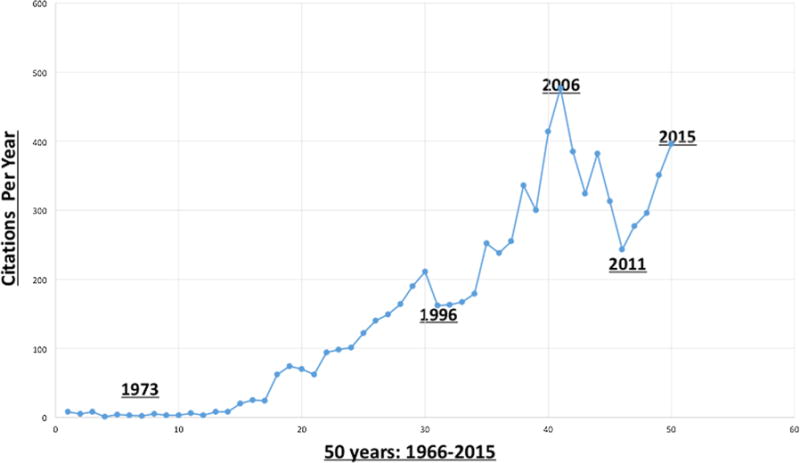
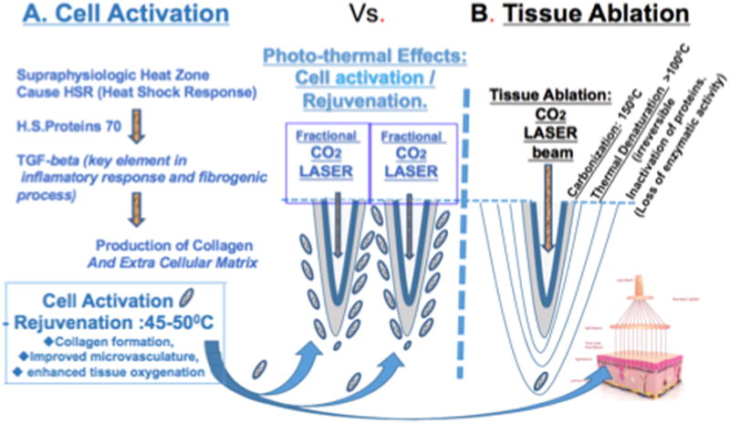
Gynecologist and plastic surgeons pioneered the application of lasers in medicine and surgery almost 5 decades ago, initially used to treat cervical and vaginal pathologies. Ever since, energy-based devices have been deployed to treat pelvic pathologies and improve fertility. Recent technological developments triggered an unprecedented wave of publications, assessing the efficacy of fractional laser, and radiofrequency on the vaginal wall in reversing natural aging processes. Studies have shown that a certain degree of thermal energy deposited on the vaginal wall stimulates proliferation of the glycogen-enriched epithelium, neovascularization, and collagen formation in the lamina propria, and improves natural lubrication and control of urination. This review aimed to review such data and to guide future research. A unique assembly of experts from around the globe, compiled and edited this manuscript based on a thorough literature review and personal experience. Lasers Surg. Med. 49:137-159, 2017. © 2017 Wiley Periodicals, Inc.

Keywords: energy based device; genitourinary syndrome of menopause (GSM); laser; lichen sclerosus; vulvodynia; radiofrequency; rejuvenation; stress urinary incontinence (SUI); vagina; vulva.


Conflict of interest statement

Conflict of Interest Disclosures: All authors have completed and submitted the ICMJE Form for Disclosure of Potential Conflicts of Interest and have disclosed the following: Yona Tadir is a scientific consultant for Alma Lasers. Red Alinsod is a consultant for Thermi, receive royalty for ThermiVa, Cooper Surgical, Lone Star Retractor, and Coloplast. Consultant, Caldera Medical: Stock owner, Monarch Medical Products: Consultant, Royalty for Alinsod Surgical Equipment. Selih-Martinec K. is an invited speaker by Alma Lasers. Alberto Calligaro, Stefano Salvatore, and Nicola Zerbinati are scientific consultants for DEKA laser. All other co-authors: nothing to disclose. No commercial source supported the collaboration and preparation of this manuscript.

Figures

Fig. 1
Fig. 1
Fifty years’ citations of the key words: “Laser & Gynecology” in peer-review articles (1964– 2015).
Fig. 2
Fig. 2
Histological sections of the vaginal wall. (A) Normal. (B) Moderately atrophic. (C) Severely atrophic. (A) Estrogenized vaginal histology. The two upper layers of the vaginal wall are shown: stratified, squamous epithelium (SSE), and the lamina propria (LP). The stratified squamous epithelium is rich in glycogen (larger cells with abundant clear cytoplasm—blue arrow) and is nonkeratinizing. The basal cell layer (black arrow) consists of a single layer of columnar cells. (B) Moderately atrophied vagina: Atrophy is shown by thinner epithelium (E) and loss of maturation (smaller cell size with less cytoplasm) on the surface. (C) Marked vaginal atrophy. (Courtesy: Lev-Sagie A).
Fig. 3
Fig. 3
(A) Genital warts (condyloma accuminata) on the uterine cervix. (B) The uterine cervix following superficial ablation of the diseased area. Carbonized crater base is visible. (Courtesy: Levavi H, Tadir Y).
Fig. 4
Fig. 4
Energy (Joule) = Power (Watt)×Time (Second). Effect on tissue is different even if the same energy is deposited depending on exposure time. Same energy may cause different effect, that is, crater shape, superficial carbonization, and thermal coagulation. Three examples of same energy, 90 J, will cause different effect on the tissue. (Courtesy: Tadir Y).
Fig. 5
Fig. 5
(Left) Laser beam fractionated by holographic lens, printing 9×9 (81 microbeams) on 10 mm2. (Right) Laser beam delivered through computer controlled, two parallel mirrors and scanning lens. (Courtesy: Alma Lasers).
Fig. 6
Fig. 6
(A) Fractional micro-ablation inducing cell activation and tissue rejuvenation at 45–50°C [42]. (B) Tissue ablation and thermal effects on adjacent layers (Courtesy: Tadir Y).
Fig. 7
Fig. 7
Energy-based probes for vaginal and vulvar treatment listed in alphabetic order. (Courtesy: manufacturers).
Fig. 8
Fig. 8
Trichrome staining of biopsies from atrophic vaginal mucosa before (A) and vaginal mucosa following the fractional CO2 treatment 2 months from the first laser application (B). Particularly appreciable are the remarkable differences: (A) thin epithelium, with small size and few layers of epithelial cells, no superficial desquamation, smooth surface of the interface epithelium-connective tissue, absence of papillae, compactness of the connective tissue. (B) Very thick epithelium, with many layers of big cells, many shedding cells at the epithelial surface, uneven interface epithelium-connective tissue, a well visible deep papilla formed by a fine fibrillar connective tissue with vessels inside (A and B same magnification). Due to the thickness of epithelium following treatment, Figure 8b is formed by two frames. (Courtesy: Calligaro A).
Fig. 9
Fig. 9
Common feature following treatment. At the center of the figure, a papilla with a small and long vessel inside is easily identifiable due to its erythrocyte content and the thin endothelial profiles. In the insert, numerous newly formed papillae following treatment are recognizable. H&E staining. (Courtesy: Calligaro A).
Fig. 10
Fig. 10
PAS staining of vaginal mucosa sections from biopsies before (A) and following treatment (B). Besides the evident difference in the epithelial thickness, the PAS signal shows (A) few small cells containing glycogen (fuchsin-stained) versus (B) many larger cells stained, starting from the low intermediate cell layer. Also of note in (B) is the superficial desquamation of intensely stained cells, which deposit glycogen at the surface of mucosal epithelium. (Courtesy: Calligaro A).
Fig. 11
Fig. 11
High-resolution, light microscope image of a CO2 laser treated-section, embedded in epoxy resin for electron microscopy, and stained with toluidine blue. In the lower part of the image, some cells of the lower intermediate layer with a very clear nucleus containing a highly visible nucleolus (as a dark blue spot), show some violet-stained masses (white arrows). These represent the first stores of newly synthesized glycogen inside epithelial cells, resulting from the restored differentiation process. In the upper layers, glycogen masses are increasingly extended to almost completely fill the cytoplasm, until superficial shedding (desquamation). (Courtesy: Calligaro A).
Fig. 12
Fig. 12
Electron microscopy of fibroblasts in samples collected following fractional CO2 treatment. (A) Connective tissue fibroblasts showing a relatively compacted cell body, with a cytoplasm appearing almost completely filled with rough endoplasmic reticulum (rER). The nucleus is euchromatic, with a well-presented nucleolus. These features support active synthesis of proteins (collagen and others) to be delivered to the surrounding extracellular matrix, promoting its renewal. (B) In fibroblasts, close to the rER profiles (cisternae formed by membranes with ribosomes attached at their surface), dilated cisternae with attached ribosomes are frequently observed as vesicles containing a fine, filamentous material, representing the molecular precursors of extracellular fibrillar components. (C) A Golgi apparatus is clearly visible. Two Golgi complexes, formed by stacks of membranes with associated dilations and vesicles, are recognizable (white arrows). (Courtesy: Calligaro A).
Fig. 13
Fig. 13
Normal vulva and histology. Left: Hair bearing vulvar skin A-labia majora, B-labia minora, C-clitoral hood, D-Hymen, E-vestibule. black arrow-clitoris, red arrow-Hart’s line. Right: Histology of the non-hair bearing vulvar skin. This is hair bearing skin, ×40 magnification. The epidermis (E) does not have estrogen receptors, so does not atrophy like the vagina when estrogen is withdrawn. D-dermis. There are superficial sebaceous glands (Fordyce spots) present (SG). Like hair bearing skin, the labia minora atrophy when estrogen is withdrawn because the dermal fibrioblasts have estrogen receptors. (Courtesy: Lev-Sagie A).
Fig. 14
Fig. 14
Vulvar lichen sclerosus—(A) LS can be diagnosed clinically by inspection of the vulva for the characteristic thin, white, wrinkled skin, and changes in vulvar architecture. Findings in LS include hypopigmentation, hemorrhages (black arrow), loss of normal architecture including disappearance of labia minora (blue arrow), buried clitoris (red arrow), and narrowing of the introital opening. Note that the distinction between the labia majora and minora is lost. The disease involves the perineal and perianal areas. (B) LS histology ×40 magnification—on histological examination, the epidermis (E) is typically thinned (accounting for the older nomenclature “lichen sclerosus et atrophicus”), although areas of thickened skin (H-hyperkeratosis) may exist. The upper dermis exhibits homogenization of collagen (black arrow) with a band of lymphocytes below this region (blue arrow). (Courtesy: Lev-Sagie A).
Fig. 15
Fig. 15
Vulvar lichen sclerosus. Histological assessment of Masson’s trichrome-stained tissue samples before versus after factional CO2 laser treatment. Left, case 1; Right, case 2. Histology: Top row: Hyperkeratosis (a), dermal atrophy, hydropic degeneration of the basal epithelial cells (b) dermo epidermal clefts (c) homogeneous papillary dermis, afibrillar with frosted glass appearance and edema (d) and inflammatory infiltrate of polymorphonuclear band and plasma cells (e) and inflammatory infiltrate of polymorphonuclear band and plasma cells. Bottom row: Trophic epithelium without superficial hyperkeratosis (a), persistence in some basal cells hydropic degeneration (b), persistence of some areas with dermo-epidermal clefts (c), and lamina propria fibrillar with irregular spaces containing translucent material (d). (Courtesy: Elias J, Galich M).

References

    1. Kaplan I, Goldman J, Ger R. The treatment of erosions of the uterine cervix by means of the CO2 laser. Obstet Gynecol. 1973;41(5):795–796. - PubMed
    1. Bellina JH, Polanyi TG. Management of vaginal adenosis and related cervico-vaginal disorders in DES-exposed pregnancy by means of carbon dioxide laser surgery. J Reprod Med. 1976;16:295–296. - PubMed
    1. Bruhat M, Mage G, Mahnes M. Use of the CO2 laser in laparoscopy. Proceedings of the 3rd international society for laser surgery. Ed. Kaplan I. 1979;24:235–238.
    1. Tadir Y, Kaplan I, Zukerman Z, Ovadia J. Laparoscopic CO2 laser sterilization. In: Semm K, Mettler L, editors. Human Reproduction. Exerpta Medica, Amsterdam: Elsevier; 1981. pp. 551pp. 423–431.
    1. Nezhat FR, Nezhat CR, Silfen SL. Videolaseroscopy for oophorectomy. Am J Obstet Gynecol. 1991;165:1323–1330. - PubMed