Review
. 2017 May;12(5):10.1002/biot.201600394.
doi: 10.1002/biot.201600394. Epub 2017 Feb 21.

Development of hydrogels for regenerative engineering

Affiliations
Review

Development of hydrogels for regenerative engineering

Xiaofei Guan et al. Biotechnol J. 2017 May.

Abstract

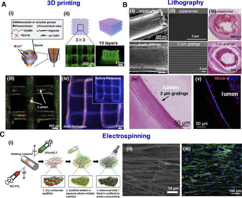
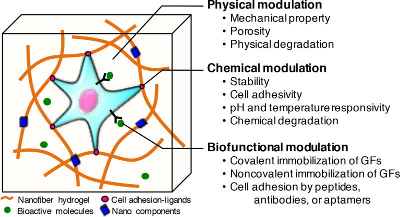
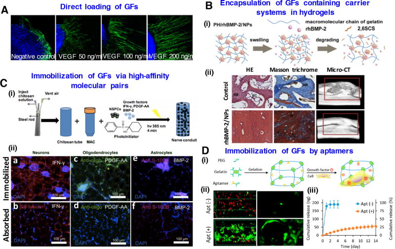
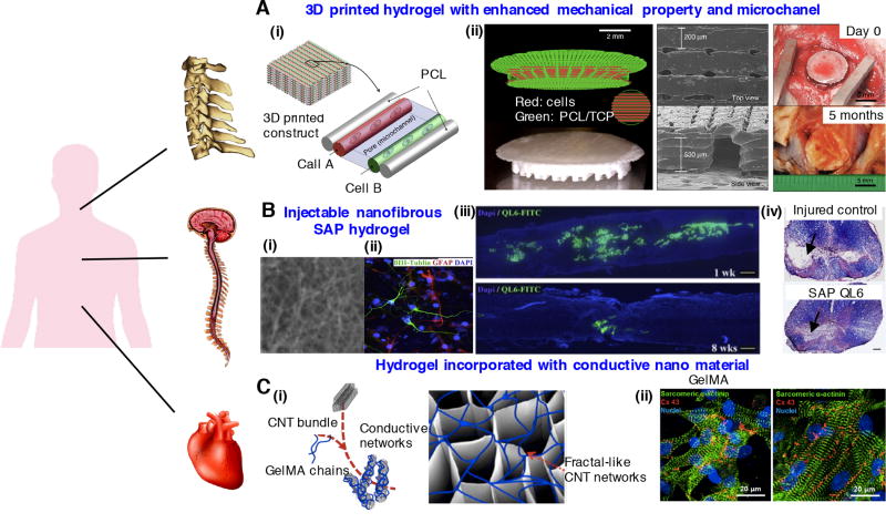
The aim of regenerative engineering is to restore complex tissues and biological systems through convergence in the fields of advanced biomaterials, stem cell science, and developmental biology. Hydrogels are one of the most attractive biomaterials for regenerative engineering, since they can be engineered into tissue mimetic 3D scaffolds to support cell growth due to their similarity to native extracellular matrix. Advanced nano- and micro-technologies have dramatically increased the ability to control properties and functionalities of hydrogel materials by facilitating biomimetic fabrication of more sophisticated compositions and architectures, thus extending our understanding of cell-matrix interactions at the nanoscale. With this perspective, this review discusses the most commonly used hydrogel materials and their fabrication strategies for regenerative engineering. We highlight the physical, chemical, and functional modulation of hydrogels to design and engineer biomimetic tissues based on recent achievements in nano- and micro-technologies. In addition, current hydrogel-based regenerative engineering strategies for treating multiple tissues, such as musculoskeletal, nervous and cardiac tissue, are also covered in this review. The interaction of multiple disciplines including materials science, cell biology, and chemistry, will further play an important role in the design of functional hydrogels for the regeneration of complex tissues.

Keywords: Biofabrication; Hydrogel; Nanotechnology; Regenerative engineering; Tissue regeneration.


Figures

Figure 1
Figure 1
Strategies for the nano- and micro-fabrication of hydrogels: (A) 3D printed vascular hydrogel constructs using a blended bioink. (i) Schematic diagram of the blended bioink, including cell-laden alginate, GelMA, and PEGTA. (ii) Schematics of the 3D printed tubular constructs and corresponding fluorescence microscopy images. (iii) 3D confocal structure of the bioprinted tubes containing green fluorescent beads. Red fluorescent beads were perfused inside the lumens. (iv) Fluorescence images before and after perfusion of red fluorescent beads in a continuous 3D printed tube. Images reprinted from [31] with permission of Elsevier. (B) Tubular-patterned PVA hydrogel graft based on nanoimprint lithography. (i) The macroscopic view of either unpatterned or patterned PVA graft. (ii) High magnification images of the tubular patterns in PVA graft. (iii) Patency of patterned PVA graft with 2 µm gratings and occlusion of unpatterned PVA graft demonstrated by hematoxylin and eosin (H&E) staining. (iv) Grating structures were observed through H&E. (v) Endothelial cells adhered to the lumen demonstrated by immunostaining of PVA graft with 2 µm gratings. Images adapted from [38] with permission of Elsevier. (C) Multilayered tendon tissue graft fabricated by dual electrospinning of methacrylated gelatin and PCL. (i) Schematic for fabrication of composite scaffold. (ii) Aligned fibers observed by scanning electron microscope (SEM). (iii) Human adipose-derived stem cells with elongated morphology grow along the fiber direction. Images were reproduced from [49] with permission of Elsevier.
Figure 2
Figure 2
Controlling cell behavior in 3D hydrogels by physical, chemical, and biofunctional modulation for regenerative engineering.
Figure 3
Figure 3
Biofunctionalization of hydrogels for regenerative engineering: (A) Direct loading of VEGF growth factors in gelatin-based hydrogels enhances axon outgrowth of neuron cells. Reproduced with permission [121]. Copyright 2014, John Wiley & Sons. (B) Encapsulation of GFs containing carrier systems in hydrogels. (i) Schematic representation of rhBMP-2 release from nanoparticles into hydrogels (i) Histological and micro-CT evaluation of bone formation of rhBMP-2 containing hydrogels at 12 weeks. Images modifies from [127] with permission of Elsevier. (C) Immobilization of GFs via high-affinity molecular pairs. (i) Creation of spinal cord regenerative conduits incorporating encapsulated neural stem cells and immobilized differentiation factors. (ii) Immunostaining results after 4-week implantation showing directed differentiation to neurons, oligodendrocytes and astrocytes. Images reprinted from [144] with permission of Elsevier. (D) Immobilization of growth factors by aptamers in PEG-gelatin hydrogels. (i) Schematic of chimeric hydrogel synthesis. (ii) Confocal microscopy images of live and dead cells and cell morphology in the hydrogels. (iii) VEGF release profile from the hydrogels with or without aptamer modification. Images reprinted from [153] with permission of American Chemical Society.
Figure 4
Figure 4
Regenerative engineering strategies for treating multiple tissues: (A) 3D printed hydrogel constructs incorporating microchannels with enhanced mechanical properties for musculoskeletal tissue regeneration. (i) Schematic of 3D patterned architecture including cell-laden hydrogels, supporting PCL polymer and microchannels. (ii) 3D printed PCL/tricalcium phosphate (TCP) mixture and cell-laden hydrogel for bone reconstruction. SEM images of the 3D printed calvarial bone constructs and photos at 5 months after in vivo implantation. Images reprinted from [60]. Copyright 2016, Nature Publishing Group. (B) Injectable nanofibrous SAP hydrogel promotes neurological recovery of spinal cord injuries. (i) Scanning electron microscopy image of K2(QL)6K2 SAP (QL6) nanofibrous hydrogel. (ii) Neural precursor cells grown on QL6 hydrogel scaffold demonstrated by immunocytochemistry. (iii) Longitudinally sectioned view of rat spinal cord, monitored 1 week and 8 weeks after implementing QL6 hydrogel at the injury site. The QL6 hydrogel almost biodegraded after 8 weeks. (iv) Luxol fast blue and H&E staining showed a greater size of the spared tissue in QL6-treated group compared to saline control group. Images reprinted from [170] with permission of Elsevier. (C) GelMA hydrogel incorporated with electrically conductive CNT for cardiac tissue regeneration. (i) Schematic of CNT-embedded GelMA hydrogel fabrication process. (ii) Enhanced alignment and elongation of cardiac cells grown on CNT-GelMA compared to pristine GelMA. Reproduced with permission [181]. Copyright 2013, American Chemical Society.

Similar articles

Cited by

References

    1. Vacanti JP, Langer R. Tissue engineering: the design and fabrication of living replacement devices for surgical reconstruction and transplantation. The Lancet. 1999;354:S32–S34. - PubMed
    1. Mason C, Dunnill P. A brief definition of regenerative medicine. Regen. Med. 2008;3:1–5. - PubMed
    1. Laurencin CT, Khan Y. Regenerative engineering. Sci. Transl. Med. 2012;4:160ed169. - PubMed
    1. Slaughter BV, Khurshid SS, Fisher OZ, Khademhosseini A, Peppas NA. Hydrogels in regenerative medicine. Adv. Mater. 2009;21:3307–3329. - PMC - PubMed
    1. Huebsch N, Arany PR, Mao AS, Shvartsman D, et al. Harnessing traction-mediated manipulation of the cell/matrix interface to control stem-cell fate. Nat. Mater. 2010;9:518–526. - PMC - PubMed