. 2017 Mar 21;56(11):1585-1595.
doi: 10.1021/acs.biochem.7b00056. Epub 2017 Mar 13.

The Cation-π Interaction Enables a Halo-Tag Fluorogenic Probe for Fast No-Wash Live Cell Imaging and Gel-Free Protein Quantification

Affiliations

The Cation-π Interaction Enables a Halo-Tag Fluorogenic Probe for Fast No-Wash Live Cell Imaging and Gel-Free Protein Quantification

Yu Liu et al. Biochemistry. .

Abstract

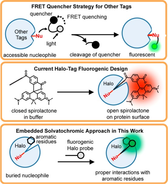
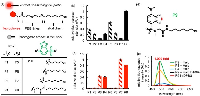
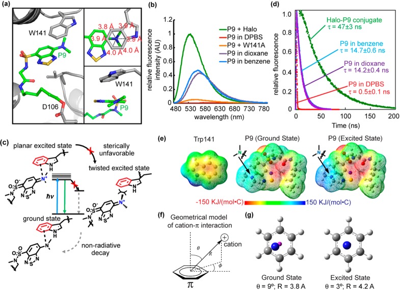
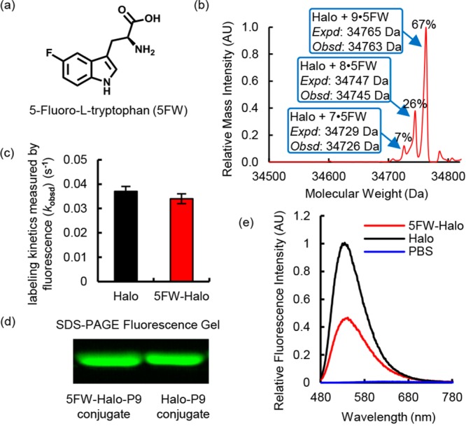
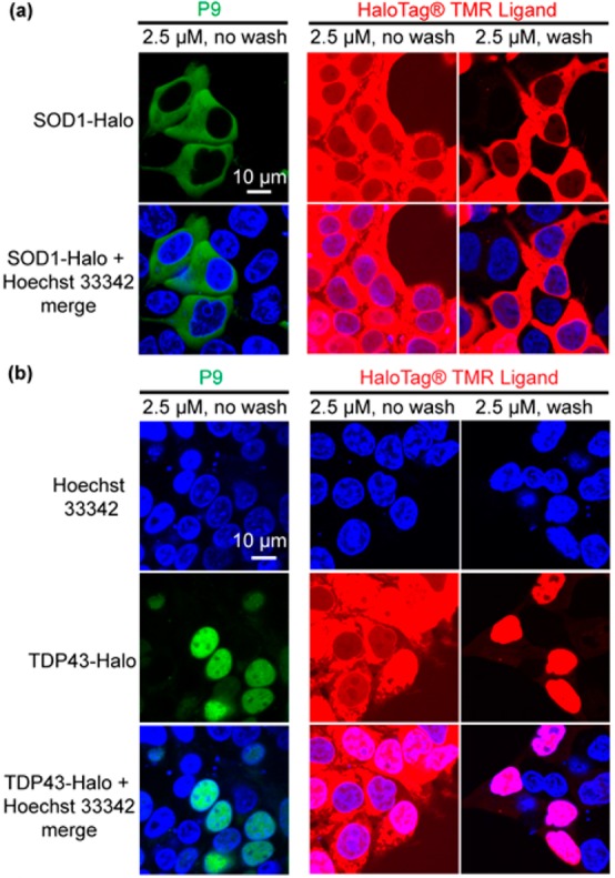
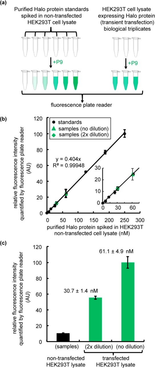
The design of fluorogenic probes for a Halo tag is highly desirable but challenging. Previous work achieved this goal by controlling the chemical switch of spirolactones upon the covalent conjugation between the Halo tag and probes or by incorporating a "channel dye" into the substrate binding tunnel of the Halo tag. In this work, we have developed a novel class of Halo-tag fluorogenic probes that are derived from solvatochromic fluorophores. The optimal probe, harboring a benzothiadiazole scaffold, exhibits a 1000-fold fluorescence enhancement upon reaction with the Halo tag. Structural, computational, and biochemical studies reveal that the benzene ring of a tryptophan residue engages in a cation-π interaction with the dimethylamino electron-donating group of the benzothiadiazole fluorophore in its excited state. We further demonstrate using noncanonical fluorinated tryptophan that the cation-π interaction directly contributes to the fluorogenicity of the benzothiadiazole fluorophore. Mechanistically, this interaction could contribute to the fluorogenicity by promoting the excited-state charge separation and inhibiting the twisting motion of the dimethylamino group, both leading to an enhanced fluorogenicity. Finally, we demonstrate the utility of the probe in no-wash direct imaging of Halo-tagged proteins in live cells. In addition, the fluorogenic nature of the probe enables a gel-free quantification of fusion proteins expressed in mammalian cells, an application that was not possible with previously nonfluorogenic Halo-tag probes. The unique mechanism revealed by this work suggests that incorporation of an excited-state cation-π interaction could be a feasible strategy for enhancing the optical performance of fluorophores and fluorogenic sensors.


Conflict of interest statement

The authors declare no competing financial interest.

Figures

Figure 1
Figure 1
Fluorogenic probe design strategies for fusion protein tags. As shown in the top box, conventional approaches involve a linked FRET quencher that is cleaved when the probe reacts with an accessible nucleophile on the surface. This strategy is not suitable for Halo tag because it contains a deeply buried nucleophile. As shown in the middle box, one current design strategy for fluorogenic probes for Halo tag involves a chemical switch between closed and open forms of a spirolactone group. As shown in the bottom box, an embedded solvatochromic strategy is employed in this work to develop a novel class of fluorogenic probes.
Figure 2
Figure 2
Fluorogenic benzoxadiazole and benzothiadiazole probes for Halo tag. (a) Current nonfluorogenic probes (top) for Halo tag harbor a six-carbon alkyl chain, a polyethylene glycol (PEG) linker, and a fluorescent fluorophore. The table shows structures of fluorogenic probes for Halo tag evaluated in this work. R1 is a chloroalkane chain of varying length, with and without a sarcosine amide linker. R2 denotes the structure of an electron donating group, which is either a methylamino or dimethylamino substituent. (b) Relative intensity of conjugates of P1–P8 with Halo and (c) its relative labeling kinetics. Halo samples (20 μM) were incubated with P1–P8 (10 μM) for 18 h prior to fluorescence intensity measurements (Ex. = 450 nm, and Em. = 530 nm). The labeling rate was recorded upon mixing 5 μM Halo protein with 0.5 μM ligand (Ex. = 450 nm, and Em. = 530 nm). (d) P9 consists of a six-carbon alkyl chain, a sarcosine amide linker, and a solvatochromic fluorophore based on a benzothiadiazole scaffold. (e) P9 exhibits ∼1000-fold fluorescence enhancement upon reaction with Halo protein (green), compared to P9 in DPBS (red). The probe is weakly fluorescent upon binding to Halo-D106A (black). A solution of the protein (20 μM) was incubated with 0.5 equiv (10 μM) of P9 (green), P8 (yellow), or P4 (blue) in DPBS buffer at 25 °C for 1 h. Fluorescence emission spectra were recorded at 450 nm excitation.
Figure 3
Figure 3
Cation−π interaction enhances P9 fluorescence. (a) The benzothiadiazole ring of P9 is embedded inside Halo, accommodated by a shift of a loop containing Trp141. The inset at the top right shows that the tertiary N of the dimethylamino group resides close to the geometrical center of the benzene ring of the Trp141 indole (3.8–4.0 Å distance between N and benzene carbons). The inset at the bottom right shows that the dimethylamino group is oriented just slightly twisted in relative to the benzothiadiazole moiety. (b) Fluorescence spectra of the Halo–P9 conjugate (green), W141A–P9 conjugate (orange), P9 in DBPS buffer (red), P9 in 1,4-dioxane (purple), and P9 in benzene (blue). For the Halo–P9 and W141A–P9 conjugates, a solution of the protein (20 μM) was incubated with 0.5 equiv (10 μM) of P9 in DPBS buffer at 25 °C for 1 h. For P9 in different solvents, 10 μM P9 was added in solvents at 25 °C for 1 h. Fluorescence emission spectra were recorded at 450 nm excitation. (c) The dimethylamino group of P9 is thought to take on a positive charge in the excited state and interact with the aromatic group in Trp141 via a cation−π interaction. (d) Fluorescence decay of the Halo–P9 conjugate (green), P9 in DBPS buffer (red), P9 in 1,4-dioxane (purple), and P9 in benzene (blue). P9 (20 μM) was incubated in DPBS buffer, benzene, 1,4-dioxane, or purified Halo protein (50 μM) for 1 h at 25 °C. (e) Electrostatic potential map of Trp141 in the ground state (left), P9 in ground state S0 (middle), and P9 in excited state S3 (right). (f) Geometric model of the cation−π interaction. (g) The left panels shows the simplified geometry of the ground state with θ = 9° and R = 3.8 Å, where the magenta “atom” represents the center of the benzene ring. The right panel shows the simplified geometry at the relaxed excited state with θ = 3° and R = 4.2 Å, exhibiting a canonical cation−π geometry.
Figure 4
Figure 4
Incorporation of 5-fluorotryptophan (5FW) reduced the fluorescence intensity of the P9–Halo conjugate. (a) Molecular structure of 5FW. (b) Mass spectroscopic evidence of incorporation of 5FW into the Halo protein (5FW-Halo). (c) 5FW-Halo reacts with P9 at an observed rate similar to that observed with wild-type Halo. The reaction mixture contained 10 μM P9 and 30 μM Halo or 5FW-Halo in DPBS buffer. The labeling reaction was monitored at 450 nm excitation and 530 nm emission at 25 °C. (d) 5FW-Halo can be covalently labeled with P9, like wild-type Halo. The labeling reaction was performed using 10 μM P9 and 30 μM Halo or 5FW-Halo for 10 min in DPBS buffer at 25 °C. The Halo–P9 conjugates were visualized on an SDS–PAGE gel using the Bio-Rad Gel Doc EZ imager. (e) The 5FW-Halo–P9 conjugate (red) exhibits an ∼50% decrease in fluorescence intensity, compared to that of the wild-type Halo–P9 conjugate (black). The samples contain 30 μM protein incubated with 10 μM P9 in DPBS buffer at 25 °C for 1 h. Fluorescence emission spectra were recorded at 450 nm excitation.
Figure 5
Figure 5
No-wash imaging of Halo-tagged proteins in live cells using P9. (a) No-wash imaging of Halo-tagged cytosolic superoxide dismutase (SOD1) in HEK293T cells after labeling with P9. SOD1-Halo in the cytoplasm of transfected cells can be selectively imaged by P9 (2.5 μM) without a wash step (left lane). The Halo-tag TMR ligand (2.5 μM) exhibits a high fluorescence background without inclusion of washing steps (middle lane). The background can be eliminated by rinsing the cell with fresh medium (right lane). (b) No-wash imaging of Halo-tagged nuclear TAR DNA binding protein 43 (TDP43) in HEK293T cells using P9. TDP43-Halo is visible in the nuclei of transfected cells prior to washing using P9, whereas TDP43-Halo cannot be visualized clearly under the same conditions using TMR ligand (middle lane). HEK293T cells were transiently transfected with SOD1-Halo or TDP43-Halo for 24 h in 35 mm poly-d-lysine-coated glass bottom dishes. P9 and the Halo-tag TMR ligand (2.5 μM) were directly dissolved in the medium. Confocal images were taken after incubation for 30 min at 37 °C. The TMR-ligand-treated samples were washed further and incubated in fresh DMEM for an additional 30 min at 37 °C prior to confocal imaging. Hoechst 33342 is a nuclear staining dye.
Figure 6
Figure 6
Gel-free quantification of Halo protein expression levels in transiently transfected HEK293T cell lysates. (a) Flowchart describing the procedure for measuring Halo protein expression levels in transfected cell lysates via P9 fluorescence. The workflow involves preparation of Halo standards at concentrations ranging from 6.25 to 250 nM and generation of a standard curve via addition of purified Halo protein to the nontransfected HEK293T cell lysate (0.2 mg/mL). Halo protein was transiently transfected in HEK293T cells for 36 h to generate the test samples. Cells were harvested and lysed by sonication. Lysates were prepared at 1× (0.2 mg/mL) and 2× (0.1 mg/mL) dilutions. To determine the concentration of Halo protein in transfected samples, the standard samples and the test samples were incubated with P9 (500 nM) for 2 h. Fluorescence intensities were recorded in a 96-well plate using a Tecan M1000Pro plate reader (Ex. = 450 nm, and Em. = 530 nm). (b) Standard curve for purified Halo protein in the nontransfected lysate (black dots) compared to the fluorescence intensity of samples containing Halo protein (green triangles and diamonds) in the transfected lysate. Green triangles denote transfected samples measured without dilution (1×), and green diamonds correspond to the 2×-diluted samples. The inset shows a close-up of the low concentration range. (c) Relative fluorescence intensity of nontransfected and transfected lysates treated with P9 (500 nM). All experiments were performed in biological triplicate. Error bars show the standard deviation of three measurements. The concentration of Halo in the transfected sample was determined by fitting the standard data points to a linear function as shown in panel b.

References

    1. Jing C. R.; Cornish V. W. (2011) Chemical Tags for Labeling Proteins Inside Living Cells. Acc. Chem. Res. 44, 784–792. 10.1021/ar200099f. - DOI - PMC - PubMed
    1. Griffin B. A.; Adams S. R.; Tsien R. Y. (1998) Specific covalent labeling of recombinant protein molecules inside live cells. Science 281, 269–272. 10.1126/science.281.5374.269. - DOI - PubMed
    1. Adams S. R.; Campbell R. E.; Gross L. A.; Martin B. R.; Walkup G. K.; Yao Y.; Llopis J.; Tsien R. Y. (2002) New biarsenical Ligands and tetracysteine motifs for protein labeling in vitro and in vivo: Synthesis and biological applications. J. Am. Chem. Soc. 124, 6063–6076. 10.1021/ja017687n. - DOI - PubMed
    1. Keppler A.; Gendreizig S.; Gronemeyer T.; Pick H.; Vogel H.; Johnsson K. (2003) A general method for the covalent labeling of fusion proteins with small molecules in vivo. Nat. Biotechnol. 21, 86–89. 10.1038/nbt765. - DOI - PubMed
    1. Miller L. W.; Sable J.; Goelet P.; Sheetz M. P.; Cornish V. W. (2004) Methotrexate conjugates: a molecular in vivo protein tag. Angew. Chem., Int. Ed. 43, 1672–1675. 10.1002/anie.200352852. - DOI - PubMed

MeSH terms