. 2017 Feb 21;13(2):e1006224.
doi: 10.1371/journal.ppat.1006224. eCollection 2017 Feb.

ALPK1 controls TIFA/TRAF6-dependent innate immunity against heptose-1,7-bisphosphate of gram-negative bacteria

Affiliations

ALPK1 controls TIFA/TRAF6-dependent innate immunity against heptose-1,7-bisphosphate of gram-negative bacteria

Milica Milivojevic et al. PLoS Pathog. .

Abstract

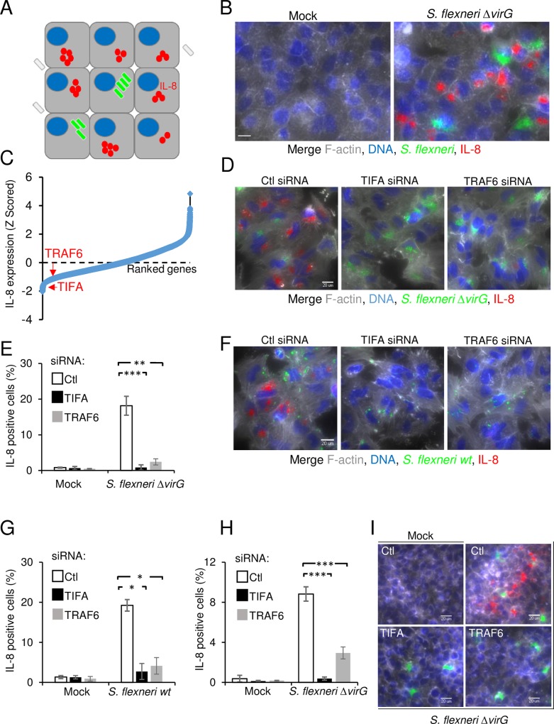
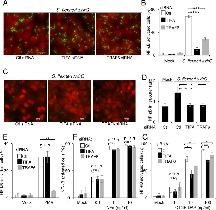
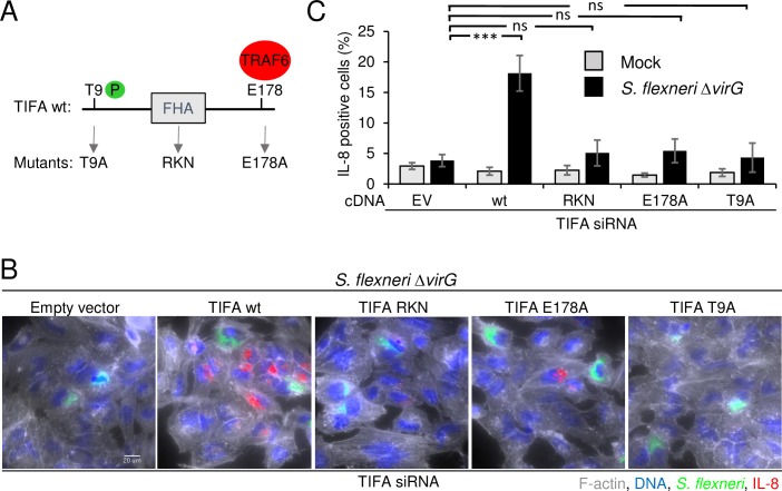
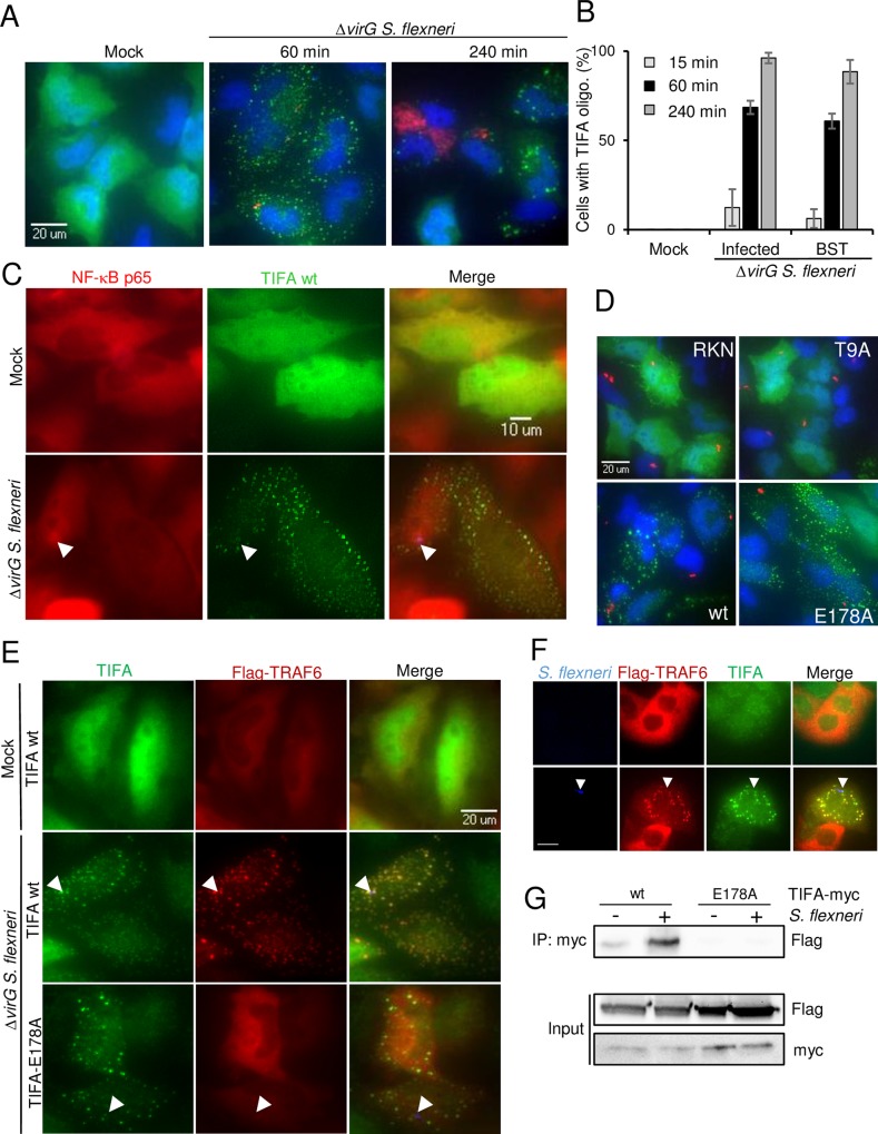
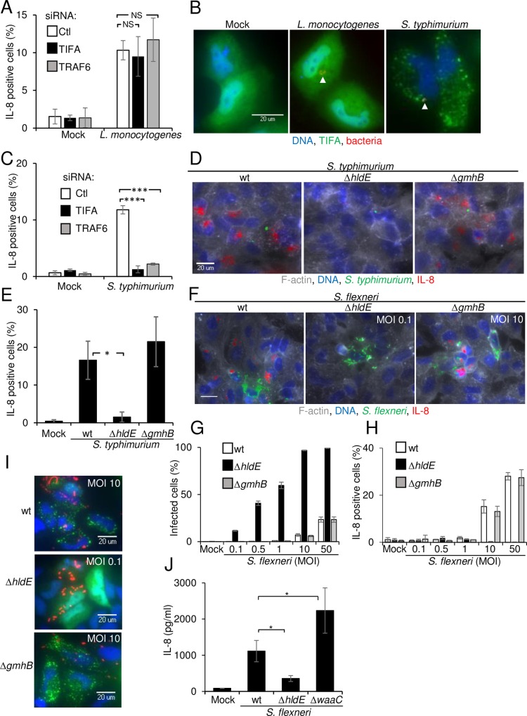
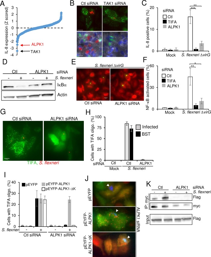
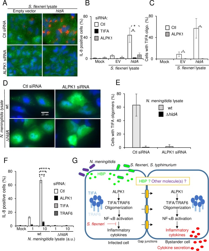
During infection by invasive bacteria, epithelial cells contribute to innate immunity via the local secretion of inflammatory cytokines. These are directly produced by infected cells or by uninfected bystanders via connexin-dependent cell-cell communication. However, the cellular pathways underlying this process remain largely unknown. Here we perform a genome-wide RNA interference screen and identify TIFA and TRAF6 as central players of Shigella flexneri and Salmonella typhimurium-induced interleukin-8 expression. We show that threonine 9 and the forkhead-associated domain of TIFA are necessary for the oligomerization of TIFA in both infected and bystander cells. Subsequently, this process triggers TRAF6 oligomerization and NF-κB activation. We demonstrate that TIFA/TRAF6-dependent cytokine expression is induced by the bacterial metabolite heptose-1,7-bisphosphate (HBP). In addition, we identify alpha-kinase 1 (ALPK1) as the critical kinase responsible for TIFA oligomerization and IL-8 expression in response to infection with S. flexneri and S. typhimurium but also to Neisseria meningitidis. Altogether, these results clearly show that ALPK1 is a master regulator of innate immunity against both invasive and extracellular gram-negative bacteria.


Conflict of interest statement

The authors have declared that no competing interests exist."

Figures

Fig 1
Fig 1. RNAi screen reveals the roles of TIFA and TRAF6 in S. flexneri infection-induced IL-8 expression.
A) Schematic representation of the assay used to monitor IL-8 expression in the S. flexneri infection assay. B) Illustration of the image-based assay developed for the screen. HeLa cells were infected for 3.5 hours with S. flexneri ΔvirG expressing dsRed under the control of the uhpT promoter (green). Cells were stained for F-actin (grey), DNA (blue) and IL-8 (red). Scale bars, 20 μm. C) Genome-wide RNAi screening data of IL-8 expression after S. flexneri infection in HeLa cells. IL-8 measurements were extracted with CellProfiler, Z-scored and ranked. D) Validation of the role of TIFA and TRAF6 in S. flexneri infection-induced IL-8. HeLa cells were transfected with control, TIFA- or TRAF6-targeting siRNAs and infected with S. flexneri ΔvirG for 3.5 hours. Cells were stained as in B. E) Impact of TIFA and TRAF6 depletion on IL-8 expression. Quantification of cells producing IL-8 as shown in D by automated image analysis (see Methods). Data show the mean +/- SD of 3 independent experiments, p**<0.005, p***<0.0005. F) TIFA and TRAF6 control inflammation after wild-type S. flexneri infection of HeLa cells. Cells were treated as in D and infected with wild-type S. flexneri for 3.5 hours. G) Impact of TIFA and TRAF6 depletion on IL-8 expression. Quantification of cells producing IL-8 as shown in F. Data show the mean +/- SD of 3 independent experiments, p*<0.05. H) TIFA and TRAF6 regulate IL-8 expression in HEK293 cells. HEK293 cells were transfected and infected as in D. IL-8 was measured by image analysis. Data correspond to the mean +/- SD of 3 independent experiments, p***<0.0005. I) Images showing the implication of TIFA and TRAF6 in HEK293 cells after infection as quantified in H.
Fig 2
Fig 2. TIFA and TRAF6 control S. flexneri-induced NF-κB activation.
A) TIFA and TRAF6 control S. flexneri-induced NF-κB activation in infected cells. HeLa cells were transfected with control, TIFA- or TRAF6-targeting siRNAs and infected with S. flexneri ΔvirG (green) at MOI 20 for 60 minutes. After fixation, cells were stained for NF-κB p65 (red). B) Quantification of NF-κB translocation in infected cells after depletion of TIFA and TRAF6. NF-κB translocation was quantified by measuring the intensity ratio between the nucleus and the cytoplasm by automated image analysis, defining a threshold ratio and quantifying the fraction of NF-κB positive cells. Data correspond to the mean +/- SD of triplicate wells from a representative of 3 independent experiments, p***<0.0005. C) TIFA and TRAF6 control NF-κB activation both in infected and bystander cells. HeLa cells were treated as in A and infected at a MOI 0.5 (bacteria in green) for 60 minutes. After fixation, cells were stained for NF-κB p65 (red). D) Quantification of NF-κB translocation in bystander cells. The fluorescence intensity ratio between the cytoplasm and the nucleus was measured in bystander cells. Data correspond to the mean +/- SD of 3 independent experiments, p*<0.05. E) Impact of TIFA and TRAF6 depletion on PMA-induced NF-κB activation. After siRNA transfection, HeLa cells were stimulated with PMA (100 ng/ml) for 60 minutes. Data correspond to the mean +/- SD of 3 independent experiments, p**<0.005, ns: non-significant p>0.05. F) Impact of TIFA and TRAF6 depletion on TNFα-induced NF-κB activation. After siRNA transfection, HeLa cells were stimulated for 30 minutes with TNFα at the indicated concentrations. Data correspond to the mean +/- SD of 3 independent experiments, ns: non-significant p>0.05. G) Impact of TIFA and TRAF6 depletion on C12-iE-DAP-induced NF-κB activation. After siRNA transfection, HeLa cells were stimulated for 60 minutes with C12-iE-DAP at the indicated concentrations. Data correspond to the mean +/- SD of 3 independent experiments, non-significant p>0.05, p*<0.05, p***<0.0005.
Fig 3
Fig 3. Residue T9, the FHA domain and residue E178 of TIFA are necessary for IL-8 expression.
A) Schematic representation of wild-type TIFA and the T9, RKN and E178A TIFA mutants. B) Only wild-type TIFA rescues IL-8 expression after siRNA-mediated depletion of TIFA. HeLa cells were transfected for 72 hours with TIFA-targeting siRNA. 24 hours prior infection, cells were transfected with empty vector, wild-type or mutated TIFA cDNA constructs. All TIFA cDNA constructs are TIFA siRNA-resistant. Cells were infected with S. flexneri ΔvirG (green) for 3.5 hours. After fixation, cells were stained for F-actin (grey), DNA (blue) and IL-8 (red). Scale bars, 20 μm. C) Quantification of IL-8 as shown in B. Data correspond to the mean +/- SD of 3 independent experiments, p***<0.0005, ns: non-significant p>0.05.
Fig 4
Fig 4. TIFA and TRAF6 form co-localizing oligomers in infected and bystander cells.
A) TIFA forms large oligomers in infected and bystander cells. HeLa cells were transfected with wild-type TIFA cDNA and infected or not with S. flexneri ΔvirG (red) at MOI 0.5. Cells were stained for TIFA (green) and DNA (blue). B) Quantification of cells showing TIFA oligomers post infection. Cells were treated as in A. Cells showing TIFA punctuates were manually quantified for infected and bystander cells. Graph shows the mean of triplicate wells with a total of n = 130 cells per condition, data representative of 3 experiments. C) TIFA oligomerization occurs within minutes of infection in infected and bystander cells. HeLa cells were transfected with wt TIFA cDNA, infected or not for 15 minutes and co-stained for TIFA (green) and NF-κB p65 (red). Arrows indicate bacteria. D) Localization of wt, T9A, RKN and E178A TIFA mutants. Cells were transfected with wt TIFA or the different mutants, infected for 1 hour and stained as in A. Images are representative of three independent experiments. E) TRAF6 oligomerization is TIFA-dependent. HeLa cells were co-transfected with wild-type TIFA or E178A TIFA and Flag-TRAF6. After infection, cells were stained for TIFA (green) and Flag (red). Arrows indicate S. flexneri. F) Co-localizing TIFA and TRAF6 oligomers after S. flexneri infection in Caco-2 cells. Arrows indicate S. flexneri. Scale bars, 20 μm G) Co-immunoprecipitation of TIFA and TRAF6 after S. flexneri infection. HeLa cells were co-transfected with wt or E178A myc-TIFA and Flag-TRAF6 and infected for 1 hour at MOI 10. Myc IP was blotted with an anti-Flag antibody and the input lysate with anti-Flag and anti-myc antibodies. Data representative of two independent experiments.
Fig 5
Fig 5. Sensing of HBP triggers TIFA/TRAF6-dependent innate immunity.
A) TIFA and TRAF6 are not involved in L. monocytogenes-induced IL-8 production. Cells were transfected with control, TIFA- or TRAF6-targeting siRNAs, infected with L. monocytogenes for 3.5 hours and stained for IL-8. Data show the mean +/- SD of 3 independent experiments, ns: p>0.05. B) S. typhimurium infection induces TIFA oligomers. Hela cells were transfected with wild-type TIFA cDNA, infected with L. monocytogenes or S. typhimurium for 45 minutes and stained for TIFA (green) and DNA (blue). Arrows indicate bacteria (red). C) TIFA and TRAF6 are involved in IL-8 expression after S. typhimurium infection. Cells were transfected as in A, infected with S. typhimurium for 3.5 hours and stained for IL-8. Data show the mean +/- SD of 3 independent experiments, p***<0.0005. D) HBP is required for IL-8 induction after S. typhimurium infection. Cells were infected with wt, ΔhldE or ΔgmhB S. typhimurium (green) and stained for IL-8 (red), F-actin (grey) and DNA (blue). E) Quantification of IL-8 after infection with wt, ΔhldE or ΔgmhB S. typhimurium. Data show the mean +/- SD of 3 independent experiments, p*<0.05. F) HBP is required for IL-8 expression after S. flexneri infection. Cells were infected with wt, ΔhldE or ΔgmhB S. flexneri (green) and stained as in D. Scale bars, 20 μm. G) Comparison of the infection rates after infection with wt, ΔhldE or ΔgmhB S. flexneri at multiple MOIs. Data show the mean +/- SD of triplicate wells, graph representative of 3 independent experiments. H) Quantification of IL-8 after infection with wt, ΔhldE or ΔgmhB S. flexneri. Data show the mean +/- SD of triplicate wells, graph representative of 3 independent experiments. I) TIFA oligomerization is HBP-dependent. Cells were transfected with TIFA cDNA and infected with wt, ΔhldE or ΔgmhB S. flexneri (red). Cells were stained for TIFA (green) and DNA (blue). J) IL-8 secretion of S. flexneri-infected Caco-2 cells is largely HBP-dependent. ELISA assay measuring the secretion of IL-8 after infection of Caco-2 cells. Cells were infected for 6 hours with wt (MOI 400), ΔhldE (MOI 4) or ΔwaaC (MOI 4) S. flexneri. Data correspond to the mean +/- SD of 3 independent experiments, p*<0.05.
Fig 6
Fig 6. ALPK1 controls TIFA-mediated innate immunity during infection with invasive bacteria.
A) Kinome RNAi screening data of IL-8 expression after S. flexneri infection in HeLa cells. IL-8 measurements were extracted with CellProfiler, Z-scored and ranked. B) Silencing TAK1 prevents S. flexneri-induced IL-8 expression but not TIFA oligomerization. Top panels show TIFA in green and S. flexneri in red. Bottom panels show F-actin in grey, DNA in blue, IL-8 in red and S. flexneri in green. Scale bars, 20 μm. C) Silencing ALPK1 inhibits IL-8 expression induced by S. flexneri infection. Cells were transfected with control, TIFA and ALPK1-targeting siRNAs, infected and stained for IL-8. Data correspond to the mean +/- SD of three independent experiments, p**<0.005, p***<0.0005. D) Silencing ALPK1 inhibits S. flexneri-induced IκBα degradation. Lysates of control or infected cells were blotted with IκBα or actin antibodies. E) ALPK1 depletion inhibits S. flexneri-induced NF-κB activation. Cells were transfected with control and ALPK1-targeting siRNAs, infected with S. flexneri ΔvirG (green) and stained for NF-κB p65 (red). Scale bars, 20 μm. F) Silencing ALPK1 inhibits NF-κB activation induced by S. flexneri infection. Cells were transfected as in E, infected for 1h and stained for NF-κB p65. Cells showing NF-κB nuclear translocation were quantified. Data show the mean +/- SD of 3 independent experiments, p**<0.005, p*<0.05. G) Silencing ALPK1 prevents the formation of TIFA oligomers. Cells were transfected as in E and with wt TIFA cDNA. After infection (S. flexneri in red) for 45 minutes, cells were stained for TIFA (green). H) Impact of ALPK1 depletion on the formation of TIFA oligomers. Cells were treated as in G. Cells showing TIFA punctuates were manually quantified (n = 130 cells per condition), BST: bystander. Data show the mean +/- SD of triplicate wells, graph representative of 3 independent experiments. I) Only full length ALPK1 rescues TIFA oligomerization in ALPK1-depleted cells. Cells were transfected with control or ALPK1 siRNAs and then with pEYFP, pEYFP-ALPK1 or pEYFP-ALPK1-ΔK cDNA constructs. The fraction of cells showing TIFA oligomers was manually quantified (n>180 cells). Data show the mean +/- SD of three independent experiments. J) Images illustrating the oligomerization of TIFA in the rescue experiment as described in I. TIFA is show in red, YFP-ALPK1 in green and S. flexneri in blue. Arrows indicate bacteria. K) ALPK1 controls the TIFA-TRAF6 interaction. Co-immunoprecipitation of TIFA-myc after S. flexneri infection. HeLa cells were transfected with control or ALPK1 siRNAs and with myc-TIFA and Flag-TRAF6. They were then infected for 1 hour at MOI 10. Myc IPs were blotted with anti-Flag and anti-myc antibodies and input lysates with an anti-Flag antibody.
Fig 7
Fig 7. ALPK1 is a master regulator of HBP-induced innate immunity.
A) ALPK1 controls IL-8 expression and the formation of TIFA oligomers induced by HBP. Cells were transfected with control or ALPK1 siRNAs and incubated with lysates from S. flexneri containing pUC19 empty vector (EV) or expressing hldA from pUC19. They were stained for IL-8 (red), F-actin (green) and DNA (blue). Scale bars, 20 μm. B) Quantification of data shown in F. Data show the mean +/- SD of 3 independent experiments, p*<0.05. C) Silencing ALPK1 prevents HBP-induced TIFA oligomerization. Cells were transfected as in A and with wt TIFA. After incubation with lysates from S. flexneri containing pUC19 or pUC19-hldA, cells were stained for TIFA. Cells showing TIFA punctuates were manually quantitated (n = 105 cells per condition). Data show the mean +/- SD of 3 independent experiments, p*<0.05. D) Silencing ALPK1 abrogates the formation of TIFA oligomers induced by N. meningitidis HBP. HeLa cells were transfected with control or ALPK1-targeting siRNA and then with wt TIFA cDNA. After incubation with lysates from wt or ΔhldA N. meningitidis, cells were stained for TIFA (green) and DNA (blue). E) Impact of ALPK1 depletion on TIFA oligomerization. Cells were treated as in D. The fraction of cells showing TIFA punctuates was manually quantitated with n = 130 cells per condition. Data show the mean +/- SD of triplicate wells and the graph is a representative of 3 independent experiments. F) Silencing ALPK1 abrogates the production of IL-8 induced by N. meningitidis lysates. Cells were transfected with control, TIFA, TRAF6 or ALPK1-targeting siRNA. Cells were then incubated with lysates from wt or ΔhldA N. meningitidis and stained for IL-8. Data show the mean +/- SD of triplicate well and the graph is a representative of 3 independent experiments, p***<0.0005. G) Schematic illustration of the ALPK1/TIFA/TRAF6 pathway controlling IL-8 expression after infection by gram-negative bacteria.

References

    1. Schroeder GN, Hilbi H. Molecular pathogenesis of Shigella spp.: controlling host cell signaling, invasion, and death by type III secretion. Clin Microbiol Rev. 2008;21(1):134–56. Epub 2008/01/19. PubMed Central PMCID: PMC2223840. 10.1128/CMR.00032-07 - DOI - PMC - PubMed
    1. Cornelis GR. The type III secretion injectisome. Nat Rev Microbiol. 2006;4(11):811–25. Epub 2006/10/17. 10.1038/nrmicro1526 - DOI - PubMed
    1. Girardin SE, Tournebize R, Mavris M, Page AL, Li X, Stark GR, et al. CARD4/Nod1 mediates NF-kappaB and JNK activation by invasive Shigella flexneri. EMBO Rep. 2001;2(8):736–42. Epub 2001/07/21. PubMed Central PMCID: PMC1083992. 10.1093/embo-reports/kve155 - DOI - PMC - PubMed
    1. Girardin SE, Boneca IG, Carneiro LA, Antignac A, Jehanno M, Viala J, et al. Nod1 detects a unique muropeptide from gram-negative bacterial peptidoglycan. Science. 2003;300(5625):1584–7. Epub 2003/06/07. 10.1126/science.1084677 - DOI - PubMed
    1. Hasegawa M, Fujimoto Y, Lucas PC, Nakano H, Fukase K, Nunez G, et al. A critical role of RICK/RIP2 polyubiquitination in Nod-induced NF-kappaB activation. EMBO J. 2008;27(2):373–83. Epub 2007/12/15. PubMed Central PMCID: PMC2234345. 10.1038/sj.emboj.7601962 - DOI - PMC - PubMed

MeSH terms