

Arctic ground squirrel hippocampus tolerates oxygen glucose deprivation independent of hibernation season even when not hibernating and after ATP depletion, acidosis, and glutamate efflux
- PMID: 28222226
- PMCID: PMC5479730
- DOI: 10.1111/jnc.13996
Arctic ground squirrel hippocampus tolerates oxygen glucose deprivation independent of hibernation season even when not hibernating and after ATP depletion, acidosis, and glutamate efflux
Abstract
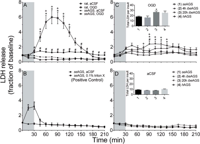
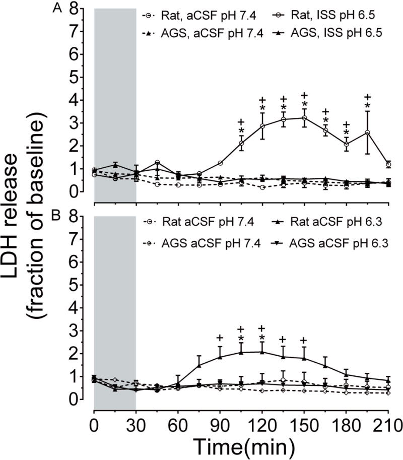
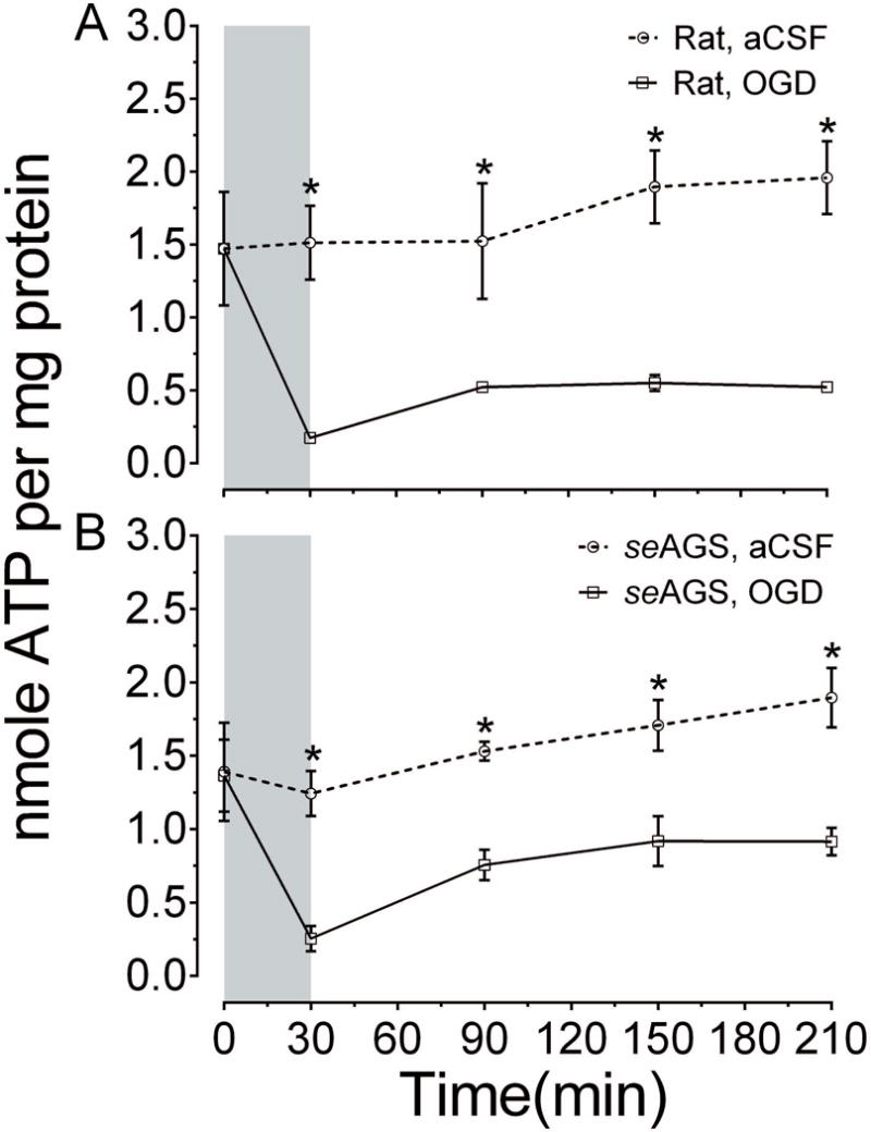
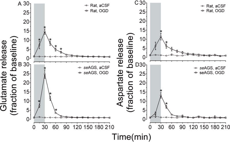
Cerebral ischemia/reperfusion (I/R) triggers a cascade of uncontrolled cellular processes that perturb cell homeostasis. The arctic ground squirrel (AGS), a seasonal hibernator resists brain damage following cerebral I/R caused by cardiac arrest and resuscitation. However, it remains unclear if tolerance to I/R injury in AGS depends on the hibernation season. Moreover, it is also not clear if events such as depletion of ATP, acidosis, and glutamate efflux that are associated with anoxic depolarization are attenuated in AGS. Here, we employ a novel microperfusion technique to test the hypothesis that tolerance to I/R injury modeled in an acute hippocampal slice preparation in AGS is independent of the hibernation season and persists even after glutamate efflux. Acute hippocampal slices were harvested from summer euthermic AGS, hibernating AGS, and interbout euthermic AGS. Slices were subjected to oxygen glucose deprivation (OGD), an in vitro model of I/R injury to determine cell death marked by lactate dehydrogenase (LDH) release. ATP was assayed using ENLITEN ATP assay. Glutamate and aspartate efflux was measured using capillary electrophoresis. For acidosis, slices were subjected to pH 6.4 or ischemic shift solution (ISS). Acute hippocampal slices from rats were used as a positive control, susceptible to I/R injury. Our results indicate that when tissue temperature is maintained at 36°C, hibernation season has no influence on OGD-induced cell death in AGS hippocampal slices. Our data also show that tolerance to OGD in AGS hippocampal slices occurs despite loss of ATP and glutamate release, and persists during conditions that mimic acidosis and ionic shifts, characteristic of cerebral I/R. Read the Editorial Comment for this article on page 10.
Keywords: in vitro; hibernation; hippocampal brain slices; ischemia; lactate dehydrogenase release; oxygen glucose deprivation.
© 2017 International Society for Neurochemistry.
Conflict of interest statement
The authors report no conflict of interest. Figure 4 (A–B) was published in preliminary form in book chapter (Drew
Figures
Comment in
-
A new insight into the ability to resist Ischemic brain injury: Does hibernation matter?: An Editorial comment for 'Arctic ground squirrel hippocampus tolerates oxygen glucose deprivation independent of hibernation season even when not hibernating and after ATP depletion, acidosis and glutamate efflux'.J Neurochem. 2017 Jul;142(1):10-13. doi: 10.1111/jnc.14022. Epub 2017 May 24. J Neurochem. 2017. PMID: 28542925
References
-
- Benveniste H, Drejer J, Schousboe A, Diemer NH. Elevation of the extracellular concentrations of glutamate and aspartate in rat hippocampus during transient cerebral ischemia monitored by intracerebral microdialysis. Journal of neurochemistry. 1984;43:1369–1374. - PubMed
-
- Bickler PE, Buck LT. Hypoxia tolerance in reptiles, amphibians, and fishes: life with variable oxygen availability. Annual review of physiology. 2007;69:145–170. - PubMed
-
- Bortner CD, Cidlowski JA. The role of apoptotic volume decrease and ionic homeostasis in the activation and repression of apoptosis. Pflugers Archiv : European journal of physiology. 2004;448:313–318. - PubMed
Publication types
MeSH terms
Substances
Grants and funding
LinkOut - more resources
Full Text Sources
Other Literature Sources
Molecular Biology Databases
