

Identification of somatic and germ-line DICER1 mutations in pleuropulmonary blastoma, cystic nephroma and rhabdomyosarcoma tumors within a DICER1 syndrome pedigree
- PMID: 28222777
- PMCID: PMC5320664
- DOI: 10.1186/s12885-017-3136-5
Identification of somatic and germ-line DICER1 mutations in pleuropulmonary blastoma, cystic nephroma and rhabdomyosarcoma tumors within a DICER1 syndrome pedigree
Abstract
Background: DICER1 syndrome is a pediatric cancer predisposition condition causing a variety of tumor types in children and young adults. In this report we studied a family with two relatives presenting a variety of neoplastic conditions at childhood.
Methods: Germ-line mutation screening of the complete coding region of the DICER1 gene in genomic DNA from the proband was performed. The presence of somatic DICER1 mutation and further alterations in driver genes was investigated in genomic DNA obtained from available tumor samples.
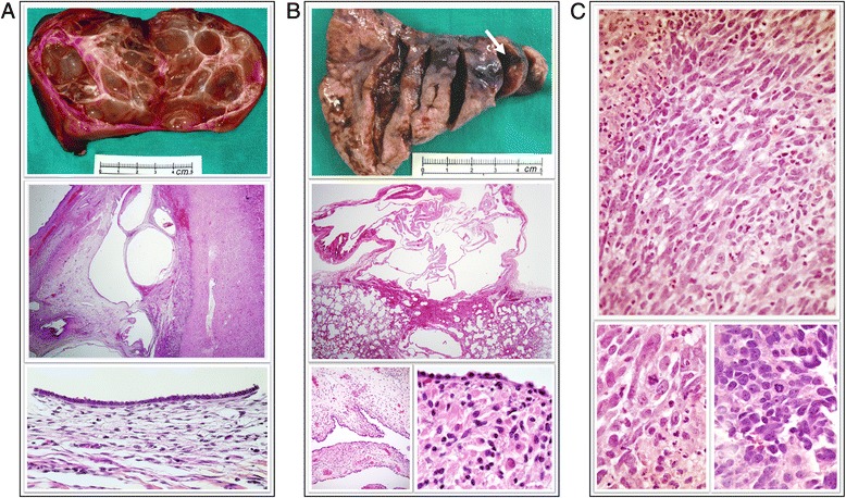
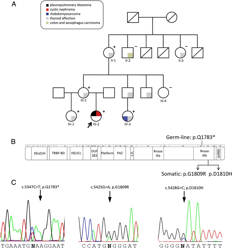
Results: A nonsense germ-line mutation in DICER1 causing a truncated protein at the IIIb domain level was identified segregating within a family including two affected relatives who developed in one case cystic nephroma and pleuropulmonary blastoma, and rhabdomyosarcoma and multinodular goiter in the other. Additional in trans DICER1 missense somatic mutations in the IIIb DICER1 domain were found both in the cystic nephroma and in the rhabdomyosarcoma, suggesting that neoplasms in this family might arise from the unusual two-hit mechanism for DICER-derived tumorigenesis in which after the presence of a truncated constitutive protein, a neomorphic DICER1 activity is somatically adquired. Additional genetic alterations, such as TP53 mutations, were identified in the rhabdomyosarcoma.
Conclusions: Besides DICER1 loss of standard activity, oncogenic cooperation of other genes, as mutated TP53, may involve developing higher grade tumors within this syndrome. Given the broad clinical spectrum that may arise, genetic counseling and close surveillance must be offered to all family members at risk of DICER1 syndrome.
Keywords: DICER1 mutations; DICER1 syndrome.
Figures
Similar articles
-
Embryonal rhabdomyosarcoma in a patient with a heterozygous frameshift variant in the DICER1 gene and additional manifestations of the DICER1 syndrome.Fam Cancer. 2017 Jul;16(3):401-405. doi: 10.1007/s10689-016-9958-5. Fam Cancer. 2017. PMID: 27896549
-
DICER1 pleuropulmonary blastoma familial tumour predisposition syndrome: What the paediatric urologist needs to know.J Pediatr Urol. 2016 Feb;12(1):5-10. doi: 10.1016/j.jpurol.2015.08.012. Epub 2015 Sep 26. J Pediatr Urol. 2016. PMID: 26454454 Review.
-
DICER1 mutation and tumors associated with a familial tumor predisposition syndrome: practical considerations.Fam Cancer. 2017 Apr;16(2):291-294. doi: 10.1007/s10689-016-9949-6. Fam Cancer. 2017. PMID: 27830405
-
Biallelic DICER1 mutations in sporadic pleuropulmonary blastoma.Cancer Res. 2014 May 15;74(10):2742-9. doi: 10.1158/0008-5472.CAN-13-2470. Epub 2014 Mar 27. Cancer Res. 2014. PMID: 24675358
-
The co-occurrence of an ovarian Sertoli-Leydig cell tumor with a thyroid carcinoma is highly suggestive of a DICER1 syndrome.Virchows Arch. 2016 May;468(5):631-6. doi: 10.1007/s00428-016-1922-0. Epub 2016 Mar 16. Virchows Arch. 2016. PMID: 26983701 Review.
Cited by
-
New roles for Dicer in the nucleolus and its relevance to cancer.Cell Cycle. 2017 Sep 17;16(18):1643-1653. doi: 10.1080/15384101.2017.1361568. Epub 2017 Aug 28. Cell Cycle. 2017. PMID: 28846478 Free PMC article. Review.
-
Rhabdomyosarcoma of the Cervix in a Post-Menopausal Woman-An Unparalleled Phenomenon.Int J Environ Res Public Health. 2021 Jul 24;18(15):7851. doi: 10.3390/ijerph18157851. Int J Environ Res Public Health. 2021. PMID: 34360144 Free PMC article.
-
DICER1 Mutational Spectrum in Intracranial CNS-Neoplasias-A Review and a Report from the CNS-InterREST GPOH Study Center.Cancers (Basel). 2025 Apr 30;17(9):1513. doi: 10.3390/cancers17091513. Cancers (Basel). 2025. PMID: 40361440 Free PMC article. Review.
-
DICER1 Syndrome: DICER1 Mutations in Rare Cancers.Cancers (Basel). 2018 May 15;10(5):143. doi: 10.3390/cancers10050143. Cancers (Basel). 2018. PMID: 29762508 Free PMC article. Review.
-
DICER1: The Argonaute Endonuclease Family Member and Its Role in Pediatric and Youth Pathology.Biology (Basel). 2025 Jan 18;14(1):93. doi: 10.3390/biology14010093. Biology (Basel). 2025. PMID: 39857323 Free PMC article. Review.
References
-
- Gómez de la Torre R, Enguix Armada A, García L, Otero J. Thyroid nodule disease in a previously endemic goiter area. An Med Interna. 1993;10:487–9. - PubMed
Publication types
MeSH terms
Substances
Supplementary concepts
LinkOut - more resources
Full Text Sources
Other Literature Sources
Research Materials
Miscellaneous
