Review
. 2017 May:58:89-114.
doi: 10.1016/j.preteyeres.2017.02.001. Epub 2017 Feb 20.

Characterizing the "POAGome": A bioinformatics-driven approach to primary open-angle glaucoma

Affiliations
Review

Characterizing the "POAGome": A bioinformatics-driven approach to primary open-angle glaucoma

Ian D Danford et al. Prog Retin Eye Res. 2017 May.

Abstract

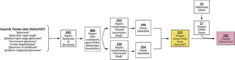
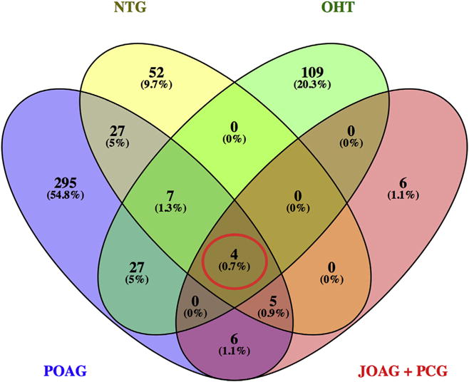
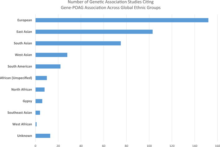
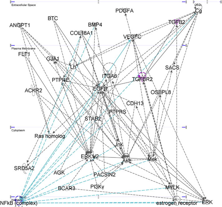
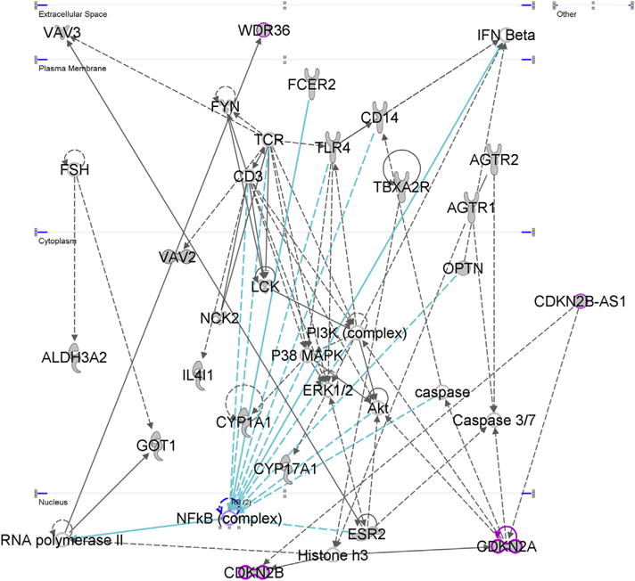
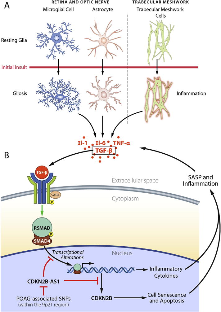
Primary open-angle glaucoma (POAG) is a genetically, physiologically, and phenotypically complex neurodegenerative disorder. This study addressed the expanding collection of genes associated with POAG, referred to as the "POAGome." We used bioinformatics tools to perform an extensive, systematic literature search and compiled 542 genes with confirmed associations with POAG and its related phenotypes (normal tension glaucoma, ocular hypertension, juvenile open-angle glaucoma, and primary congenital glaucoma). The genes were classified according to their associated ocular tissues and phenotypes, and functional annotation and pathway analyses were subsequently performed. Our study reveals that no single molecular pathway can encompass the pathophysiology of POAG. The analyses suggested that inflammation and senescence may play pivotal roles in both the development and perpetuation of the retinal ganglion cell degeneration seen in POAG. The TGF-β signaling pathway was repeatedly implicated in our analyses, suggesting that it may be an important contributor to the manifestation of POAG in the anterior and posterior segments of the globe. We propose a molecular model of POAG revolving around TGF-β signaling, which incorporates the roles of inflammation and senescence in this disease. Finally, we highlight emerging molecular therapies that show promise for treating POAG.

Keywords: Gliosis; NFκB; Neurodegeneration; Neuroinflammation; Primary open-angle glaucoma (POAG); Senescence; Senescence-associated secretory phenotype (SASP); TGF-β.


Figures

Fig. 1
Fig. 1
Schematic showing the anatomy of POAG. Sub-image A reflects the abnormally high pressure transmitted to the posterior segment of the globe containing the retina and optic disc where RGCs and their axons reside. Sub image B reflects aqueous outflow pathways.
Fig. 2
Fig. 2
Flowchart summarizing the findings of our comprehensive literature review of 904 studies (892 from DisGeNET and 12 from additional GWAS). 542 genes were confirmed to be associated with POAG and related phenotypes.
Fig. 3
Fig. 3
Venn diagram summarizing POAG-associated genes from 891 studies. Among the 522 genes found to be associated with a POAG phenotype, 4 genes (MYOC, CYP1B1, OPTN, WDR36–red circle) were found to be associated with all POAG phenotypes. [POAG = Primary Open Angle Glaucoma, NTG = Normal Tension Glaucoma, OHT = Ocular Hypertension, JOAG = Juvenile Open Angle Glaucoma, PCG = Primary Congenital Glaucoma].
Fig. 4
Fig. 4
This summarizes the number of studies with a confirmed gene-glaucoma association with subjects from several different ethnic populations. We binned ethnic groups roughly according to the United Nations geoscheme. The vast majority of studies were performed in European, Chinese, Japanese, and Indian populations.
Fig. 5
Fig. 5
Top associated network function for ONH astrocyte genes: “Cardiovascular System Development and Function, Cellular Movement, Cellular Development.” Many of the genes converge onto the NFκB complex (within the nucleus) suggesting that inflammation and cell survival are important biological processes within ONH astrocytes during POAG’s pathogenesis.
Fig. 6
Fig. 6
“Cell Death and Survival, Nervous System Development and Function, Tissue Morphology.” This was the top most associated network according to IPA core analysis of the RGC gene input set. Molecules within the ERK and MAP families seem to play an important role, as do the Ap1 and Creb transcription factors. The ERK1/2 family is highlighted above with the blue arrows highlighting interactions with its immediate neighbors, including VEGFA, TNF, NGF, and NRTK2.
Fig. 7
Fig. 7
“Tissue Development, Cardiovascular System Development and Function, Organismal Development.” This was the top most associated network according to IPA core analysis of the TM gene input set which included the microarray study by Fan et al. The TGF-β signaling pathway played an important role in this network.
Fig. 8
Fig. 8
“Cell Cycle, Skeletal and Muscular System Development and Function, Cellular Development.” This was the top most associated network according to IPA core analysis of the POAG gene input set which included all 360 genes from functional and genetic association studies investigating the POAG phenotype.
Fig. 9
Fig. 9
“Free Radical Scavenging, Glomerular Injury, Organismal Injury and Abnormalities.” This was the second most associated network according to IPA core analysis of the NTG gene input set which included all 95 genes from functional and genetic association studies investigating the NTG phenotype. We highlighted CDKN2A, CDKN2B, and CDKN2B-AS1 in magenta, all of which have been highlighted in POAG GWAS’. Other important POAG GWAS loci in this network include TLR4, WDR36, and OPTN.
Fig. 10
Fig. 10
TGF-β and the senescent-inflammatory phenotype in action during POAG. In panel A of the figure we see important cells of the posterior (astrocytes and microglia) and anterior segments (TM cells) of the globe. In healthy eyes, these cells are in a quiescent state. However, an initial insult, such as elevated IOP, triggers inflammatory (or gliotic) responses from these cells causing the release of inflammatory cytokines, including TGF-β. In panel B we see TGF-β interaction with its receptor on the surface of a non-descript cell, which could represent other glia, TM cells, or even RGCs. This triggers SMAD activation which eventually results in the transcription of other inflammatory cytokines as well as senescence-inducing molecules such as CDKN2B. Senescence also contributes to the inflammatory milieu through the SASP. CDKN2B-AS1 may modulate inflammation and senescence by inhibitory interactions with the SMAD complex or CDKN2B directly–however the exact mechanism of action of CDKN2B-AS1 is still unclear. SNPs in the 9p21 region which are associated with POAG may impair the function or expression of CDKN2B-AS1 thus contributing to development and progression of the senescent-inflammatory phenotype.

Similar articles

Cited by

References

    1. Abu-Amero K, Kondkar AA, Chalam KV. An updated review on the genetics of primary open angle glaucoma. Int J Mol Sci. 2015;16:28886–28911. - PMC - PubMed
    1. Acott TS, Kelley MJ. Extracellular matrix in the trabecular meshwork. Exp Eye Res. 2008;86:543–561. - PMC - PubMed
    1. Aga M, Bradley JM, Wanchu R, Yang Y-f, Acott TS, Keller KE. Differential effects of caveolin-1 and -2 knockdown on aqueous outflow and altered extracellular matrix turnover in caveolin-silenced trabecular meshwork cells. Invest Ophthalmol Vis Sci. 2014;55:5497–5509. - PMC - PubMed
    1. AGIS Investigators. The advanced glaucoma intervention study (AGIS): 7. The relationship between control of intraocular pressure and visual field deterioration The AGIS investigators. Am J Ophthalmol. 2000;130:429–440. - PubMed
    1. Alm A, Nilsson SF. Uveoscleral outflow–a review. Exp Eye Res. 2009;88:760–768. - PubMed

Publication types

MeSH terms