. 2017 Apr 7;292(14):5655-5664.
doi: 10.1074/jbc.M116.768374. Epub 2017 Feb 21.

Peptide recognition by heterochromatin protein 1 (HP1) chromoshadow domains revisited: Plasticity in the pseudosymmetric histone binding site of human HP1

Affiliations

Peptide recognition by heterochromatin protein 1 (HP1) chromoshadow domains revisited: Plasticity in the pseudosymmetric histone binding site of human HP1

Yanli Liu et al. J Biol Chem. .

Abstract

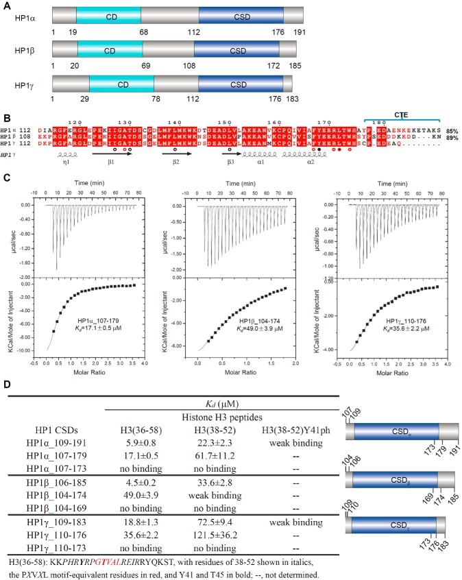
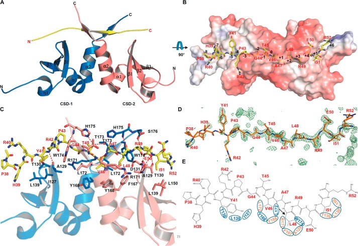
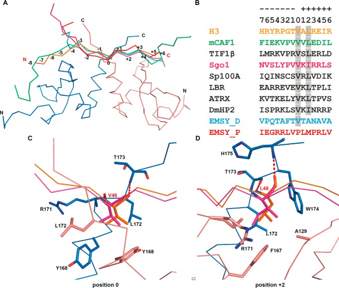
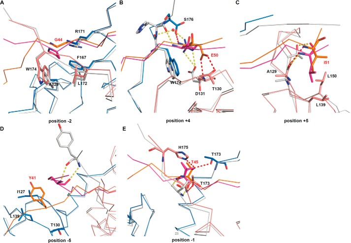
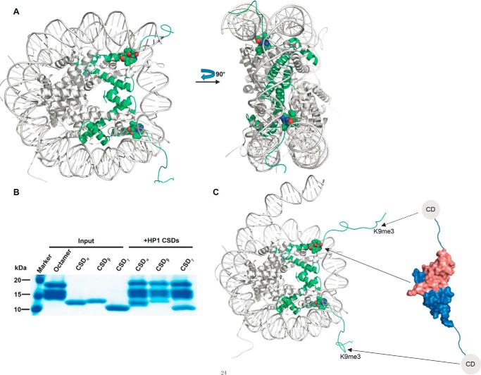
Heterochromatin protein 1 (HP1), a highly conserved non-histone chromosomal protein in eukaryotes, plays important roles in the regulation of gene transcription. Each of the three human homologs of HP1 includes a chromoshadow domain (CSD). The CSD interacts with various proteins bearing the PXVXL motif but also with a region of histone H3 that bears the similar PXXVXL motif. The latter interaction has not yet been resolved in atomic detail. Here we demonstrate that the CSDs of all three human HP1 homologs have comparable affinities to the PXXVXL motif of histone H3. The HP1 C-terminal extension enhances the affinity, as does the increasing length of the H3 peptide. The crystal structure of the human HP1γ CSD (CSDγ) in complex with an H3 peptide suggests that recognition of H3 by CSDγ to some extent resembles CSD-PXVXL interaction. Nevertheless, the prolyl residue of the PXXVXL motif appears to play a role distinct from that of Pro in the known HP1β CSD-PXVXL complexes. We consequently generalize the historical CSD-PXVXL interaction model and expand the search scope for additional CSD binding partners.

Keywords: chromatin regulation; chromatin structure; heterochromatin; histone; peptide interaction.


Conflict of interest statement

The authors declare that they have no conflicts of interest with the contents of this article

Figures

Figure 1.
Figure 1.
CSDs of HP1α/β/γ similarly bind to H3(36–58). A, schematic representation of the domain structure of human HP1 homologs. B, sequence alignment of human CSDs with structural annotation. Secondary structure elements and residue numbers of the CSDγ are indicated below or above the sequence alignment. Residues interacting with certain positions of the H3 peptide are marked as follows: position 0 (closed black circles), positions +2 and −2 (open red circles), and positions +5 and −5 (open black circles). The identities of CSDs of HP1α and HP1β to HP1γ are indicated at the end of the associated sequence. The alignments were constructed with ClustalW (56) and refined with ESPript (57). C, ITC binding curves of histone H3 peptide H3(36–58) to CSDα/β/γ. D, ITC binding affinities of histone H3 peptides to CSDα/β/γ. All Kd values were calculated from a single measurement, and errors were estimated by curve fitting. A schematic representation of the recombinant HP1 constructs expressed in E. coli for this assay is shown on the right.
Figure 2.
Figure 2.
Recognition of the H3(38–52) peptide by CSDγ. A, overall structure of CSDγ-H3 complex. CSDγ dimer and H3 peptide are shown in schematic mode and colored in marine blue (CSD-1), salmon (CSD-2), and yellow (H3). B, surface representation of CSDγ-H3 complex. The electrostatic potential surface was calculated with APBS (58) using coordinates prepared with PDB entry 2PQR (59) at a pH setting of 7 and colored in a red-blue spectrum in the ±7kT/e range. A model of the histone peptide is shown in yellow sticks. C, detailed interactions of the histone H3 peptide with CSDγ. The H3 peptide and H3-interacting CSD residues are shown in stick mode. Hydrogen bonds are shown as red dashes. CSDγ residues are labeled in black, and H3 residues are labeled in red. H3 positions <−3 were poorly resolved by electron density. D, electron density for the H3 peptide. Omit map coefficients were calculated with PHENIX, and a slab of the mFo − DFc map was contoured in PyMOL at level 3. E, schematic view of how hydrophobic pockets of the CSDγ homodimer recognize H3 residues. Interacting residues of CSDγ are contained within ovals and labeled in colors corresponding to CSDs (A). Molecular representations were generated with PyMOL.
Figure 3.
Figure 3.
Structural comparison of different CSD-PXVXL complexes. A, structural superimposition of CSDγ-H3 complex (PDB code 5T1I (this work); peptide colored in orange) with the CSDβ-CAF1 complex (PDB code 1S4Z; green), the CSDβ-EMSY complex (PDB code 2FMM, ENT-distal site (cyan), and ENT-proximal site (red)), and the CSDβ-Sgo1 complex (PDB code 3Q6S; pink). For clarity, only the CSDγ is shown in a ribbon and colored as in Fig. 2A because the structures of CSDγ and CSDβ are highly conserved. B, sequence alignment of canonical and variant PXVXL motifs, adapted from Lechner et al. (32). The peptides are colored as in A, and conserved interacting residues are highlighted in gray. EMSY_D and EMSY_P represent the ENT-distal and -proximal site peptides of EMSY, respectively (28). C and D, highly conserved binding pockets of position 0 (residue V/P) (C) and position +2 (residue L/I) (D), respectively. CSDβ-Sgo1 complex is taken as a representative of CSDβ-PXVXL complexes. For clarity, only the interaction-associated residues in CSDγ-H3 complex are shown.
Figure 4.
Figure 4.
Partially conserved and specific interactions of CSD-PXVXL complexes. A–C, partially conserved interactions of different CSD-PXVXL complexes. D and E, specific interactions of the CSDγ-H3 complex. The CSDβ-Sgo1 complex is taken as a representative of CSDβ-PXVXL complexes and colored in gray. The interaction-associated residues in both complexes are shown, and only the residues of the CSDγ-H3 complex are labeled for clarity. Red and yellow dashed lines represent hydrogen bonds in CSDγ-H3 and CSDβ-Sgo1 complexes, respectively.
Figure 5.
Figure 5.
A proposed working model of the interaction between HP1 and histone H3 within the nucleosome. A, location of CSD binding site within the nucleosome (PDB code 1KX5). Histone H3 is colored in green, and the GTVAL motif is shown in a sphere model. B, CSDs bind to the histone H3 in the context of the octamer. Tag-removed HP1 CSDs are pulled down by His-tagged octamer. C, possible model of the interaction between HP1 and histone H3 within the nucleosome.

References

    1. Will H., and Bautz E. K. F. (1980) Immunological identification of a chromocenter-associated protein in polytene chromosomes of Drosophila. Exp. Cell Res. 125, 401–410 - PubMed
    1. Chandra T., and Narita M. (2013) High-order chromatin structure and the epigenome in SAHFs. Nucleus 4, 23–28 - PMC - PubMed
    1. Li Y., Kirschmann D. A., and Wallrath L. L. (2002) Does heterochromatin protein 1 always follow code? Proc. Natl. Acad. Sci. U.S.A. 99, 16462–16469 - PMC - PubMed
    1. Eissenberg J. C., and Elgin S. C. (2000) The HP1 protein family: getting a grip on chromatin. Curr. Opin. Genet. Dev. 10, 204–210 - PubMed
    1. Maison C., and Almouzni G. (2004) HP1 and the dynamics of heterochromatin maintenance. Nat. Rev. Mol. Cell Biol. 5, 296–304 - PubMed

MeSH terms

Substances

LinkOut - more resources