. 2017 Mar 7;114(10):E1968-E1976.
doi: 10.1073/pnas.1616070114. Epub 2017 Feb 21.

Serum-borne bioactivity caused by pulmonary multiwalled carbon nanotubes induces neuroinflammation via blood-brain barrier impairment

Affiliations

Serum-borne bioactivity caused by pulmonary multiwalled carbon nanotubes induces neuroinflammation via blood-brain barrier impairment

Mario J Aragon et al. Proc Natl Acad Sci U S A. .

Abstract

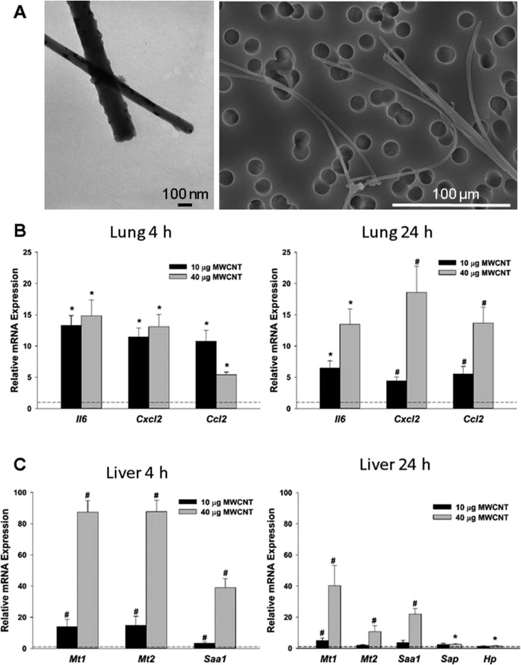
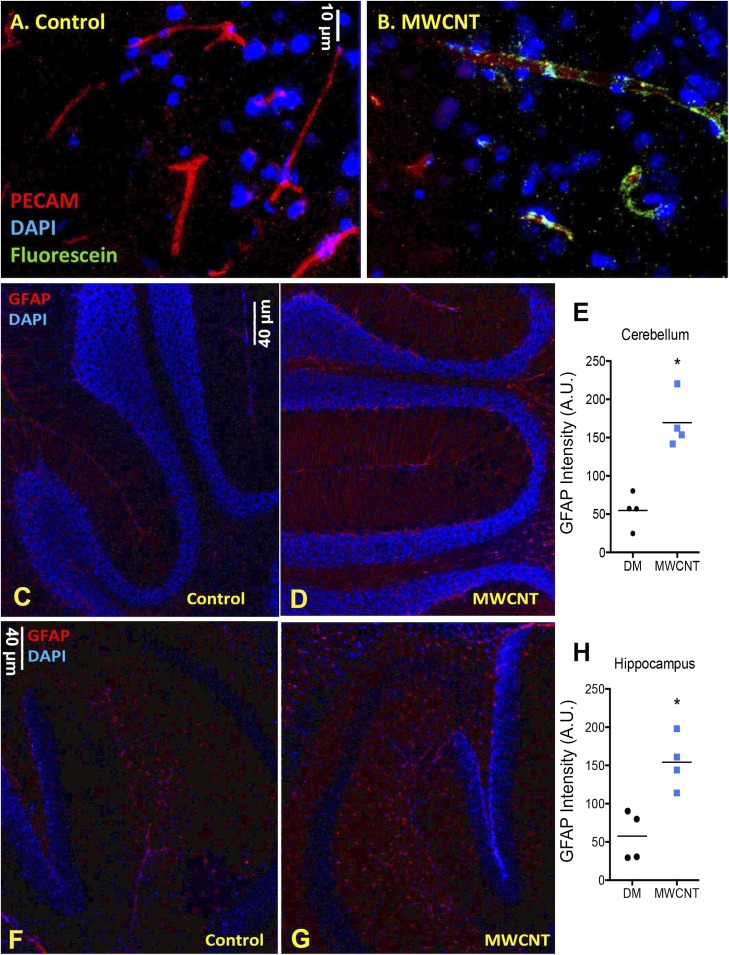
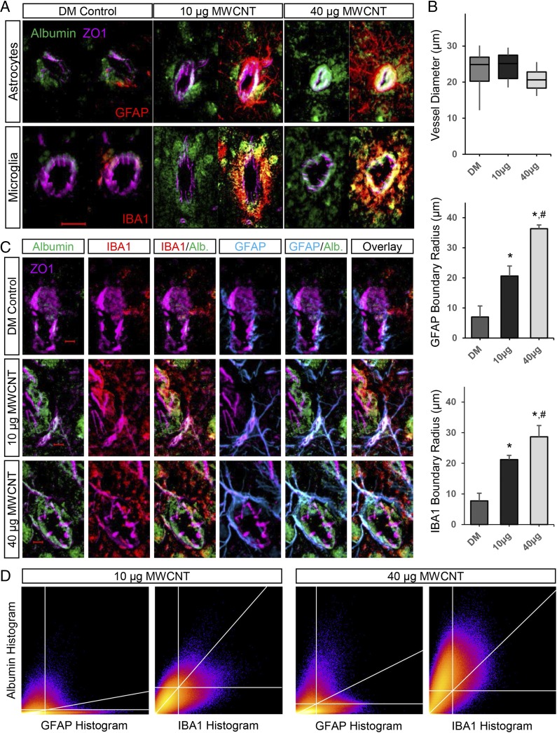
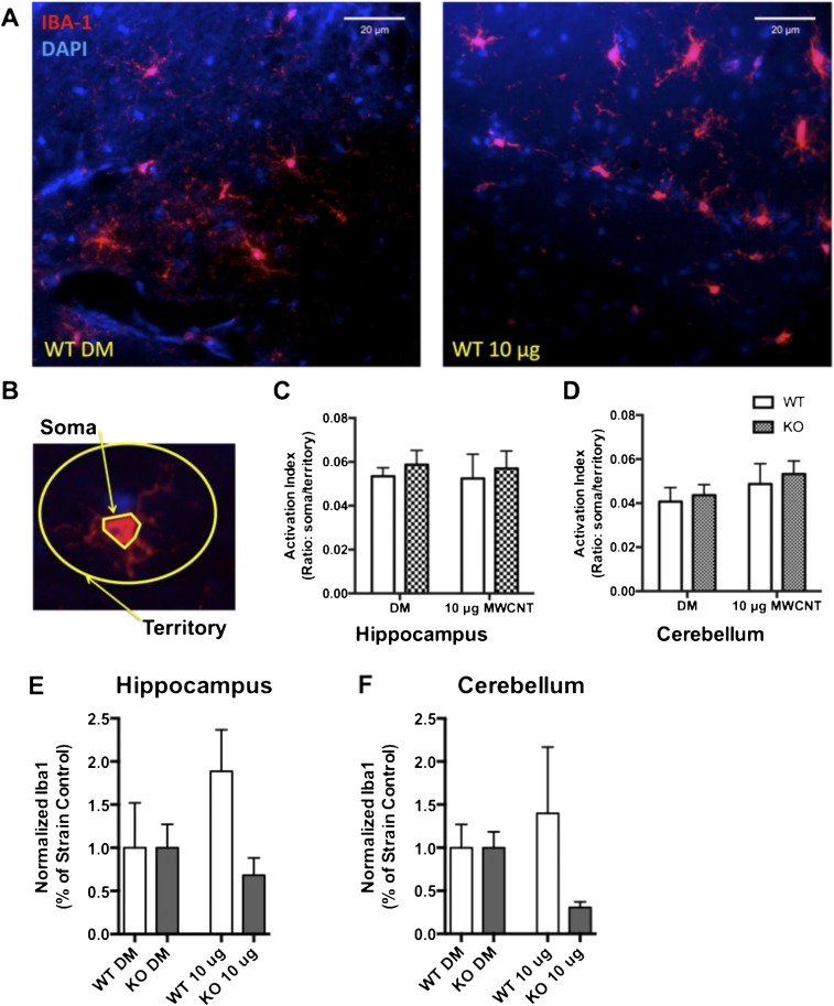
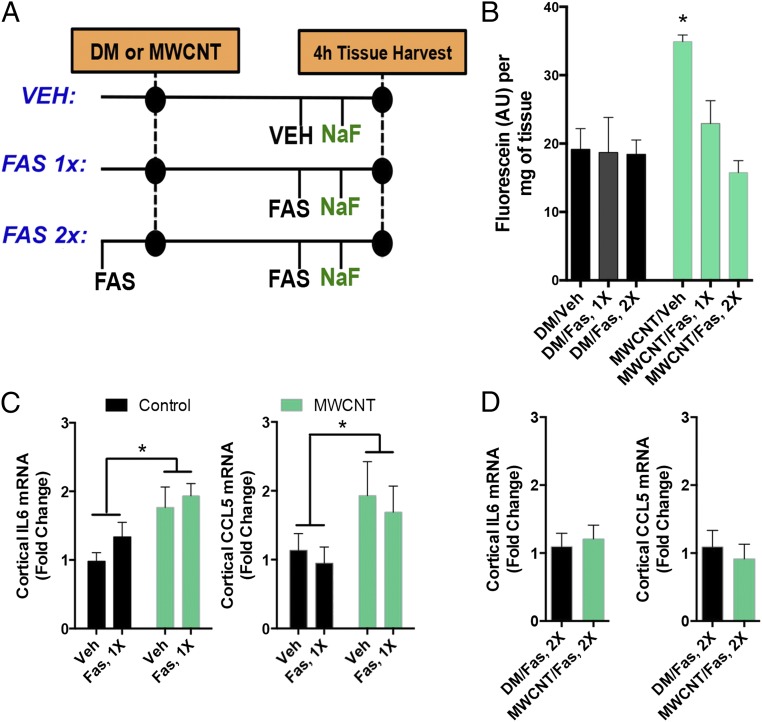
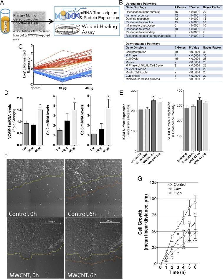
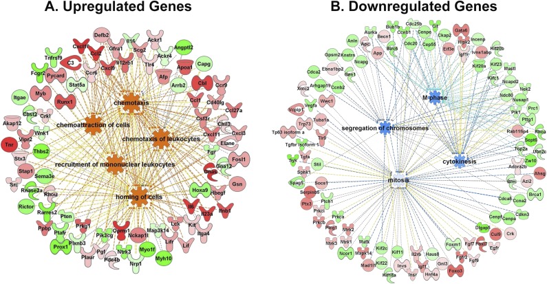
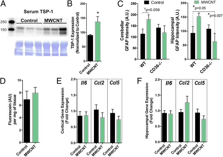
Pulmonary exposure to multiwalled carbon nanotubes (MWCNTs) causes indirect systemic inflammation through unknown pathways. MWCNTs translocate only minimally from the lungs into the systemic circulation, suggesting that extrapulmonary toxicity may be caused indirectly by lung-derived factors entering the circulation. To assess a role for MWCNT-induced circulating factors in driving neuroinflammatory outcomes, mice were acutely exposed to MWCNTs (10 or 40 µg/mouse) via oropharyngeal aspiration. At 4 h after MWCNT exposure, broad disruption of the blood-brain barrier (BBB) was observed across the capillary bed with the small molecule fluorescein, concomitant with reactive astrocytosis. However, pronounced BBB permeation was noted, with frank albumin leakage around larger vessels (>10 µm), overlain by a dose-dependent astroglial scar-like formation and recruitment of phagocytic microglia. As affirmed by elevated inflammatory marker transcription, MWCNT-induced BBB disruption and neuroinflammation were abrogated by pretreatment with the rho kinase inhibitor fasudil. Serum from MWCNT-exposed mice induced expression of adhesion molecules in primary murine cerebrovascular endothelial cells and, in a wound-healing in vitro assay, impaired cell motility and cytokinesis. Serum thrombospondin-1 level was significantly increased after MWCNT exposure, and mice lacking the endogenous receptor CD36 were protected from the neuroinflammatory and BBB permeability effects of MWCNTs. In conclusion, acute pulmonary exposure to MWCNTs causes neuroinflammatory responses that are dependent on the disruption of BBB integrity.

Keywords: blood-brain barrier; microglia; multiwalled carbon nanotube; nanoparticle; thrombospondin-1.


Conflict of interest statement

The authors declare no conflict of interest.

Figures

Fig. S1.
Fig. S1.
Exposure to MWCNTs induces dose-dependent pulmonary and systemic inflammation. (A) MWCNT characterization by electron microscopy demonstrates the relative size and adequacy of dispersion. (B) Relative mRNA expression of pulmonary inflammatory markers was increased at 4 h and 24 h postexposure. (C) Relative mRNA expression of stress response and acute-phase response genes were dose-dependently increased at 4 h and 24 h postexposure. n = 6 per group. *P < 0.05 vs. DM; #P < 0.05 vs. all groups, one-way ANOVA.
Fig. 1.
Fig. 1.
MWCNT (10 µg) aspiration acutely reduces BBB integrity and induces GFAP expression on astrocytes. (A and B) Fluorescein staining (green) is not apparent in control-treated mouse brains (A), but is clearly adjacent to vascular structures (red; PECAM) in MWCNT-treated mice (B). (C–H) Enhanced GFAP staining (red) in the cerebellum (C–E) and hippocampus (F–H) of control and MWCNT-treated mice, indicative of astrocyte activation. *P <0.05, Student’s t test.
Fig. 2.
Fig. 2.
MWCNT-induced BBB disruption promotes astrocytic and microglial reactivity selectively around larger cerebrovasculature. (A) Distinct extravascular accumulation of serum albumin (green) is observed following 10 µg and 40 µg MWCNT exposures relative to control-treated mouse brains in larger cerebral vessels (>10-µm diameter) stained for ZO1 (magenta). These vessels are further enveloped by a dense accumulation of astrocytes (GFAP-stained) and microglia (IBA1-stained) that are reactive to MWCNT exposure, distinct from basal glial associations with vessels in DM control mouse brains. (Magnification: 200×.) (Scale bar: 20 µm.) (B) Boxplot depicting the diameter distribution in the assessed vessels. Bar graphs show the boundary thickness for accumulated extravascular reactive glia extending out from the vessel wall (ZO1) in a dose-dependent manner. *P < 0.05 compared with DM control; #P < 0.05 compared with 10 µg MWCNT dose. (C) Optical sectioning (0.5-µm thickness) revealing a predominant colocalization of microglial (IBA1) staining encircling and engulfing leaked serum albumin, but astrocytes (GFAP) costaining with serum albumin only when in tight association with the vessel wall, indicative of the functional difference for the two glial responses. (Magnification: 400×.) (Scale bar: 5 µm.) (D) A 2D heatmap of results from colocalization analysis (Costes method), showing the correlation between albumin and either GFAP or IBA1 histogram pixel data. The upper right quadrant includes pixels demonstrating significant positive correlation between channels, the diagonal calculated linear regression line.
Fig. S2.
Fig. S2.
Exposure to 10 μg of MWCNTs does not induce microglial activation broadly throughout the brain. Activation of microglia was assessed at 24 h after exposure to 10 μg of MWCNTs in WT and KO animals. (A) Representative images obtained with a 40× objective of the hippocampus for DM WT animals (Left) and WT 10 μg MWCNT-exposed animals (Right). Iba-1 immunohistochemistry (red) indicates microglial cells in this region. (B) Microglial activation was determined by assessing the perimeter of the soma relative to the territory occupied by the ramified processes for each cell and is presented as the ratio of soma to territory. (C and D) Exposure to 10 μg of MWCNTs did not alter the activation index of microglia in the hippocampus (C) or in the cerebellum (D) of animals, as assessed by two-way ANOVA. (E and F) Total Iba-1 immunofluorescence, suggesting that whereas microglia proximal to MWCNT-affected neurovascular units are responding to BBB deficits, the overall population of microglia does not appear to be activated.
Fig. 3.
Fig. 3.
MWCNT-induced neuroinflammatory responses are dependent on BBB disruption. (A) General study design, incorporating a single (1×) fasudil treatment after MWCNT to reduce brain fluorescein uptake and a preventative (2×) fasudil treatment to prevent MWCNT-induced BBB disruption throughout the 4-h time course. (B) Fluorescein uptake in brains at 4 h following MWCNT aspiration with vehicle, 1× fasudil, or 2× fasudil treatment. * P < 0.05 compared with control, ANOVA. (C) Inflammatory markers Il6 and Ccl5 mRNA in the cortex at 4 h after DM or MWCNT aspiration. *A significant effect of MWCNT compared with control in two-way ANOVA (P < 0.05) with no influence of the single post-MWCNT fasudil treatment. (D) Preventative (2×) fasudil administration abrogated inflammatory marker mRNA expression (Il6, Ccl5) in the cortex.
Fig. 4.
Fig. 4.
Inflammatory responses of cerebrovascular endothelial cells treated with serum from MWCNT-exposed mice. (A) General depiction of inflammatory potential assay and wound healing protocols, with serum from exposed mice incubated on primary murine cerebrovascular endothelial cells. (B) Microarray results indicating numerous significantly altered transcripts (n = 84) from endothelial cells incubated with serum from MWCNT-exposed mice, compared with cells incubated with serum from control mice (at 4 h after aspiration). (C) Elevated transcripts (in red) were ontologically related to inflammatory and/or cellular defense response, whereas down-regulated transcripts (blue) were related to proliferation and migration pathways, listed in the table. (D) Confirmation of key inflammatory mRNA responses by PCR showing that endothelial Vcam1, Ccl2, and Ccl5 mRNA were significantly up-regulated by serum from MWCNT-treated mice. (E) Endothelial cell surface Icam-1 and Vcam-1 protein expression was elevated by serum from mice exposed to the 40 μg dose of MWCNTs at 4 h compared with controls. *P < 0.05 compared with DM control serum. (F) Live cell images of wound recovery in mCECs treated with serum from control and 40 µg MWCNT-treated mice, showing the initial edge of endothelial cells (yellow dashed line) and the edge after 6 h (orange dashed line). (G) Mean cell regrowth following wounding in primary cerebrovascular endothelial cells incubated with serum from DM- and MWCNT-exposed mice. *P < 0.05; **P < 0.01; ***P < 0.001 compared with DM control serum effects, two-way repeated-measures ANOVA.
Fig. S3.
Fig. S3.
Gene ontology graphs showing the relationships between up-regulated, predominantly inflammatory genes (A) and down-regulated pathways (B).
Fig. 5.
Fig. 5.
Serum TSP-1 levels are increased by MWCNT treatment, and CD36 mice are largely protected from the BBB disruption and neuroinflammatory effects of pulmonary MWCNT exposure. (A) TSP-1 protein levels in serum from control (left three lanes) and MWCNT-treated mice (right three lanes) normalized to Coomassie blue loading controls. (B) Quantification of TSP-1 protein levels from all study subjects. n = 5–6. *P < 0.05, Student’s t test. (C) GFAP staining in WT and CD36−/− mice treated with MWCNTs. Cortex (Left) and hippocampus (Right) were imaged as described for Fig. 1. WT mice displayed similar findings as shown previously, whereas CD36−/− mice showed no impact of MWCNT exposure on GFAP staining intensity. *P < 0.05, two-way ANOVA with Fisher’s least significant difference post hoc comparison test. (D) Fluorescein uptake in the brain of CD36−/− mice at 4 h after treatment with vehicle (DM) or MWCNTs. (E and F) Neuroinflammatory mRNA markers in the cortex (E) and hippocampus (F) of control and MWCNT-treated CD36−/− mice.

References

    1. Kioumourtzoglou MA, et al. Long-term PM2.5 exposure and neurological hospital admissions in the northeastern United States. Environ Health Perspect. 2016;124(1):23–29. - PMC - PubMed
    1. Loane C, Pilinis C, Lekkas TD, Politis M. Ambient particulate matter and its potential neurological consequences. Rev Neurosci. 2013;24(3):323–335. - PubMed
    1. Volk HE, Hertz-Picciotto I, Delwiche L, Lurmann F, McConnell R. Residential proximity to freeways and autism in the CHARGE study. Environ Health Perspect. 2011;119(6):873–877. - PMC - PubMed
    1. Kafa H, et al. The interaction of carbon nanotubes with an in vitro blood-brain barrier model and mouse brain in vivo. Biomaterials. 2015;53:437–452. - PMC - PubMed
    1. De Pittà M, Brunel N, Volterra A. Astrocytes: Orchestrating synaptic plasticity? Neuroscience. 2016;323:43–61. - PubMed

Publication types

MeSH terms