. 2017 Mar 7;114(10):2562-2567.
doi: 10.1073/pnas.1701529114. Epub 2017 Feb 21.

Distinct conformations of GPCR-β-arrestin complexes mediate desensitization, signaling, and endocytosis

Affiliations

Distinct conformations of GPCR-β-arrestin complexes mediate desensitization, signaling, and endocytosis

Thomas J Cahill 3rd et al. Proc Natl Acad Sci U S A. .

Abstract

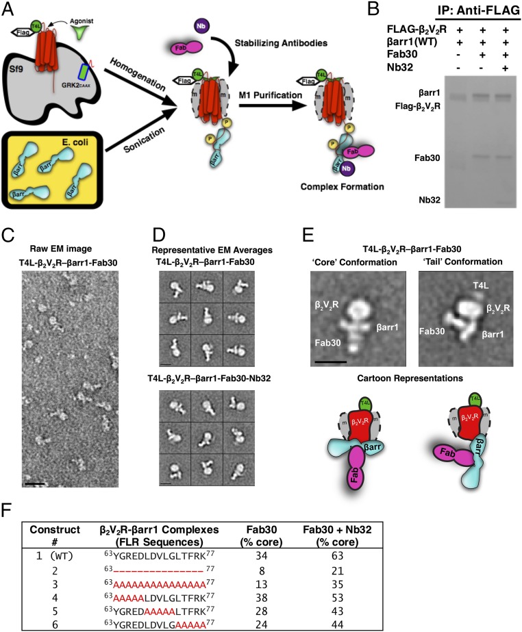
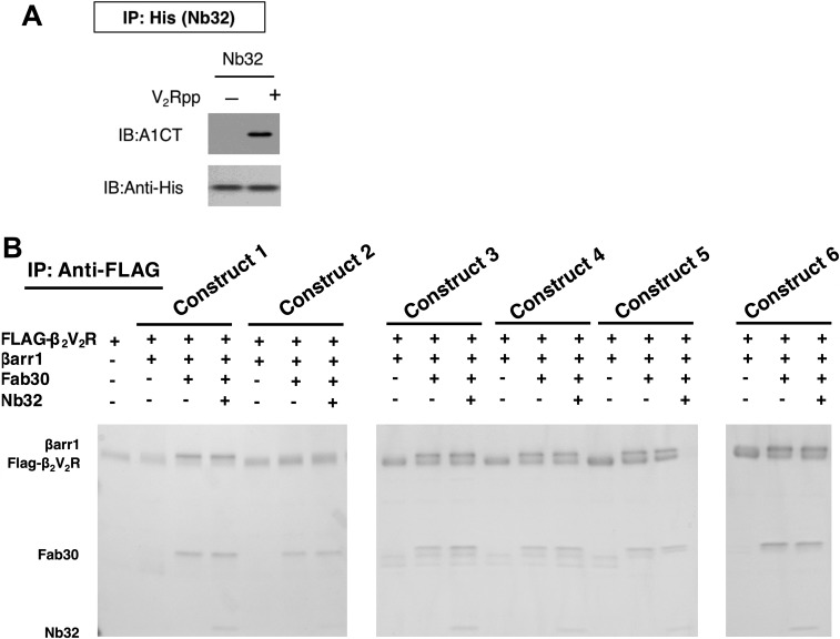
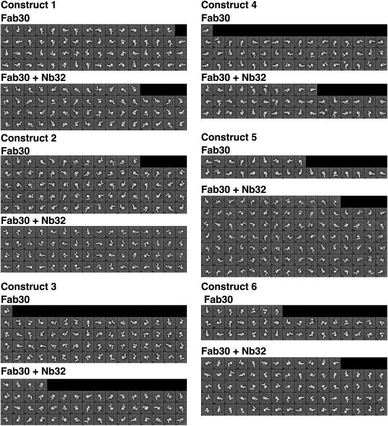
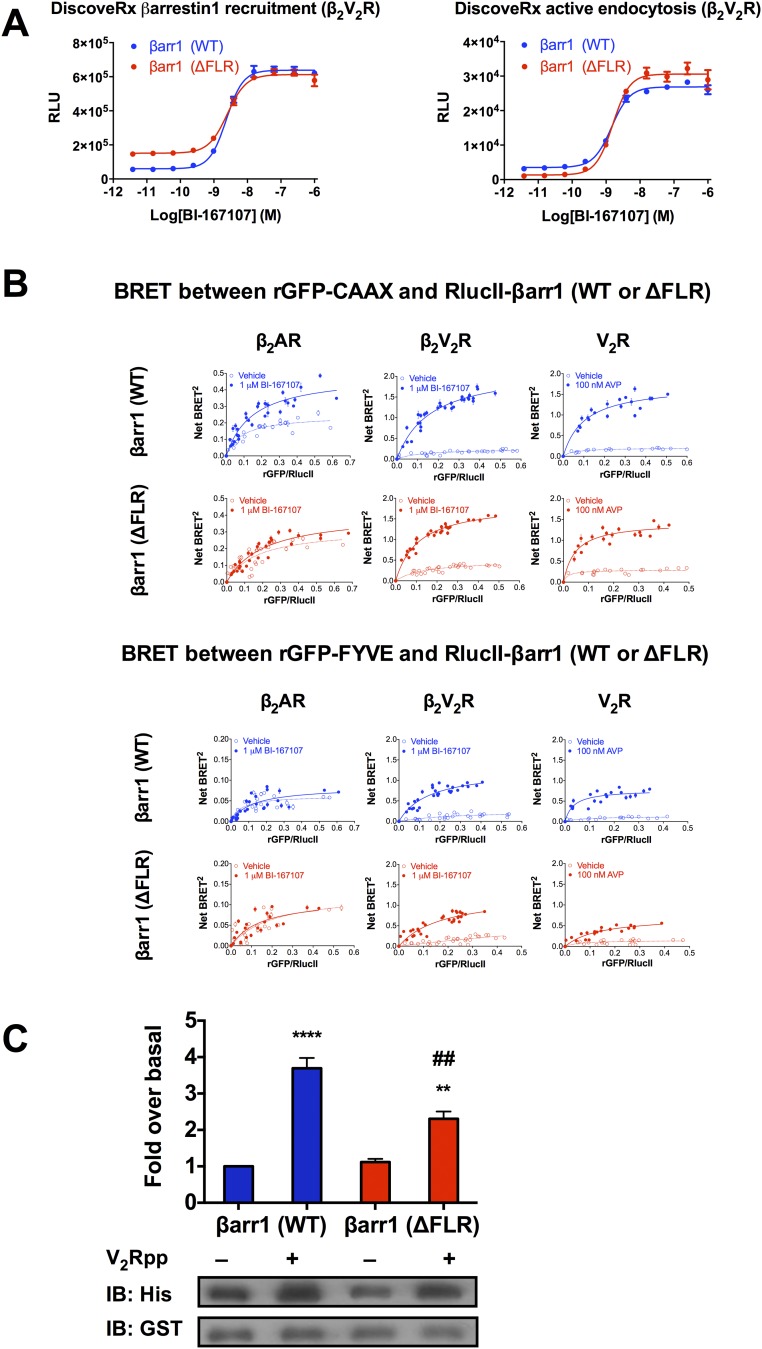
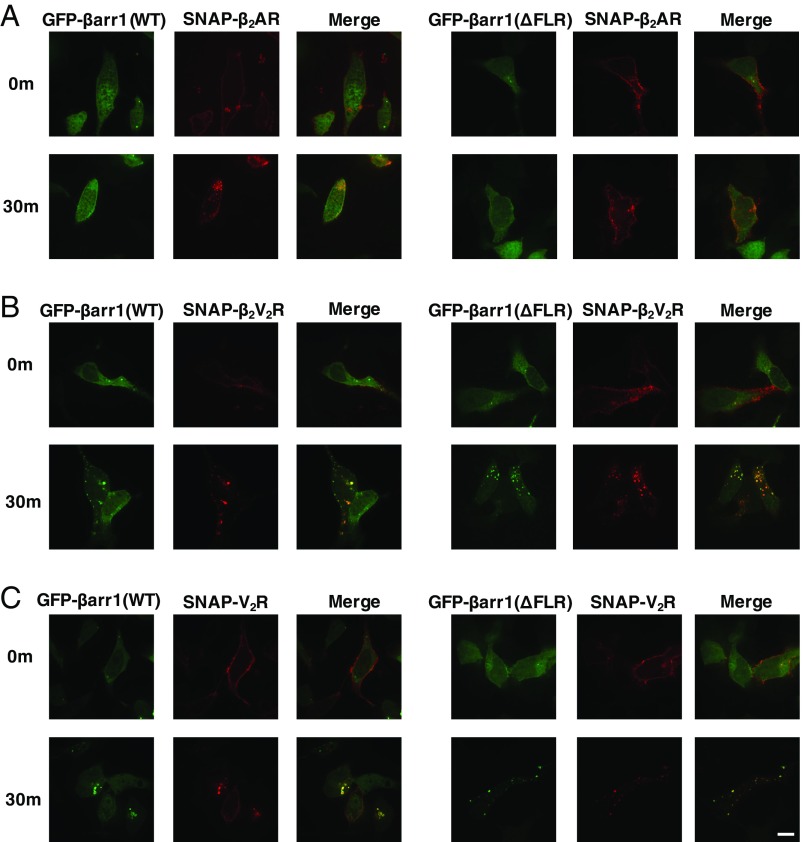
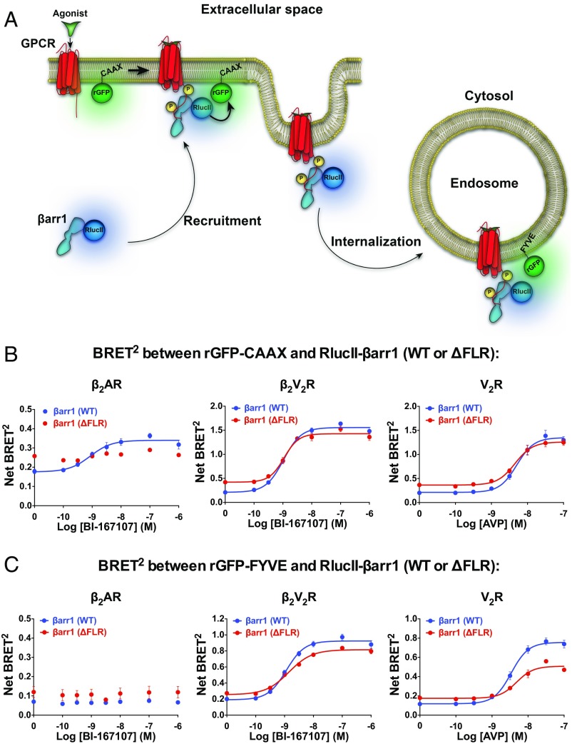
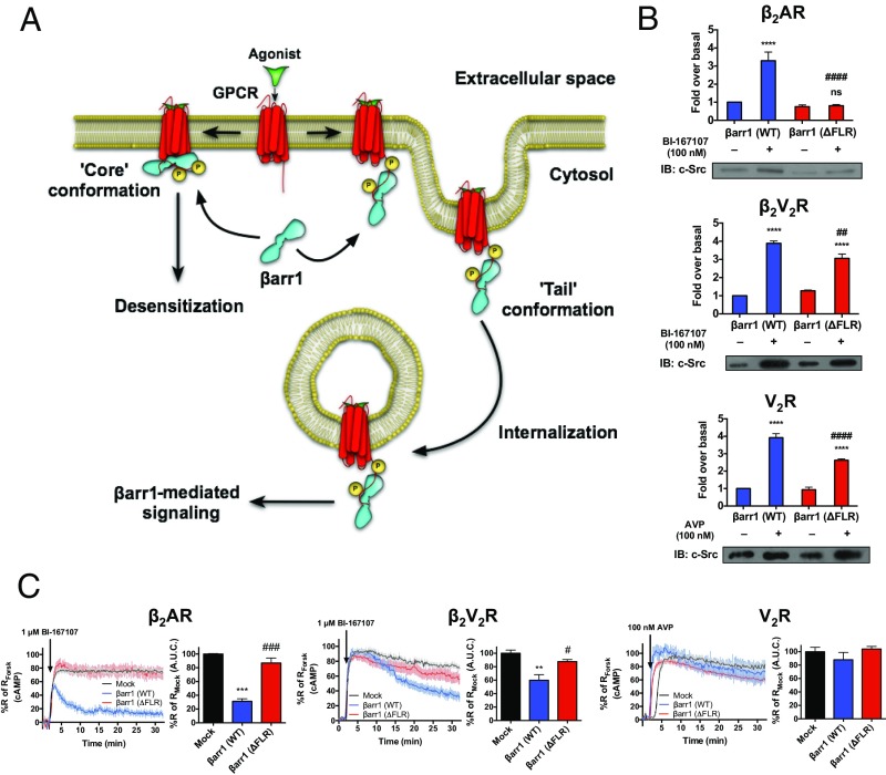
β-Arrestins (βarrs) interact with G protein-coupled receptors (GPCRs) to desensitize G protein signaling, to initiate signaling on their own, and to mediate receptor endocytosis. Prior structural studies have revealed two unique conformations of GPCR-βarr complexes: the "tail" conformation, with βarr primarily coupled to the phosphorylated GPCR C-terminal tail, and the "core" conformation, where, in addition to the phosphorylated C-terminal tail, βarr is further engaged with the receptor transmembrane core. However, the relationship of these distinct conformations to the various functions of βarrs is unknown. Here, we created a mutant form of βarr lacking the "finger-loop" region, which is unable to form the core conformation but retains the ability to form the tail conformation. We find that the tail conformation preserves the ability to mediate receptor internalization and βarr signaling but not desensitization of G protein signaling. Thus, the two GPCR-βarr conformations can carry out distinct functions.

Keywords: GPCR; arrestin; desensitization; endocytosis; signaling.


Conflict of interest statement

Conflict of interest statement: R.J.L. is a cofounder and shareholder of Trevena.

Figures

Fig. 1.
Fig. 1.
T4 lysozyme (T4L)β2V2R–βarr1 complexes formed and analyzed via EM with a newly developed functional purification method. (A) Schematic representation of the purification method to generate (T4L)β2V2R–βarr1 complexes. (B) Coomassie gel showing WT βarr1 interaction with (T4L)β2V2R in the absence or presence of conformation-stabilizing antibodies (Fab30, Nb32). IP, immunoprecipitation. (C) Representative negative-stain raw EM image of (T4L)β2V2R–βarr1–Fab30 complexes. (D) Class averages of the (T4L)β2V2R–βarr1–Fab30 complexes (Top) and (T4L)β2V2R–βarr1–Fab30–Nb32 complexes (Bottom) from negative-stain EM classification analysis. (E) Representative class averages (with cartoon representations) of the (T4L)β2V2R–βarr1–Fab30 complex in the tail and core conformations. (Scale bars: C, 20 nm; D and E, 10 nm.) (F) Summarized results of the different βarr1 FLR constructs tested for their ability to form the (T4L)β2V2R–βarr1–Fab30 core conformation in the presence or absence of Nb32. Note that the tail conformation encompasses all those (T4L)β2V2R–βarr1 complexes that are not in the core conformation.
Fig. S1.
Fig. S1.
(A) Immunoblot (IB) verifying the ability of nanobody (Nb32) to interact with the active V2Rpp-bound βarr1 in vitro. IP, immunoprecipitation. (B) Coomassie gel showing different βarr1 constructs and their interaction with (T4L)β2V2R in the absence or presence of conformation-stabilizing antibodies (Fab30, Nb32).
Fig. S2.
Fig. S2.
Class averages of the different preparations of (T4L)β2V2R–βarr1–Fab30 complexes and (T4L)β2V2R–βarr1–Fab30–Nb32 complexes, using different βarr1 constructs, from negative-stain EM analysis.
Fig. S3.
Fig. S3.
(A) Concentration–response curves of BI-167107–stimulated βarr1 (WT or ΔFLR) recruitment to the β2V2R and βarr1 (WT or ΔFLR)-mediated β2V2R internalization. RLU, relative luminescence units. (B) Interaction between βarr1 (WT or ΔFLR) and either rGFP-CAAX (Top, plasma membrane marker) or rGFP-FYVE (Bottom, early endosomal marker) upon agonist stimulation of β2AR, β2V2R, or V2R. BRET titration curves were obtained using a constant amount of rGFP-CAAX and with increasing amounts of RlucII-βarr1 (WT or ΔFLR). BRET titration curves were obtained using a constant amount of rGFP-FYVE and with increasing amounts of RlucII-βarr1 (WT or ΔFLR). BRET was measured 35 min following addition of agonist or vehicle. To stimulate the GPCRs, 1 μM BI-167107 was applied for the SNAP-β2AR and SNAP-β2V2R, and 100 nM AVP was applied for the SNAP-V2R. Data are expressed as net BRET absolute values, represent the mean ± SE, and are pooled from six experiments. (C) Two hundred nanomolar 6×His-βarr1 (WT or ΔFLR) was incubated with equal concentrations of GST-Src-3D and with either control buffer or V2Rpp. The complexes were pulled down using Glutathione Sepharose beads, and the amount of 6×His-βarr1 (WT or ΔFLR) bound to GST-Src-3D was determined by immunoblotting using an anti-His antibody. Data represent the mean ± SE of three experiments. One-way ANOVA was performed to determine statistical differences between basal and V2Rpp-stimulated states (**P < 0.01, ****P < 0.0001) or V2Rpp-stimulated states of βarr1 (WT) and βarr1 (ΔFLR) (##P < 0.01).
Fig. 2.
Fig. 2.
Cellular localization of SNAP-β2AR (A), SNAP-β2V2R (B), or SNAP-V2R (C), prelabeled with SNAP-surface 649 fluorescent substrate (red) and GFP-βarr1 (WT or ΔFLR) (green), visualized by confocal microscopy. Cellular localization of fluorescently tagged proteins is shown before agonist addition (0 min) or 30 min after agonist stimulation. To stimulate the GPCRs, 1 μM BI-167107 was applied for the SNAP-β2AR and SNAP-β2V2R, and 100 nM arginine vasopressin was applied for the SNAP-V2R (100× objective, n = 3 independent experiments, n = 20–50 cells per experiment). (Scale bar: 10 μm.)
Fig. 3.
Fig. 3.
Interaction between βarr1 (WT or ΔFLR) and either rGFP-CAAX (plasma membrane marker) or rGFP-FYVE (early endosomal marker) upon agonist stimulation of β2AR, β2V2R, or V2R. (A) Schematic representation of the experimental design used to monitor agonist-promoted BRET between RlucII-βarr1 (WT or ΔFLR) and rGFP-CAAX or rGFP-FYVE. (B) BRET concentration–response experiments assessing the agonist-stimulated RlucII-βarr1 (WT or ΔFLR) recruitment to plasma membrane-located rGFP-CAAX. Upon agonist addition, a difference in BRET was detected between βarr1 (WT) and β2AR (P = 0.0022), but not between βarr1 (ΔFLR) and β2AR (P = 0.4306). Agonist-mediated changes in net BRET between βarr1 (WT) and βarr1 (ΔFLR) were detected for both the β2AR (P = 0.0015) and β2V2R (P < 0.0001), but not for V2R (P = 0.0820). (C) BRET concentration–response experiments assessing the agonist-stimulated RlucII-βarr1 (WT or ΔFLR) localization to early endosomal-located rGFP-FYVE. Upon agonist addition, no BRET difference was detected between either βarr1 (WT) or βarr1 (ΔFLR) and β2AR (P = 0.4188 or P = 0.9016, respectively). Agonist-mediated changes in net BRET between βarr1 (WT) and βarr1 (ΔFLR) were detected for β2V2R (P = 0.0034) and V2R (P = 0.0014), but not for β2AR (P = 0.9057). In all experiments, BRET was measured 30 min following addition of agonist or vehicle. To stimulate the GPCRs, 1 μM BI-167107 was applied for the β2AR and β2V2R, and 100 nM arginine vasopressin (AVP) was applied for the V2R. Data are expressed as net BRET absolute values, represent the mean ± SE, are pooled from four to six experiments, and are analyzed using either a paired t test (two conditions) or one-way ANOVA with Tukey’s multiple comparisons post hoc test (three or more conditions).
Fig. 4.
Fig. 4.
Functional outcomes of different GPCR–βarr1 complex conformations. (A) Schematic representation of the functional outcomes mediated by GPCR–βarr1 complex tail conformation and GPCR–βarr1 complex core conformation. (B) βarr1-mediated scaffolding of c-Src upon activation of β2AR, β2V2R, or V2R. HEK293 DKO cells were transfected with plasmids for β2AR, β2V2R, or V2R, c-Src, and HA-βarr1 (WT or ΔFLR). Serum-starved cells were stimulated with or without agonist BI-167107 (1 μM) or AVP (100 nM) for 10 min and then cross-linked using dithiobis(succinimidyl propionate); finally, anti-HA beads were used to pull down βarr1 (WT or ΔFLR). The amount of total c-Src bound to HA-βarr1 (WT or ΔFLR) was determined by immunoblotting (IB). Data represent the mean ± SE of four to five experiments. One-way ANOVA was performed to determine statistical differences between basal and agonist-stimulated states (****P < 0.0001), or agonist-stimulated states in βarr1 (WT)- and βarr1 (ΔFLR)-transfected cells (##P < 0.01, ####P < 0.0001). (C) βarr1-mediated desensitization of Gs-promoted cAMP generation by the β2AR, β2V2R, or V2R. Real-time cAMP measurements, using ICUE2-expressing HEK293 cells, in response to agonist stimulation of β2AR, β2V2R, and V2R are shown. For the β2AR and β2V2R, 1 μM BI-167107 was used to stimulate cells. For V2R, 100 nM AVP was used to stimulate cells. For each GPCR, control plasmid (Mock, black), βarr1 (WT) (blue), or βarr1 (ΔFLR) (red) was transfected. Surface expression of each GPCR was matched within each βarr1 transfection condition. Data represent the mean ± SE of three to four experiments and n ≥ 44 cells. Area under the curve (A.U.C.) from 2 min after agonist stimulation to the end of the experiment was used to calculate desensitization of the cAMP response for each GPCR, and one-way ANOVA was performed to determine statistical differences relative to Mock (**P < 0.01, ***P < 0.001) and βarr1 (WT) (#P < 0.05, ###P < 0.001) responses. Forsk, forskolin.

References

    1. Lefkowitz RJ. The superfamily of heptahelical receptors. Nat Cell Biol. 2000;2(7):E133–E136. - PubMed
    1. Lefkowitz RJ. A brief history of G-protein coupled receptors (Nobel Lecture) Angew Chem Int Ed Engl. 2013;52(25):6366–6378. - PubMed
    1. Gilman AG. G proteins: Transducers of receptor-generated signals. Annu Rev Biochem. 1987;56:615–649. - PubMed
    1. Moore CAC, Milano SK, Benovic JL. Regulation of receptor trafficking by GRKs and arrestins. Annu Rev Physiol. 2007;69:451–482. - PubMed
    1. Goodman OB, Jr, et al. Beta-arrestin acts as a clathrin adaptor in endocytosis of the beta2-adrenergic receptor. Nature. 1996;383(6599):447–450. - PubMed

Publication types

MeSH terms